Acute rheumatic fever remains a serious problem for public health which is common in developing countries. Rheumatic valvar heart disease, an important sequel to rheumatic fever, remains the most common acquired cardiac disease worldwide.1, 2 One of the most characteristic disturbances of conduction in acute rheumatic fever is first-degree heart block. Prolongation of the PR interval relative to the heart rate is a nonspecific finding, present in more than one-third of the patients. The PR interval usually returns to normal after the disease becomes inactive, and this can occur with or without carditis. Low-voltage QRS complexes, and changes in the ST segments, may be found in the presence of pericarditis and pericardial effusion.3 A measurement that may be useful in this context, and one that has been identified as a marker of electrical myocardial instability, is the variation in the duration of the QT interval between the leads, so-called QT dispersion. Such dispersion reflects variations in the repolarization in different regions of the myocardium. These result from reentrant mechanisms due to the existence of areas of slow conduction.4, 5 The dispersion has been measured in electrocardiograms obtained in various clinical populations, ranging from healthy volunteers6, 7 to patients with the long QT syndrome,8 survivors of acute myocardial infarction,9 and patients with hypertrophic10 and dilated cardiomyopathies,11 as well as many other well defined groups.4, 12, 13 The extent of QT dispersion in patients with acute rheumatic fever, however, has not yet, to the best of our knowledge, been elucidated. In this study, therefore, we have analyzed prospectively the variations of the QT dispersion in the surface electrocardiogram of children with acute rheumatic fever.
Methods
Patients and Groups
We studied 88 consecutive patients presenting with their first acute episode of rheumatic fever to the cardiac unit of the Siyami Ersek Training and Research Hospital over a period of 24-months. Diagnosis of acute rheumatic fever in these patients was based on the revised Jones' criterions.14–17 A routine blood count, erythrocyte sedimentation rate, level of C-reactive protein, titers of antistreptolysin-O, chest radiograph, and electrocardiogram were obtained in all patients.
The age of the patients ranged from 5 to 14 years. There were 53 male and 35 female patients. Patients with acute rheumatic fever were divided into two groups based on the absence or presence of carditis. Because the population included subjects with and without carditis, we also enrolled a group of control subjects. The control group consisted of 36 healthy children, free of any disease, and matched for age with both groups.
Of the patients, 48 presented with acute rheumatic fever and evidence of carditis, with 38 presenting with typical migratory polyarthritis, and the remaining 10 patients having at least two minor manifestations of rheumatic fever, along with evidence of recent streptococcal infection as manifested by elevated titers of antistreptolysin-O. Carditis, evidenced by the presence of murmurs suggestive of valvar regurgitation, constituted a major manifestation in all these patients. Isolated mitral regurgitation was present in 39, combined mitral and aortic regurgitation in 6, and isolated aortic regurgitation in 3 patients. In addition, congestive heart failure was present in 8 patients.
Acute rheumatic fever without evidence of carditis occurred in 40 patients. All presented with typical migratory polyarthritis, and each had at least two minor manifestations of rheumatic fever, along with evidence of recent streptococcal infection as manifested by elevated titers of antistreptolysin-O.
Patients with rheumatic carditis and congestive heart failure were treated with steroids, along with dietary salt restriction, digoxin, diuretics, and inhibitors of angiotensin converting enzyme, whereas patients with carditis but without congestive heart failure were treated with aspirin. All patients without carditis were treated with aspirin. Injectable benzathine penicillin G for secondary prophylaxis was initiated or regulated at the time of the index attack in all patients.18
Repeat echocardiographic examinations were routinely scheduled in all patients at 3 months after the index attack to study the evolution of valvar lesions. All the repeat echocardiographic examinations were performed by the same operator, with the same ultrasound machine, and with similar gain settings.
Colour flow imaging demonstrated that valvar regurgitation greater than two or more grades was present in 21, and mild valvar regurgitation was present in 27 patients at the time of index attack. None of these patients had evidence of any significant valvar regurgitation at the end of 3 months.
Echocardiographic examination
All patients underwent echocardiographic examination within 24 to 48 hours of establishment of the diagnosis of acute rheumatic fever and before starting anti-inflammatory treatment. All echocardiographic studies were interpreted by the same observer with a random sequencing of studies. The observer was blinded to all clinical information. All ultrasound examinations were performed with a commercially available echocardiographic machine, specifically a Vivid 3 System form Vingmed–General Electric, equipped with 5- and 7-Megahertz transducers. A standardized cross-sectional and Doppler echocardiographic examination was performed with multiple orthogonal parasternal, apical, and subcostal views with the patient in the left lateral decubitus position. Valvar regurgitation was diagnosed when colour Doppler flow mapping demonstrated reversed flow away from the valve when the valve was closed. Signals of less than 100 milliseconds detected at the time of valvar closure were not regarded as true regurgitation. To differentiate abnormal from physiological regurgitation,19 the high-velocity turbulent jet had to extend by more than 1 centimetre beyond the paravalvar region, and had to be confirmed by colour-guided pulsed Doppler spectral analysis.20
On-line computerized planimetry of the maximal regurgitant jet areas was performed, and the mean of three cardiac cycles was taken. The severity of mitral and aortic regurgitation was graded on the basis of the maximal distance of the regurgitant jet from the valvar orifice, using the criterions established by Helmcke et al.21 and Perry et al.,22 respectively.
12-lead surface electrocardiogram
Electrocardiogram tracings were blindly analyzed in all patients and their controls by 2 independent investigators, initially and at 3 months after the index attack. We calculated heart rate, PR interval, QT dispersion, and QTc dispersion, in 4 successive complexes for each lead. The QT interval was measured starting from the onset of the QRS complex until the end of the T wave, which is the return of the T wave to the baseline. When the U wave was present, the QT interval ended at the middle point between the T and U waves, which was obtained at the intersection of a line taken tangentially to the repolarization line with the isoelectric line.4 Correction of the QT interval was obtained using Bazett's formula.23 QT dispersion, defined as the difference between maximum and minimum QT, was calculated based on the QT intervals obtained in the 12 leads. The same was done for the QTc dispersion, which was corrected using the RR interval.
Statistical analysis
The results obtained were statistically analyzed, and the mean values of each variable were compared in the initial and follow-up examination, using a paired t-test, and a value for p of less than 0.05 was considered significant.
Results
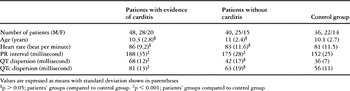
None of 88 patients with acute rheumatic fever died, and none of them underwent cardiac surgery during the episode of acute rheumatic fever. Table 1 shows the baseline characteristics of the subjects for whom QT dispersion was obtained.
Table 1. Baseline demographics and electrocardiographic variations of study groups and matched control group.

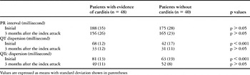
Patients with evidence of carditis had a greater QT and QTc dispersion than their controls, but there was no difference in QT and QTc dispersion between patients without evidence of carditis and the controls. A significant increase in the PR interval was found in both patients with or without evidence of carditis compared to the controls. QTc dispersion calculated in all patients also showed the same correlation with QT dispersion (Table 1). There was also a significant difference in QT and QTc dispersion between patients with evidence of carditis and without carditis, albeit that there was no difference in PR interval between patients with and without carditis (Table 2).
Table 2. Comparison of patient groups.

At follow-up examination, a clear reduction on the PR interval, QT dispersion, and QTc dispersion was the main finding, reflecting an electrophysiological improvement. There was no statistically difference in PR interval, QT dispersion, QTc dispersion between the three groups 3 months after the index attack (Table 2).
Figure 1 shows the receiver operating characteristic curve for the link between QT and QTc dispersion values and acute rheumatic carditis. A QT dispersion of greater than 55 milliseconds had an 85% sensitivity and a 70% specificity in predicting acute rheumatic carditis. A QTc dispersion of greater than 78 milliseconds had a 75% sensitivity and a 75% specificity in predicting acute rheumatic carditis.

Figure 1. Receiver operating curve plot of all QT and QTc dispersion measurements in respect to predict acute rheumatic carditis. Area under the ROC curve equal to 0.87; standard error equal to 0.040; 95% confidence interval equal to 0.793 to 0.950 and area under the ROC curve equal to 0.78; standard error equal to 0.052; 95% confidence interval equal to 0.679 to 0.882, respectively.
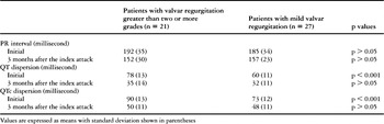
According to the cardiac involvement, QT dispersion and QTc dispersion were observed to be significantly increased in patients with valvar regurgitation greater than two or more grades when compared to patients with mild regurgitation. But there was no difference in PR interval between these two subgroups of patients with evidence of carditis (Table 3).
Table 3. Comparison of two subgroups of patients with evidence of carditis.

Sensitivity and specificity of measurements of QT and QTc dispersion in predicting severe valvar lesion were also estimated using receiver operating characteristic curves (Fig. 2). A QT dispersion of greater than 65 milliseconds had 81% sensitivity and 85% specificity in predicting severe valvar lesions, whereas a value of greater than 85 milliseconds had 76% sensitivity and 86% specificity.

Figure 2. Receiver operating curve plot of all QT and QTc dispersion measurements carried out to predict severe valvar lesion in acute rheumatic carditis. Area under the ROC curve equal to 0.84; standard error equal to 0.064; 95% confidence interval equal to 0.712 to 0.964 and area under the ROC curve equal to 0.85; standard error equal to 0.058; 95% confidence interval equal to 0.733 to 0.960, respectively.
Discussion
Although Sanyal et al.24 reported that the extension of the PR interval may prove to be a marker of myocardial involvement in patients with acute rheumatic fever; we found that such prolongation is a nonspecific finding. The PR interval in patients with acute rheumatic fever was significantly prolonged when compared to normal controls, but did not show any significant difference according to the cardiac involvement.
There is evidence that increased QT dispersion on the electrocardiogram represents regional differences in myocardial recovery of excitability.25 As far we are aware, however, there are few published limits of normal dispersion in children.26, 27 The largest study to date showed normal ranges from 10 to 44 milliseconds. A value of 50 milliseconds provides a highly specific estimate of the normal dispersion in adults and children.26 In our study, QT dispersion was observed to be significantly increased in patients with acute rheumatic fever when compared with the normal controls. QTc dispersion calculated in all patients also showed a significant increased, indicating that the variation in the QT dispersion does not depend on the heart rate. Using a cutoff point of 55 milliseconds, we found a higher sensitivity in predicting relevant rheumatic carditis as compared to use of the corrected value. It is noteworthy that the specificity in predicting relevant rheumatic carditis was slightly lower using the cutoff point of 55 milliseconds compared to the value of 78 milliseconds for the corrected measurements. We also found a significant difference according to presence of absence of cardiac involvement in those with acute rheumatic fever. A value of greater than 65 milliseconds has a greater sensitivity and specificity for detecting the severity of valvar lesions in rheumatic carditis. This indicates that ventricular inhomogeneity may increase in patients with acute rheumatic fever and evidence of carditis. Changes in the collagen interstitial matrix of the left ventricle may be associated with reduced action potential amplitude and membrane potential, shortened action potential duration, or electrical quiescence.28 Either of these features could result in an increased QT dispersion if the changes in different parts of the ventricle are nonhomogeneous.
QT dispersion in patients with significant valvar regurgitation at the time of the index attack, or on short-term follow-up, was significantly increased when compared with the patient with mild valvar regurgitation, or regurgitation that had disappeared at follow-up examination. The PR interval, however, failed to reveal any significant difference according to the cardiac state. The increased QT dispersion, therefore, is associated with the severity of valvar lesion rather than lengthening of the PR interval.
We observed reductions in the high values of the PR interval, QT and QTc dispersion concomitant with clinical evolution of patients with acute rheumatic fever at follow-up examination. This improvement would seem to be mediated with conventional anti-inflammatory treatments, like other temporary disturbances of conduction known to occur in patients with acute rheumatic fever.29
This study has limitations of that we included patients with congestive heart failure. QT dispersion is known to be heterogeneously increased with clinical evidence of heart failure and left ventricular systolic dysfunction.30, 31 But in our study, the majority of patients with rheumatic carditis have normal left ventricular systolic function, and because of the small number of patients with heart failure, it was not possible to perform a comparative analysis between groups with and without failure. These observations concur with recent reports32, 33 showing that congestive heart failure was less common in patients suffering their first attack of rheumatic carditis.
We submit, therefore, that assessment of the regular and corrected values for QT dispersion in patients with acute rheumatic fever provides an inexpensive and useful method of predicting carditis. Such increase in QT dispersion may be a more important parameter than lengthening of the PR interval because it may reflect cardiac involvement in patients with acute rheumatic fever.