Introduction
Brain imaging studies have generally shown that patients with schizophrenia have smaller global and regional brain volumes compared to typically developing individuals (Harrison, Reference Harrison1999; Shenton et al. Reference Shenton, Dickey, Frumin and McCarley2001; Jayakumar et al. Reference Jayakumar, Venkatasubramanian, Keshavan, Srinivas and Gangadhar2006; Chua et al. Reference Chua, Cheung, Cheung, Tsang, Chen, Wong, Cheung, Yip, Tai, Suckling and McAlonan2007). Smaller cortical size has been demonstrated in the prefrontal, entorhinal and anterior cingulate cortices (Harrison, Reference Harrison1999; Tamminga et al. Reference Tamminga, Vogel, Gao, Lahti and Holcomb2000). By contrast, enlargement of subcortical regions after antipsychotic treatment has also been demonstrated (Chakos et al. Reference Chakos, Lieberman, Bilder, Borenstein, Lerner, Bogerts, Wu, Kinon and Ashtari1994). Never-medicated patients have smaller caudate nuclei, putamen and nucleus accumbens (Gur et al. Reference Gur, Cowell, Turetsky, Gallacher, Cannon, Bilker and Gur1998; Corson et al. Reference Corson, Nopoulos, Andreasen, Heckel and Arndt1999; Dazzan et al. Reference Dazzan, Morgan, Orr, Hutchinson, Chitnis, Suckling, Fearon, Salvo, McGuire, Mallett, Jones, Leff and Murray2004; Girgis et al. Reference Girgis, Diwadkar, Nutche, Sweeney, Keshavan and Hardan2006; Chua et al. Reference Chua, Cheung, Cheung, Tsang, Chen, Wong, Cheung, Yip, Tai, Suckling and McAlonan2007), the size of which increases following months of typical antipsychotic treatment (Gur et al. Reference Gur, Cowell, Turetsky, Gallacher, Cannon, Bilker and Gur1998; Corson et al. Reference Corson, Nopoulos, Andreasen, Heckel and Arndt1999) and may be partly explained by increased striatal blood flow (Corson et al. Reference Corson, O'Leary, Miller and Andreasen2002). The phenomenon is regarded as evidence of antipsychotic-induced hypertrophy (Gur et al. Reference Gur, Cowell, Turetsky, Gallacher, Cannon, Bilker and Gur1998).
This raises the question of whether treatment-related changes hold clues to the pathophysiology. Antipsychotic drugs markedly reduce symptoms within the first 3 weeks of treatment (Johnstone et al. Reference Johnstone, Crow, Frith, Carney and Price1978), even as early as week 2, with 68% of the improvement at 1 year achieved within the first month (Leucht et al. Reference Leucht, Busch, Hamann, Kissling and Kane2005). Aberrant development of prefrontal–striatal circuitry during adolescence is thought to be related to the pathophysiology of schizophrenia (Laruelle, Reference Laruelle2000). According to the dopamine hypothesis of schizophrenia, an excess of dopamine in this brain system is associated with psychosis (Carlsson, 1978) and antipsychotic drugs that block dopamine receptors are able to oppose this, a process referred to as ‘endogenous dopamine sensitization’ (Laruelle, Reference Laruelle2000).
Forty years after antipsychotic medication revolutionized the therapeutic armory against schizophrenia, the reason why there is a time lag before antipsychotic drug treatment takes effect (Johnstone et al. Reference Johnstone, Crow, Frith, Carney and Price1978) remains obscure. It may be partly explained by the time taken for an interaction between dopamine receptors and antipsychotics to alter gene expression, leading to neurogenesis and synaptic changes (Konradi & Heckers, Reference Konradi and Heckers2001). It has been suggested that the clinical lag period coincides with the time course of an underlying neural response to treatment (Leucht et al. Reference Leucht, Busch, Hamann, Kissling and Kane2005).
Neuroplastic modulation of the caudate nucleus, part of the dopamine-rich striatum, is supported by a volumetric increase after 3 (Massana et al. Reference Massana, Salgado-Pineda, Junque, Perez, Baeza, Pons, Massana, Navarro, Blanch, Morer, Mercader and Bernardo2005) to 18 months of antipsychotic treatment (Chakos et al. Reference Chakos, Lieberman, Bilder, Borenstein, Lerner, Bogerts, Wu, Kinon and Ashtari1994). However, after shorter periods of treatment, this effect may be less marked. For example, increases in grey matter were reported in the superior temporal gyrus and middle temporal gyrus, but not the basal ganglia, after 15 antipsychotic-naive patients underwent 6 weeks of treatment with the atypical antipsychotic risperidone (Girgis et al. Reference Girgis, Diwadkar, Nutche, Sweeney, Keshavan and Hardan2006). Another study of 13 patients suffering a relapse of schizophrenia found no effects on the basal ganglia, despite a diffuse volumetric increase throughout the cortex after 4 weeks of treatment with second-generation atypical antipsychotics, but not with haloperidol (Garver et al. Reference Garver, Holcomb and Christensen2005). Conceivably, the caudate nuclear volume increase was not demonstrated because of weak study power. An alternative explanation is that the effect depends upon the duration of exposure to antipsychotics.
To address this issue, we conducted a pilot study of 22 patients newly diagnosed with schizophrenia scanned for a median period of 3 weeks after starting antipsychotic medication for the first time and 26 control patients also newly diagnosed with schizophrenic but never medicated. A voxel-by-voxel whole-brain grey-matter volumetric comparison (voxel-based morphometry, VBM) of the two patient groups enabled the early effects of antipsychotic treatment in first-episode schizophrenia to be evaluated. In addition, a region-of-interest (ROI) measurement of the caudate nuclei was undertaken. An important aspect of this study was its naturalistic design for medication selection, in that the choice of antipsychotic did not conform to a fixed protocol but instead was an entirely clinician-led decision.
Method
Subjects
All patients presenting to the hospital with psychosis were screened for eligibility to join the study. Inclusion criteria were: age 18–50 years; no previous history of any antipsychotic medication; and first experience of psychotic symptoms (i.e. hallucinations and/or delusions and/or thought disorder, with decline in daily functioning) according to DSM-IV (APA, 1994) confirmed by two independent specialists in psychiatry. Duration of psychosis was assessed using the Interview for the Retrospective Assessment of Schizophrenia (IRAOS; Hafner et al. Reference Hafner, Riecher-Rossler, Hambrecht, Maurer, Meissner, Schmidtke, Fatkenheuer, Loffler and van der Heiden1992) and has been described in detail elsewhere (Chen et al. Reference Chen, Dunn, Miao, Yeung, Wong, Chan, Chen, Chung and Tang2005). Exclusion criteria were: any history of neurological problems; loss of consciousness; persistent headaches; head trauma; electroconvulsive therapy; psychostimulant use; and special school attendance. The study received the approval of the Institutional Review Board of the hospital concerned. All subjects were Chinese. Patients were screened within the first day of presenting to hospital and all gave full informed written consent to participate according to the Declaration of Helsinki. These patients were part of a larger brain imaging project of first-episode psychosis patients to evaluate brain morphology compared to typically developing matched controls (results available in Chua et al. Reference Chua, Cheung, Cheung, Tsang, Chen, Wong, Cheung, Yip, Tai, Suckling and McAlonan2007). The Positive and Negative Syndrome Scale (PANSS; Kay et al. Reference Kay, Fiszbein and Opler1987) was used to assess patients' psychopathology when they presented to hospital. The antipsychotic-treated group received a magnetic resonance imaging (MRI) brain scan 3 weeks after antipsychotic treatment had started. The antipsychotic-naive group had an MRI brain scan just before antipsychotic treatment was initiated.
MRI data acquisition
Patients were scanned on a GE Signa 1.5 T system (General Electric, Milwaukee, WI, USA) in Queen Mary Hospital, Hong Kong. The scan lasted for 20 minutes. A consultant radiologist (K.S.T.), blind to diagnosis, reviewed each MRI scan for any gross anomaly. Scans aligned to the AC–PC line were acquired across the whole brain as follows. PD/T2 sequence: dual-echo fast spin–echo data sets with TR 5–6 s, anterior–posterior direction 0.86 mm, right–left direction 1.15 mm, slice direction 3 mm, matrix size 256×192, contiguous, phase inversion time 10 ms, two echoes with TE=20/80 ms, number of excitations=1, echo train length=8. PD/T2 scans were used for computerized VBM analysis. T1 sequence: fast SPGR 3D oblique scans with 128 slices, contiguous, 1.2 mm thick, Nex=1, TR 11.4 s, TE 4.2 ms, flip angle 15°, FOV 24×24, matrix 25×256. T1 scans were used for manual (ROI) tracing.
Preprocessing and analysis
Group differences in grey matter were mapped using Brain Activation and Morphological Mapping (BAMM) software version 2.5 (www.bmu.psychiatry.cam.ac.uk/BAMM/index.html) on a SPARC workstation (Sun Microsystems Europe Inc., Surrey, UK) as described previously (Sigmundsson et al. Reference Sigmundsson, Suckling, Maier, Williams, Bullmore, Greenwood, Fukuda, Ron and Toone2001; McAlonan et al. Reference McAlonan, Daly, Kumari, Critchley, van Amelsvoort, Suckling, Simmons, Sigmundsson, Greenwood, Russell, Schmitz, Happe, Howlin and Murphy2002, Reference McAlonan, Cheung, Cheung, Suckling, Lam, Tai, Yip, Murphy and Chua2005; Chua et al. Reference Chua, Cheung, Cheung, Tsang, Chen, Wong, Cheung, Yip, Tai, Suckling and McAlonan2007). In brief, PD/T2 images were processed to remove extracerebral tissues (Suckling et al. Reference Suckling, Brammer, Lingford-Hughes and Bullmore1999a) and then segmented into grey and white matter, cerebrospinal fluid (CSF), dura and other non-cerebral tissues, which were subsequently ignored (Suckling et al. Reference Suckling, Sigmundsson, Greenwood and Bullmore1999b). The segmented images were mapped into the standard space of Talairach and Tournoux (Talairach & Tournoux, Reference Talairach and Tournoux1988) by affine transformation of each proton density image to a group-specific template (Suckling et al. Reference Suckling, Sigmundsson, Greenwood and Bullmore1999b; McAlonan et al. Reference McAlonan, Cheung, Cheung, Suckling, Lam, Tai, Yip, Murphy and Chua2005; Chua et al. Reference Chua, Cheung, Cheung, Tsang, Chen, Wong, Cheung, Yip, Tai, Suckling and McAlonan2007), applying the derived mappings to the tissue maps and followed by smoothing with a 4.4 mm Gaussian kernel. The effect of diagnostic group was estimated at each voxel by regression of a general linear model to the estimates of grey and white matter occupancy separately. Maps of the corresponding standardized coefficient were subject to an inference procedure in which the significance of three-dimensional cluster statistics was assessed using non-parametric (permutation) methods (Bullmore et al. Reference Bullmore, Suckling, Overmeyer, Rabe-Hesketh, Taylor and Brammer1999). The statistical thresholds were corrected for multiple comparisons by setting the p value such that <1 false positive cluster was expected in each map under the null hypothesis (Suckling & Bullmore, Reference Suckling and Bullmore2004).
ROI analysis
Caudate and lateral ventricular volumes were evaluated using the T1 dataset as described previously (Murphy et al. Reference Murphy, DeCarli, Schapiro, Rapoport and Horwitz1992; Chua et al. Reference Chua, Cheung, Cheung, Tsang, Chen, Wong, Cheung, Yip, Tai, Suckling and McAlonan2007). One independent operator who was blind to the groups aligned the axial scans along the AC–PC line using MEASURE software (Barta et al. Reference Barta, Dhingra, Royall and Schwartz1997). Left and right caudate nuclei were traced according to standard anatomical boundaries. Volumes were calculated by multiplying the summed pixel cross-sectional areas by slice thickness. As all measurements were performed blind by a single operator, only the intra-class correlation coefficient (ICC) was calculated.
Results
Demographic and clinical comparison of groups
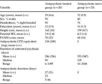
In total, 50 first-episode psychotic patients were recruited for the study; of these, two patients declined the MRI scan. Of the 48 patients who underwent MRI brain scanning, 22 were antipsychotic medicated and 26 antipsychotic naive. However, the scans for three patients had movement artefacts and were not suitable for further processing. Thus, the final numbers for image analysis were 20 antipsychotic-treated and 25 antipsychotic-naive patients. Two independent clinicians gave the diagnosis of schizophrenia to 16 (80%) of the antipsychotic-treated and 21 (84%) of the antipsychotic-naive patients; for the remaining four patients in each group, diagnoses of acute and transient psychosis or schizophreniform psychosis were given. The diagnoses were also confirmed by the responsible clinician upon case-note review, 6 months after presentation. Antipsychotic medication prescribed followed a naturalistic approach, that is it was entirely clinician led according to local practice. The treated group received either typical (haloperidol in 13 patients, trifluoperazine in one patient, and sulpiride in one patient) or atypical medication (amisulpride in five patients). Both groups were balanced for age, sex, handedness, ethnicity, paternal socio-economic status (SES) and height (see Table 1).
Characteristics of all patients with psychosis (n=45)

SES, Socio-economic status; PANSS, Positive and Negative Syndrome Scale; CPZ, chlorpromazine; s.d., standard deviation.
Brain morphological differences
Grey-matter volume differences
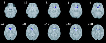
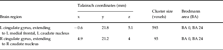
In the antipsychotic-treated group, compared to the antipsychotic-naive patient group, significant clusters of volume excess in cerebral grey matter were detected after 3–4 weeks of antipsychotic treatment, involving bilateral regions of the caudate, putamen, anterior cingulate and medial prefrontal cortex (see Fig. 1 and Table 2). When the analysis was restricted to subjects with schizophrenia, the results did not change significantly.
Grey-matter differences in the antipsychotic-naive (n=25) and antipsychotic-treated groups (n=20). The blue colour represents regions where grey-matter volumes were significantly greater in the antipsychotic-treated group compared to the antipsychotic-naive group. The right side of the brain corresponds to the right side of the figure. The z coordinate for each axial slice in standard Talairach and Tournoux space is given in millimetres. Statistical threshold is p<0.002 corrected.

Grey-matter differences in the antipsychotic-naive (n=25) and antipsychotic-treated groups (n=20) (p<0.002 corrected)Footnote a

L, Left; R, right.
a A sample Talairach coordinate (x, y, z) is given for the approximate centre of each three-dimensional (3D) cluster but, as shown in Fig. 1, the 3D cluster is not confined to this area alone.
ROI measurements
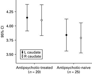
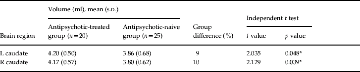
We used ROI planimetry to trace the caudate nuclei of the striatum. The ICC of the rater was 0.94 for both left and right caudate on 10 scans performed 1 month apart. In patients treated for a median of 3 weeks, the left caudate was larger by 9% (two-tailed p<0.048) and the right caudate by 10% (two-tailed p<0.039) (see Fig. 2 and Table 3). When the analysis was restricted to subjects with schizophrenia, the antipsychotic-treated group also had significantly larger left and right caudate nuclei by 10.7% on each side (independent t test, p<0.03, one-tailed).
Caudate volume differences between antipsychotic-treated and antipsychotic-naive groups.

Region-of-interest analysis of the caudate nuclei

L, Left; R, right; s.d., standard deviation.
* Two-tailed significance, p<0.005.
Discussion
We report that discrete brain morphological differences related to antipsychotic treatment occur as early as 3 weeks post-exposure. In particular, we have demonstrated for the first time that the caudate nucleus appears enlarged in patients who have undergone just a few weeks of antipsychotic treatment for first-episode schizophrenia. In this study of 48 patients, we found that, compared to the antipsychotic-naive patient group, the antipsychotic-treated group had significantly greater grey-matter volumes in the bilateral caudate nuclei and cingulate gyri, extending to the left medial frontal gyrus. ROI analysis confirmed larger striatal volumes in the treated group of the order of 9–10% in both caudate nuclei.
The present finding has an intriguing symmetry with our previous work showing that caudate volumes are as much as 11% smaller in untreated patients with first-episode schizophrenia when compared to matched healthy volunteers (Chua et al. Reference Chua, Cheung, Cheung, Tsang, Chen, Wong, Cheung, Yip, Tai, Suckling and McAlonan2007). This indicates that caudate volume might be ‘normalized’ or increased after antipsychotic drug administration in the very early weeks of treatment. We did not attempt to correlate caudate volumes with PANSS ratings because the latter were performed at first presentation, whereas the brain scan was conducted at two time-points (i.e. at presentation or 3 weeks after drug treatment). The groups did not differ significantly on baseline PANSS ratings, yet after 3 weeks of treatment caudate volumes were significantly greater than those never treated. It is now known that the antipsychotic effect may take place as early as week 2 (Leucht et al. Reference Leucht, Busch, Hamann, Kissling and Kane2005), and even manifest as early as some hours post-intramuscular antipsychotic, without being due to simple reduction in agitation (Agid et al. Reference Agid, Kapur, Warrington, Loebel and Siu2008). Conceivably, treatment effectiveness may depend on ‘rewiring’ neuronal circuitry (Konradi & Heckers, Reference Konradi and Heckers2001), and would partly account for the time needed for clinical efficacy. The mechanism by which antipsychotic drugs cause hypertrophy of the caudate nucleus (Chakos et al. Reference Chakos, Lieberman, Bilder, Borenstein, Lerner, Bogerts, Wu, Kinon and Ashtari1994; Shenton et al. Reference Shenton, Dickey, Frumin and McCarley2001) and possibly also of the putamen and thalamus (Gur et al. Reference Gur, Cowell, Turetsky, Gallacher, Cannon, Bilker and Gur1998) remains to be fully elucidated. However, it has already been shown that antipsychotic drugs can lead to genetic modulation of neuronal function in the striatum or prefrontal cortex (Weinberger & Lipska, Reference Weinberger and Lipska1995), the putative sites of action of typical and atypical antipsychotic drugs (Pilowsky, Reference Pilowsky2001). We were intrigued that our treated group had grey-matter excess in the medial frontal and cingulate gyri because these regions are implicated in behavioural monitoring and error detection (Rushworth et al. Reference Rushworth, Buckley, Behrens, Walton and Bannerman2007). Animal studies have shown that dopamine blockade with olanzapine (Wang et al. Reference Wang, Dunnavant, Jarman and Deutch2004) or haloperidol (Kippin et al. Reference Kippin, Kapur and van der Kooy2005) can stimulate new cell proliferation and therefore, taken together, the data support neuroplastic change in the fronto-limbic circuitry in the early phase of drug treatment.
The coincidence of structural abnormality and antipsychotic target in psychosis adds to the evidence that effective medication modifies those prefrontal-temporolimbic cortical networks most vulnerable to progressive tissue loss in the first 5 years after illness onset (Thompson et al. Reference Thompson, Vidal, Giedd, Gochman, Blumenthal, Nicolson, Toga and Rapoport2001). Although others have shown that antipsychotic medication may increase caudate size (Chakos et al. Reference Chakos, Lieberman, Bilder, Borenstein, Lerner, Bogerts, Wu, Kinon and Ashtari1994; Shenton et al. Reference Shenton, Dickey, Frumin and McCarley2001), placed with our previous findings of smaller caudate volume in first-episode schizophrenia (Chua et al. Reference Chua, Cheung, Cheung, Tsang, Chen, Wong, Cheung, Yip, Tai, Suckling and McAlonan2007), we suggest that this constitutes a ‘reversal’ of pathology as early as 3 weeks into treatment. We previously proposed that reduced caudate volumes might provide a useful biomarker for psychosis and it has been argued that the excess of dopamine in patients with schizophrenia has a toxic effect on neurons, potentially inducing an increase in oxidative stress (Keshavan, Reference Keshavan1999). The results of the present study lead us to extend our proposal and we speculate that normalized caudate volumes early in treatment might mirror the clinical response.
However, there is controversy in the field. Although patients with first-episode psychosis have been shown to have reduced caudate volumes in many recent MRI studies (Harrison, Reference Harrison1999; Shenton et al. Reference Shenton, Dickey, Frumin and McCarley2001; Jayakumar et al. Reference Jayakumar, Venkatasubramanian, Keshavan, Srinivas and Gangadhar2006; Chua et al. Reference Chua, Cheung, Cheung, Tsang, Chen, Wong, Cheung, Yip, Tai, Suckling and McAlonan2007), others have reported no changes in caudate volume (Crespo-Facorro et al. Reference Crespo-Facorro, Perez-Iglesias, Ramirez-Bonilla, Martinez-Garcia, Llorca and Vazquez-Barquero2006). The question of whether patients were already medicated, however briefly, on entry to each study is a salient point, as is the type of medication administered. Girgis et al. (Reference Girgis, Diwadkar, Nutche, Sweeney, Keshavan and Hardan2006) reported no increase in caudate nucleus volume despite increases in various cortical regions following risperidone treatment. In our study the medication patients received was entirely clinician led: 2/3 received typical antipsychotics and 1/3 were treated with atypical antipsychotics reflecting the changes in treatment guidelines over time. Differences in the effect of typical and atypical antipsychotic medication on neuroplasia have been observed previously and discussed (McClure et al. Reference McClure, Phillips, Jazayerli, Barnett, Coppola and Weinberger2006). In ongoing work, first-episode patients are usually treated with atypical antipsychotics, therefore we plan to repeat the present study in these patients.
A limitation of our study was the use of two separate patient groups. This design does not allow for the exclusion of a possible confound of inherent group differences. A longitudinal study of a single cohort of patients, each serving as their own control, would be the optimal to unequivocally demonstrate drug treatment effects on brain morphology and is currently in progress. The results presented here motivate further research into antipsychotic drug actions in patients with first-episode schizophrenia early in the course of treatment and sharpen a focus on the precise mechanisms by which medication may increase grey-matter volume and thereby reduce symptoms of psychosis.
In conclusion, our data indicate that greater striatal volumes in patients treated with antipsychotic drugs compared to never-treated patients are present after just 3 weeks. This extends our previous report of striatal deficits in untreated patients with first-episode psychosis (Chua et al. Reference Chua, Cheung, Cheung, Tsang, Chen, Wong, Cheung, Yip, Tai, Suckling and McAlonan2007). We propose that, in addition to acting as a useful disease marker (Chua et al. Reference Chua, Cheung, Cheung, Tsang, Chen, Wong, Cheung, Yip, Tai, Suckling and McAlonan2007), caudate nuclear volume may represent a useful indicator of early recovery in schizophrenia.
Acknowledgments
Dr S. E. Chua was supported by the University of Hong Kong Committee for Conference and Research Grants (CRCG). We thank the patients for their participation and staff of the Radiology Department, Queen Mary Hospital for MRI scanning.
Declaration of Interest
None.