INTRODUCTION
Schistosomiasis (caused by chronic infection with blood-dwelling flukes of the genus Schistosoma) and soil-transmitted helminthiasis (caused by infections with nematode worms Ascaris lumbricoides, Trichuris trichiura and hookworm) remain important public health issues, particularly in the tropics and subtropics (Hotez et al. Reference Hotez, Brindley, Bethony, King, Pearce and Jacobson2008; Murray et al. Reference Murray, Vos, Lozano, Naghavi, Flaxman and Michaud2012). More than 90% of the estimated 207 million individuals infected with Schistosoma reside in Africa (Steinmann et al. Reference Steinmann, Keiser, Bos, Tanner and Utzinger2006). Chronic infection with Schistosoma mansoni leads to morbidity and mortality from portal hypertension, while chronic Schistosoma haematobium infection is associated with genitourinary pathology which may result in hydronephrosis, renal failure and squamous cell bladder cancer (Ross et al. Reference Ross, Bartley, Sleigh, Olds, Li, Williams and McManus2002; Gryseels et al. Reference Gryseels, Polman, Clerinx and Kestens2006). Over a billion people are infected with soil-transmitted helminths, with school-aged children and marginalized communities in resource-constrained settings disproportionately affected. Chronic soil-transmitted helminth infection may result in anaemia, malnutrition and impaired physical and cognitive development (Bethony et al. Reference Bethony, Brooker, Albonico, Geiger, Loukas, Diemert and Hotez2006). In the developing world, infection with multiple species is the norm rather than the exception (Keiser et al. Reference Keiser, N'Goran, Traoré, Lohourignon, Singer, Lengeler, Tanner and Utzinger2002; Steinmann et al. Reference Steinmann, Utzinger, Du and Zhou2010).
The diagnosis of S. mansoni and soil-transmitted helminth infections traditionally and functionally relies on stool microscopy, with the Kato–Katz technique being widely used, although the method has important shortcomings (Katz et al. Reference Katz, Chaves and Pellegrino1972; Kongs et al. Reference Kongs, Marks, Verlé and Van der Stuyft2001; Booth et al. Reference Booth, Vounatsou, N'Goran, Tanner and Utzinger2003; Speich et al. Reference Speich, Knopp, Mohammed, Khamis, Rinaldi, Cringoli, Rollinson and Utzinger2010). Indeed, it is a relatively inexpensive and simple method of stool preparation, however multiple Kato–Katz thick smears are required to increase diagnostic sensitivity, particularly for low-intensity infections (de Vlas and Gryseels, Reference de Vlas and Gryseels1992; Knopp et al. Reference Knopp, Mgeni, Khamis, Steinmann, Stothard, Rollinson, Marti and Utzinger2008). Schistosoma haematobium is traditionally diagnosed by urine microscopy, using filtration or sedimentation techniques (Knopp et al. Reference Knopp, Becker, Ingram, Keiser and Utzinger2013). Indirect methods such as urine reagent strips assessing for microhaematuria have been widely used in endemic settings, albeit with variable estimates of sensitivity and specificity (Mott et al. Reference Mott, Dixon, Osei-Tutu and England1983; Robinson et al. Reference Robinson, Picon, Sturrock, Sabasio, Lado, Kolaczinski and Brooker2009; Kosinski et al. Reference Kosinski, Bosompem, Stadecker, Wagner, Plummer, Durant and Gute2011; Bogoch et al. Reference Bogoch, Andrews, Dadzie Ephraim and Utzinger2012).
Most individuals infected with Schistosoma and soil-transmitted helminths live in rural, resource-constrained settings where facilities to perform the aforementioned diagnostic tests are often non-existent. Additional barriers to care include the transport of patients or biological samples to functional diagnostic centres and a constant electricity supply for light microscopy. Hence, alternative diagnostic approaches are necessary to better care for affected individuals living in endemic settings. Portable light microscopy for S. mansoni diagnosis has been used with some success in a pilot study carried out in Uganda (Stothard et al. Reference Stothard, Kabatereine, Tukahebwa, Kazibwe, Mathieson, Webster and Fenwick2005). In addition, mobile phone microscopy was recently shown to have modest diagnostic yield for the diagnosis of soil-transmitted helminth infections in school-aged children in a proof-of-concept study on Pemba Island, Tanzania (Bogoch et al. Reference Bogoch, Andrews, Speich, Utzinger, Ame, Ali and Keiser2013). These diagnostic innovations have the benefit of being portable, inexpensive, easy to use, point-of-care tests that do not require a constant electricity supply.
The purpose of this study was to assess the utility of a novel commercial, portable light microscope and a simple mobile phone microscope for the diagnosis of S. mansoni, S. haematobium and soil-transmitted helminths. The diagnostic performance is compared with microscopy using standard Kato–Katz and urine filtration methods considered as reference-standard.
MATERIALS AND METHODS
Ethics statement
This study was integrated into a cross-sectional survey in Côte d'Ivoire, and was approved by the ethics committee of the Ministry of Health and Public Hygiene in Côte d'Ivoire (reference no. 7706/MSLS/CNER-dkn). Written informed consent was obtained from parents or legal guardians on behalf of children, and children assented orally. At the end of the study, anthelmintic treatment was offered to all participants regardless of their infection status (i.e. praziquantel, 40 mg kg−1 of body weight against schistosomiasis; albendazole, 400 mg against soil-transmitted helminthiasis).
Population census
The study was conducted among school-aged children residing in Azaguié Makouguié, a village of the district of Azaguié, south Côte d'Ivoire, where S. mansoni, S. haematobium and soil-transmitted helminths are co-endemic (Coulibaly et al. Reference Coulibaly, Fürst, Silué, Knopp, Hauri, Ouattara, Utzinger and N'Goran2012, Reference Coulibaly, N'Gbesso, N'Guessan, Winkler, Utzinger and N'Goran2013). A census was carried out in May 2013. All households were visited and demographic characteristics were collected from school-aged children, including name, age and sex.
Study design and procedures
Children were invited for multiple stool and urine sample collection over a 3-day period. Urine sample collection was done between 10:00 and 12:00 h, and fresh stool samples were collected from the early morning on the date of analysis in the laboratory from the cohort. Samples were transferred to a nearby laboratory in the town of Azaguié and processed the same day using standard protocols.
In brief, stool samples were subjected to the Kato–Katz technique (Katz et al. Reference Katz, Chaves and Pellegrino1972), preparing five 41·7 mg thick smears on microscope slides which were examined under a CX21LEDFS1 Olympus microscope (Olympus; Volketswil, Switzerland) by experienced laboratory technicians with 20× and 40× objective lenses. The number of S. mansoni, A. lumbricoides, hookworm and T. trichiura eggs were counted and recorded for each species separately. Urine samples were processed by a filtration technique for evaluation and quantification of S. haematobium eggs (Peters et al. Reference Peters, Mahmoud, Warren, Ouma and Siongok1976). Briefly, urine samples were shaken prior to extracting and pressing 10 mL through a meshed filter (20 μ m pores) measuring 13 mm in diameter (Sefar AG; Heiden, Switzerland). One drop of Lugol's iodine solution was placed over the filter prior to microscopic examination. For quality control, 10% of all Kato–Katz thick smears and urine filtrations were re-examined by a senior microscopist.
For the purpose of the current embedded study, we selected the first out of five Kato–Katz thick smears obtained from the third-day stool sample collection (n = 164) for further microscopic evaluation. Given the low prevalence of S. haematobium in the study village (approximately 5%), we selected all positive samples from each day of collection, resulting in a total of 14 positive slides. Then, randomly selected negative slides were added to total up to 60 slides from each day of collection, such that there were 180 slides overall. Our evaluation with experimental microscopic techniques occurred 10–12 days after initial sample collection and slide preparation.
Microscopic techniques
A mobile phone microscope was constructed and utilized as described previously (Smith et al. Reference Smith, Chu, Espenson, Rahimzadeh, Gryshuk, Molinaro, Dwyre, Lane, Matthews and Wachsmann-Hogiu2011; Bogoch et al. Reference Bogoch, Andrews, Speich, Utzinger, Ame, Ali and Keiser2013). Briefly, a 3 mm glass ball lens (Edmund Optics; Barrington, NJ, USA) was mounted over the camera lens of an iPhone 4S (Apple; Cupertino, CA, USA), with 1/16″ thick foam tape (AM Rubber; Canada). Slides were placed up against the mobile phone, such that the tape provided approximately a 1 mm buffer zone between the slide and the ball lens. Slides were illuminated from below with a hand-held incandescent flashlight powered by one AA battery. The iPhone was manually manoeuvred over the entire slide to systematically search for helminth eggs using the camera function and with the aid of auto focus and digital zoom functions.
Kato–Katz thick smears and filtered urine slides were also evaluated with the Newton Nm1-600 XY Portable Field Microscope (Newton Microscopes; Cambridge, UK). This is a commercially available, hand-held light microscope weighing 480 g with 10×, 40× and 60× objective lenses (Newton Microscopes). The objective lenses are modular such that the device can accommodate lenses with greater magnification as well. The same experienced microscopist evaluated all slides for the presence or absence of helminth eggs with the Newton portable microscope and mobile phone microscope. Infection intensity was not quantified. The microscopist was blinded to prior results from conventional microscopy.
Statistical analysis
Data were entered into a Microsoft Excel file (Microsoft; Redmond, WA, USA) and analysed with Stata version 10.1 (StataCorp.; College Station, TX, USA). The sensitivity, specificity, positive predictive value (PPV) and negative predictive value (NPV) were calculated for each helminth species using the iPhone microscope and the Newton microscope and compared with conventional light microscopy, with the latter serving as diagnostic reference standard. We then evaluated the diagnostic characteristics of the experimental microscopes for low, moderate and heavy infection intensity as per the cut-offs described by the World Health Organization (WHO, 2002), with moderate- and high-intensity infections grouped together. Two-sample tests of proportion were conducted to first compare the sensitivities and specificities of the experimental diagnostic techniques with reference-standard light microscopy and then compare the sensitivities between moderate/heavy infection intensities from light infections.
RESULTS
Stool examination
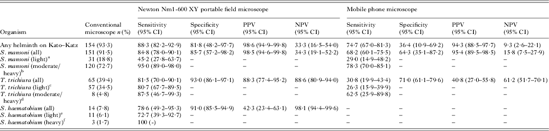
Data from the evaluation of 164 Kato–Katz thick smears with mobile phone microscopy and portable field microscopy vs standard microscopy are presented in Table 1. The observed prevalence of S. mansoni and T. trichiura in this cohort based on a single Kato–Katz thick smear examined under traditional light microscopy was 91·5% and 39·4%, respectively. No other helminths were visualized during the immediate evaluation with conventional light microscopy and subsequent evaluation with portable microscopic devices. Images can be viewed in Fig. 1. S. mansoni, S. haematobium and T. trichiura eggs can be easily visualized with conventional microscopy and the Newton portable microscope, but are more challenging to see with the mobile phone microscope.
Trichuris trichiura imaged by (A) conventional light microscopy, (B) Newton portable microscopy and (C) mobile phone microscopy; Schistosoma mansoni, imaged by (D) conventional light microscopy, (E) Newton portable microscopy and (F) mobile phone microscopy. Schistosoma haematobium imaged by (G) conventional light microscopy, (H) Newton portable microscopy and (I) mobile phone microscopy. All images were captured using the camera function of the iPhone 4S (Apple; Cupertino, CA, USA).

Operating characteristics of the mobile phone microscope and Newton portable hand-held microscope compared with conventional light microscopy for the diagnosis of S. mansoni, T. trichiura and S. haematobium (n = 164 Kato–Katz thick smears; n = 180 slides with filtered urine)
CI = confidence interval; PPV = positive predictive value; NPV = negative predictive value.
a Eggs per 1 g of stool (EPG): 1–99.
b EPG⩾100.
c EPG 1–999.
d EPG⩾1000.
e Eggs per 10 mL urine: 1–49.
f Eggs per 10 mL urine: ⩾50.
For S. mansoni and T. trichiura, the Newton portable microscope showed sensitivities of 84·8% and 81·5%, respectively, and specificities of 85·7% and 93·0%, respectively (Table 1). For S. mansoni and T. trichiura the mobile phone microscope showed sensitivities of 68·2% and 30·8%, respectively, and specificities of 64·3% and 71·0%, respectively. Compared with the iPhone microscope, the Newton portable microscope had greater sensitivity (P = 0·001) but not specificity (P = 0·383) for the diagnosis of S. mansoni. For T. trichiura infection, the Newton portable microscope had both greater sensitivity and specificity (both P values <0·001) compared with the iPhone.
For the evaluation of infection intensities, the iPhone demonstrated greater sensitivity for moderate/heavy infections compared with light infections of S. mansoni (P<0·001) and T. trichiura (P = 0·019). The Newton portable microscope's sensitivity fared better for moderate/heavy infection intensity compared with light infections with S. mansoni (P<0·001) and similarly with all infection intensities for T. trichiura infections (P = 0·323).
Urine examination
Overall, 8·3% of urine slides included in this comparison were positive for S. haematobium by light microscopy. The iPhone mobile microscope was used only in the evaluation of Kato–Katz thick smears, and was abandoned for urine filtration slides as it was unable to reliably detect eggs of S. haematobium during the pilot study, despite multiple attempts (see Discussion for details). Evaluation of samples with the Newton portable field microscope resulted in sensitivity and specificity of 78·6% and 91·0%, respectively, for S. haematobium diagnosis (Table 1). This device did not confirm any statistically significant differences in diagnostic sensitivity for light or moderate/heavy infection intensity (P = 0·154).
DISCUSSION
Scale-up of diagnostic coverage in rural areas of Africa will be necessary for a more accurate assessment of the impact of preventive chemotherapy campaigns against common parasitic worm infections and may help with direct patient care in under-resourced clinics. We demonstrate the utility of portable, hand-held microscopic techniques for the diagnosis of S. mansoni, S. haematobium and soil-transmitted helminths in a cohort of school-aged children from Côte d'Ivoire.
These diagnostic tools were evaluated in a village that has previously benefited from country-wide helminth control efforts targeting school-aged children as per WHO guidelines. Despite these efforts, there was still a very high prevalence of S. mansoni, with lower prevalences of T. trichiura and S. haematobium, while neither A. lumbricoides nor hookworm infection were found during the initial immediate evaluation with conventional light microscopy or subsequent portable microscopic devices. The prevalence of these infections would likely be higher if multiple slides were examined (Utzinger et al. Reference Utzinger, Booth, N'Goran, Müller, Tanner and Lengeler2001; Booth et al. Reference Booth, Vounatsou, N'Goran, Tanner and Utzinger2003; Knopp et al. Reference Knopp, Mgeni, Khamis, Steinmann, Stothard, Rollinson, Marti and Utzinger2008) (e.g. five slides were prepared from each stool sample in this study), however the purpose of this embedded study was to compare microscopic techniques on the same slide set. The Newton portable light microscope performed with sufficient diagnostic sensitivity and specificity for S. mansoni, S. haematobium and T. trichiura such that it could be considered a valuable diagnostic device in future clinical and epidemiological studies. The mobile phone microscope performed modestly well with moderate- to heavy-intensity infections of S. mansoni and T. trichiura, but did not have acceptable diagnostic accuracy with light infections. The specificity for both S. mansoni and T. trichiura was unacceptably low in the current configuration.
We found the Newton portable microscope to be an effective diagnostic tool. It is compact, weighs less than 500 g, and has an easy-to-use design for evaluating slides under objective lenses of differing magnification. The device requires three AAA batteries that can provide over 300 h of use. Images were easily captured by using a mobile phone with a camera function, and placing the lens of the camera directly up to the optical tube (Fig. 1B, E and H), however a specific mobile phone attachment designated for this purpose can be used as well. Also helpful was the ability to move slides to examine multiple fields, a feature not seen on previous portable microscopes used for field diagnoses of schistosomiasis (Stothard et al. Reference Stothard, Kabatereine, Tukahebwa, Kazibwe, Mathieson, Webster and Fenwick2005). This allows for the complete evaluation of a Kato–Katz thick smear or a urine filtration sample, and should decrease the number of false-negative results as eggs on the periphery of the slide will likely be visualized. Negative aspects of this device include its cost. The Newton microscope costs £580 (~US$950) in high-income countries and £250 (~US$410) in low- and middle-income countries at the time of writing. Fortunately, the prices of these microscopes and smartphones with a suitable camera function have declined and will do so in the future to make these technologies more affordable, perhaps helping the adoption and coverage of this technology in helminth-endemic settings. In contrast, quality light microscopes in laboratory settings as used in the current study are considerably more expensive (~US$1750 for the CX21SEDFS1 model of Olympus microscope employed here). In addition, the diagnostic sensitivity was low for light-intensity infections, which may pose problems for evaluating the affects of preventative chemotherapy campaigns. Another disadvantage is that slides must be placed with the specimen side facing downward. While we had no issues with downward facing slides in this study, problems could theoretically arise related to sample loss and associated hygienic complications, such as biological material contaminating the microscope, or even worse, contaminating subsequent slides placed on the microscope. Lastly, digitization of the images is an important component of portable diagnostic devices (Tuijn et al. Reference Tuijn, Hoefman, van Beijma, Oskam and Chevrollier2011) and the Newton portable microscope requires an additional piece of equipment such as a camera or a mobile phone with camera function to capture images, adding to the overall costs. We did not quantify egg counts on slides, and future studies evaluating this device should do so as this information is of critical importance from clinical, epidemiological and public health points of view. Additional studies should also evaluate the diagnostic capacity of the device when used by laboratory technicians and other medical personnel (e.g. nurses, primary care physicians) in routine practice, rather than expert microscopists.
In a previous study, mobile phone microscopy demonstrated an overall diagnostic sensitivity for T. trichiura of 54·4%, and sensitivities of 76·5% and 43·9% for moderate-to-heavy infections and light infections, respectively (Bogoch et al. Reference Bogoch, Andrews, Speich, Utzinger, Ame, Ali and Keiser2013). The sensitivities for T. trichiura are comparable in this study, albeit slightly lower, which may be related to a greater prevalence of lighter-intensity infections in the current study. As per previous studies with mobile phone microscopy, the diagnostic utility was greater with moderate-to-heavy infection intensities regardless of the helminth species. In addition, the larger eggs of S. mansoni may be easier to identify than T. trichiura, perhaps accounting for the relative greater sensitivity for this infection. The Newton portable microscope demonstrated no significant differences between light and moderate/heavy infections for T. trichiura or S. haematobium as the sensitivities were already quite high for light-intensity infections and because sample size was relatively small.
We had considerable difficulty obtaining clear images of S. haematobium with the mobile phone microscope during the pilot phase of this project such that data were not collected due to our inability to reliably capture images (e.g. Fig. 1I). Images appear to be ‘washed out’ perhaps from light refracting off the fine-meshed filter paper used to process urine. We used meshed filter paper with 20 μm pores (Sefar AG; Heiden, Switzerland), which are less expensive than the more widely used Millipore filters (Millipore Intertech; Bedford, MA) (Gyorkos et al. Reference Gyorkos, Ramsan, Foum and Khamis2001). We attempted different strategies of illuminating the specimen (e.g. varying intensities of light from different angles and using sunlight) but could not reliably capture detailed images. These issues did not arise with the Newton portable microscope. We also attempted to use a commercially available mobile phone microscope attachment (the American 3B Scientific iPhone Microscope; American 3B Scientific, Tucker, GA, USA) on both stool and urine samples during the pilot period, but found the device to be too cumbersome to reliably provide images of sufficient diagnostic quality at an appropriate magnification (3B Scientific).
The diagnostic yield of our mobile phone microscope in its current formulation is not sufficient for routine use in clinical or epidemiological settings, but perhaps can serve as a platform for future innovative devices. Several newer mobile phone technologies have shown promise for point-of-care diagnostic testing, including lens-free devices that provide a wide field of view for image analysis (Lee et al. Reference Lee, Yaglidere and Ozcan2011; Greenbaum et al. Reference Greenbaum, Sikora and Ozcan2012; Isikman et al. Reference Isikman, Greenbaum, Lee, Bishara, Mudanyali, Su and Ozcan2012; Linder et al. Reference Linder, Grote, Varjo, Linder, Lebbad, Lundin, Diwan, Hannuksela and Lundin2013). Such devices may not be far from routine use in clinical or epidemiological settings as they are relatively inexpensive and have decent operating characteristics, but will require rigorous validation outside of laboratory settings prior to scale-up. Similarly, commercially available mobile phone microscope attachments may also be useful for global health applications, although in our experience several of the devices currently on the market either do not have the appropriate magnification or are too cumbersome to use in routine practice. Digitization of samples, and tagging these with geographical coordinates may identify regions with a higher prevalence of infection, and may facilitate the rapid and real-time reporting of cross-sectional surveys in endemic regions. It may also play a role in medical education as images may be shared readily with experts or an automated device such that real-time feedback is provided to the microscopist (Tuijn et al. Reference Tuijn, Hoefman, van Beijma, Oskam and Chevrollier2011). Other low-cost diagnostic devices may be applicable for global health applications, such as the urine circulating cathodic antigen test for S. mansoni, a point-of-care test with excellent sensitivity (Coulibaly et al. Reference Coulibaly, Knopp, N'Guessan, Silué, Fürst, Lohourignon, Brou, N'Gbesso, Vounatsou, N'Goran and Utzinger2011; Colley et al. Reference Colley, Binder, Campbell, King, Tchuem Tchuenté, N'Goran, Erko, Karanja, Kabatereine, van Lieshout and Rathbun2013).
Portable diagnostic devices hold promise for enabling care in remote and resource-constrained settings. The Newton portable microscope demonstrated sufficient diagnostic yield of S. mansoni, S. haematobium and T. trichiura compared with standard light microscopy and should be studied further in different clinical and epidemiological settings. Our mobile phone microscope performed only modestly well at moderate-to-high infection intensities for S. mansoni and T. trichiura, however newer, low-cost technologies are in the pipeline for global health applications.
ACKNOWLEDGEMENTS
We thank the village authorities of Azaguié Makouguié and the participating children for providing stool and urine samples for detailed laboratory work-up. J. T. Coulibaly is grateful to the Swiss Tropical and Public Health Institute for a postdoctoral fellowship.
FINANCIAL SUPPORT
The research of B. Speich is financially supported by the Medicor Foundation, and that of J. Keiser, E. K. N'Goran and J. Utzinger by the Swiss National Science Foundation.