The surgical management of common arterial trunk has evolved over decades to a complete repair performed in the neonatal period with excellent outcomes. Beginning with the first successful series of infant repairs reported by Ebert et al,Reference Ebert, Turley, Stanger, Hoffman, Heymann and Rudolph 1 surgeons progressively pushed the operation to earlier stages, and now neonatal repair is considered the standard of care in most institutions.Reference Bove, Beekman and Snider 2 , Reference Hanley, Heinemann and Jonas 3 Despite the technical success in achieving a complete repair in infancy, significant truncal valve insufficiency remains an important problem for a subset of patients, and when present has been shown to contribute to mortality and morbidity.Reference Rajasinghe, McElhinney, Reddy, Mora and Hanley 4 , Reference Vohra, Whistance and Chia 5
Initial attempts to treat truncal insufficiency by means of aortic homograft replacement were met with disappointing results.Reference Elkins, Steinberg, Razook, Ward, Overholt and Thompson 6 Truncal valve replacement has been required in as many as 20% of patients with common arterial trunk,Reference Vohra, Whistance and Chia 5 and has been shown to be a significant risk factor for poorer survival.Reference Thompson, McElhinney, Reddy, Petrossian, Silverman and Hanley 7 Initial attempts to treat truncal valve insufficiency by means of aortic homograft replacement were met with disappointing results due to the early failure rate necessitating multiple root replacements and the difficulties of densely calcified adhesions between the aortic and pulmonary conduits. This prompted an interest in truncal valve repair techniques.Reference Imamura, Drummond-Webb, Sarris and Mee 8 – Reference Henaine, Azarnoush and Belli 10 Early results of truncal valve repair have been promising; however, there have been few studies evaluating long-term durability.Reference Tlaskal, Chaloupecky and Hucin 11 – Reference Kaza, Burch, Pinto, Minich, Tani and Hawkins 13 The purpose of this paper is to review our results with truncal valve repair over the past 20 years.
Patients and methods
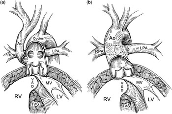
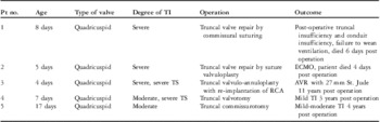
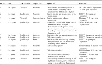
Institutional Review Board approval was obtained from Ann & Robert H. Lurie Children's Hospital of Chicago for retrospective chart review. Requirement for informed consent was waived. Medical records of all patients who underwent truncal valve repair between 1979 and 2011 were reviewed. Truncal anatomy was recorded on the basis of the simplified, aortic versus pulmonic dominant classification system of Anderson,Reference Russell, Jacobs and Anderson 14 described in Figure 1. In all, 15 of the 16 patients had aortic dominant anatomy, whereas one patient had pulmonic dominant anatomy. Of these, nine patients (56%) were female. Clinical characteristics are summarised in Tables 1 and 2.
The illustration shows the essential features of pulmonary versus aortic dominance as observed in our autopsied specimens with common arterial trunk. Panel ( a ) (pulmonary dominant) shows interruption of the aortic arch. Only in this setting, and in hearts with severe aortic coarctation, did we find origin of the pulmonary arteries from either side of the intrapericardial pulmonary trunk. Panel ( b ) (aortic dominant) shows the salient features of aortic dominance, with the pulmonary arteries arising separately but next to each other from the leftward and dorsal aspect of the common trunk. We also found pulmonary arteries arising more anteriorly, and then crossing as they extended towards the pulmonary hilums.Reference Russell, Jacobs and Anderson 14 Ao = aorta; IVS = interventricular septum; LPA = left pulmonary artery; LV = left ventricle; MV = mitral valve; RPA = right pulmonary artery; RV = right ventricle; VSD = ventricular septal defect.

Clinical characteristics of patients undergoing truncal valve repair/replacement at initial complete common arterial trunk.

AVR = atrioventricular replacement; ECMO = extracorporeal membrane oxygenation; RCA = right coronary artery; TI = truncal insufficiency; TS = truncal stenosis
Clinical characteristics of patients undergoing truncal valve repair/replacement at time of conduit replacement.

AI = aortic insufficiency; AVR = atrioventricular replacement; TI = truncal insufficiency
We evaluated our patients within two eras: one before the common practice of neonatal repair for common arterial trunk (1979–1989, n = 3) and the other after (1989–2011, n = 13). The median age of repair in the earlier era was 174 months, with significant variation owing to early palliative procedures such as pulmonary artery banding. The median age of repair for the later era was 7 days in those patients who had concomitant valve repair during their initial repair for common arterial trunk (n = 5, 39.5%). An additional eight patients underwent valve repair at the time of their conduit change at a median of 95 months. The type of repair procedure varied according to anatomic and physiologic considerations and included valvulo-annuloplasty (8/16, 50%), suture valvuloplasty (5/16, 31%), and valvotomy (2/16, 12.5%). Failed valvuloplasty occurred in one patient and prompted truncal valve replacement.
Charts were analysed for patient demographics including age, valve morphology, additional cardiac operations or malformations, valve repair techniques used, and intra-operative details. Death, conduit reintervention, valve reoperation, and valve replacement were designated end points.
Operative technique
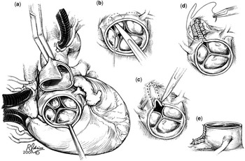
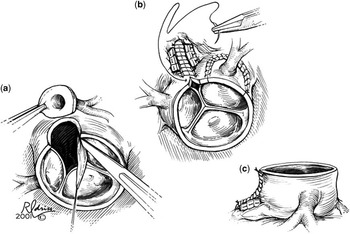
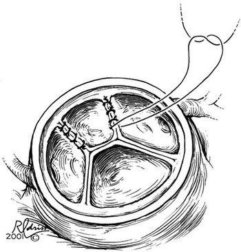
A total of 16 patients underwent truncal valve repair or replacement in association with primary repair or right ventricular to pulmonary artery conduit replacement for common arterial trunk. All procedures were performed with similar anaesthesia, perfusion circuits, myocardial preservation strategies, and operative team. Our techniques have been previously reported.Reference Mavroudis and Backer 9 , Reference Backer 15 In brief, we use aortobicaval cardiopulmonary bypass, systemic cooling to 28°C, left ventricular venting through the right superior pulmonary vein, and combinations of antegrade and retrograde cold blood cardioplegia. Figures 2–4 demonstrate the different reparative truncal valve techniques. Intra-operative transesophageal echocardiography was used on all patients to evaluate the repairs following separation from cardiopulmonary bypass.
This is a series of diagrams that show truncal valve repair by leaflet excision and annular remodeling. ( a ) The common arterial trunk is transected showing the incompetent, quadricuspid truncal valve. Usually, there is one leaflet that is grossly prolapsed. In this patient, the small prolapsed leaflet does not involve the coronary sinus of Valsalva. ( b ) The prolapsed leaflet is removed with care being taken to leave the neighbouring leaflets attached. ( c ) The sinus of Valsalva truncal wall is resected in preparation for the remodeling procedure. ( d ) Pledgeted sutures are used to tighten the annulus, thereby bringing the remaining leaflets together. The remnants of the resected leaflets are sutured together (not shown) to align the newly formed commissure. The rest of the truncal wall is approximated, thereby remodelling the truncal valve into a smaller, competent, and non-stenotic neoaortic valve. ( e ) Lateral view of the remodelled neoaortic valve.Reference Mavroudis and Backer 9

This series demonstrates truncal valve repair by leaflet excision and annular remodelling where the prolapsed leaflet involves the coronary orifice. ( a ) The coronary button has been harvested and the prolapsed leaflet excised. ( b ) The coronary button has been transferred to the adjacent sinus of Valsalva (a la arterial switch) and the annulovalvuloplasty is being sutured. ( c ) Finished lateral view.Reference Mavroudis and Backer 9

The suture valvuloplasty technique. This is performed by attaching the edges of the prolapsed leaflet to the adjacent leaflets using fine sutures, thereby creating a functional bicuspid semilunar valve.Reference Mavroudis and Backer 9

Echocardiography and follow-up data
Intra-operative data were obtained through transesophageal echocardiography, whereas follow-up data for all patients were obtained through routine transthoracic echocardiography. Follow-up data on truncal valve insufficiency were obtained from the medical record and by contact with the patient's cardiologist. At least yearly echocardiograms were available for all patients, with a median follow-up time of 5.5 years.
Results
There were two operative deaths (12.5%) from low cardiac output syndrome. A summary of the results of truncal valve repair is noted in Tables 1 and 2. There was one patient who had an acute repair failure that required truncal valve replacement.
The median age for primary repair of common arterial trunk in the first era was 7 months (n = 3), whereas for the second era the median age was 2 weeks. The median age of valvar intervention in the era before 1989 was 14.5 years (n = 3), whereas the median age of valvar intervention in the present era is 7 days in those who have valve repair concomitantly with common arterial trunk repair (n = 5), and is 8 years for those who have valve repair at the time of conduit change (n = 8). The median age of first conduit change is 5.1 years (n = 10), whereas the median age of second conduit change is 14.5 years (n = 5). There were no deaths during conduit replacement. The mean cardiopulmonary bypass time for common arterial trunk repair with valvar intervention was 185.6 minutes and the mean cross-clamp time was 73.6 minutes. The mean length of hospital stay was 12 days. In all, 12 patients (75%) had quadricuspid valves and the mean degree of truncal insufficiency at the time of operation was 3+, whereas the mean degree of post-operative neo-aortic insufficiency was 1+.
In those patients for whom eventual truncal valve replacement was necessary (n = 2), the mean age was 16.4 years. This corresponded with 9 and 10 years, respectively, post-initial repair. The one patient in our series who underwent valvulo-annuloplasty is being followed up with 3+ aortic insufficiency 8 years after repair. The remaining 10 patients have 2+ or less truncal insufficiency, with a median follow-up time of 5.5 years.
Discussion
Early attempts at truncal valve replacement were associated with a high mortality and frequent reoperations; thus, the development of various techniques to repair these valves has become the favoured approach. This has led to improved short- and mid-term outcomes.Reference Imamura, Drummond-Webb, Sarris and Mee 8 , Reference Bahrami, Mitropoulos, Leong, Levi, Laks and Plunkett 12 Henaine et alReference Henaine, Azarnoush and Belli 10 showed that mild or moderate truncal valve insufficiency is relatively well tolerated in infancy, and that it tends to improve after complete repair of common arterial trunk without valvar intervention. In addition, concomitant truncal valve repair in neonates has been shown to confer markedly increased mortality.Reference Russell, Pasquali and Jacobs 16 For these reasons, many centres, including our own, favour delaying truncal valve repair if at all possible. Those patients who required valvar intervention at the time of initial repair of common arterial trunk had a higher mortality than those who were delayed; however, durable results were obtained in those for whom the procedure was successful.
Although our series is too small to permit meaningful statistical analysis, there does not appear to be any correlation between the type of valve repair performed and outcome. Our preferred method of repair is the valvulo-annuloplasty, first proposed by Imamura et al,Reference Imamura, Drummond-Webb, Sarris and Mee 8 and we have shown it to be a favourable technique in the short term.Reference Mavroudis and Backer 9 In this technique (Fig 2), the neoaortic valve is downsized and remodelled without having to place sutures in the valve leaflets. The resulting valve maintains its competence without undue stress on valvar suture lines, which are prone to disruption. Our contribution to this technique has been the coronary artery transfer in those cases where the target prolapsing leaflet contains a coronary artery origin from the associated sinus of Valsalva.Reference Mavroudis and Backer 9 We have previously reported short-term outcomes for this type of repair both with and without coronary artery transfer, and these additional data support this technique as a durable treatment for truncal insufficiency.
Valve morphology is a key determinant of patient selection. This technique is particularly applicable in a patient with a quadricuspid truncal valve, removing the most dysplastic leaflet and creating a trileaflet valve. In our series, 75% of the patients who underwent truncal valve repair had a quadricuspid valve. Trileaflet truncal valves can be repaired with the same technique to create a bicuspid valve; however, achieving a symmetrical valve with good coaptation is more technically challenging.
Published long-term outcomes after truncal valve repair are limited. Most reports are confined to small case series, making it difficult to assess outcomes with any statistical power. Our data demonstrate that truncal valve repair can be performed with an acceptably low mortality and that a durable outcome can be achieved. When recurrent aortic insufficiency does progress to the point of requiring valve replacement, it appears to do so over a long period of time allowing for sufficient somatic growth to occur such that an adult-sized prosthesis can be used. Given the recent development of transcatheter aortic valve-in-valve replacement options, the initial valve replacement size is an important consideration for later reinterventions.
Truncal valve repair is an effective operation with reasonable durability. It should be considered in appropriately selected patients with common arterial trunk and greater than moderate truncal valve insufficiency.