INTRODUCTION
Gastrointestinal (GI) nematodes typically cause local pathology (Tu et al. Reference Tu, Koski and Scott2007) and stimulate a local Th2 immune and inflammatory response (Gause et al. Reference Gause, Urban and Stadecker2003; Reynolds et al. Reference Reynolds, Filbey and Maizels2012) that creates conditions that lead to expulsion of adult worms from the intestine (Vallance and Collins, Reference Vallance and Collins1998; Shea-Donohue et al. Reference Shea-Donohue, Sullivan, Finkelman, Madden, Morris, Goldhill, Piñeiro-Carrero and Urban2001; Madden et al. Reference Madden, Whitman, Sullivan, Gause, Urban, Katona, Finkelman and Shea-Donohue2002; Cliffe et al. Reference Cliffe, Humphreys, Lane, Potten, Booth and Grencis2005). Furthermore, even for those nematodes such as Heligmosomoides bakeri whose development occurs entirely within the intestine, there is increasing evidence of extra-intestinal organ pathology. Features of chronic inflammation are evident in the spleen and mesenteric lymph nodes of H. bakeri-infected mice (Cywińska et al. Reference Cywińska, Czumińska and Schollenberger2004), eosinophils and apoptotic Th2 cells have been shown to accumulate in the liver and lungs during H. bakeri infection (Mohrs et al. Reference Mohrs, Harris, Lund and Mohrs2005), and fetal linear growth is impaired and maternal bone re-modelling occurs in H. bakeri-infected pregnant dams (Odiere et al. Reference Odiere, Koski, Weiler and Scott2010a ). Such extra-intestinal pathologies have been largely ignored even though they may have important consequences especially when superimposed on conditions such as protein deficiency and lactation.
Protein deficiency has impacts on both visceral and lymphoid organs. For example, size and cellularity of the liver are reduced (Camargo et al. Reference Camargo, Angeleli, Burini and Campana1978) and cell populations in the spleen are decreased (Manhart et al. Reference Manhart, Vierlinger, Bergmeister, Boltz-Nitulescu, Spittler and Roth2000) under protein deprivation. Protein deficiency (3%) has also been shown to impair several of the Th2 responses against H. bakeri, including IgE antibody production, eosinophilia and mast cell hyperplasia (Boulay et al. Reference Boulay, Scott, Conly, Stevenson and Koski1998; Ing et al. Reference Ing, Su, Scott and Koski2000; Tu et al. Reference Tu, Koski and Scott2007), as well as circulating and lymphoid-derived Th2 cytokine responses not only in the intestine (Ing et al. Reference Ing, Su, Scott and Koski2000), where reduced IL-4 and IL-13 inhibit physiological changes in the gut that normally mediate parasite expulsion (Vallance and Collins, Reference Vallance and Collins1998; Madden et al. Reference Madden, Whitman, Sullivan, Gause, Urban, Katona, Finkelman and Shea-Donohue2002), but also systemically (Ing et al. Reference Ing, Su, Scott and Koski2000). Interestingly, however, not all infection-induced responses are blunted by protein deficiency. For example, H. bakeri-induced myeloperoxidase activity in the duodenum (Tu et al. Reference Tu, Koski and Scott2007) and elevation of pro-inflammatory cytokines IL-1β and IL-6 (Odiere et al. Reference Odiere, Scott, Weiler and Koski2010b ) are not affected by protein deficiency. Nevertheless, the overall consequence is prolonged parasite survival that presumably increases both local intestinal and extra-intestinal pathology.
During lactation, protein requirements are elevated due to increased demands associated with milk production (NRC, 1995). In addition, resources are allocated to growth and development and away from processes such as maternal immunity (Coop and Kyriazakis, Reference Coop and Kyriazakis1999), an explanation used to account for both the higher intestinal worm burdens in pregnant compared with non-pregnant mice (Odiere et al. Reference Odiere, Koski, Weiler and Scott2010a ) and the periparturient increase in susceptibility to GI nematodes and GI fecal egg counts typically observed in livestock (Kyriazakis and Houdijk, Reference Kyriazakis and Houdijk2006). This relaxation of immunity has a suspected nutritional basis because increased protein intake is able to halt the onset of periparturient immunosuppression during lactation at times of protein scarcity (Jones et al. Reference Jones, Houdijk, Sakkas, Bruce, Mitchell, Knox and Kyriazakis2011). Furthermore, reduction of reproductive investment by reducing litter size has been shown to restore immune function and reduce nematode numbers in lactating rats (Jones et al. Reference Jones, Sakkas, Houdijk, Knox and Kyriazakis2012). Thus, the partitioning of resources during lactation is likely to increase both intestinal and extra-intestinal histopathologies resulting from H. bakeri infection, and even more so when combined with protein deficiency.
Using biobanked tissues from an experiment designed to examine the impact of protein deficiency and H. bakeri infection during late pregnancy and lactation on neonatal growth and immune development (Odiere et al. Reference Odiere, Scott, Weiler and Koski2010b , Reference Odiere, Scott, Leroux, Dzierszinski and Koski2013), we investigated the potential interactive effects of the two factors on architecture of maternal lymphoid and visceral organs. We hypothesized that pathologies resulting from infection in the lactating dam would extend beyond the intestine to visceral organs, including the liver, pancreas and kidney, and be reflected by relevant serum analytes, but that protein deficiency would dampen these effects. We also hypothesized that changes in relative spleen size in response to infection and protein deficiency would be due to changes in the lymphoid compartment of the spleen, and that protein deficiency but not infection would reduce the lymphoid compartment of the thymus.
MATERIALS AND METHODS
Experimental design, mice and parasites
Tissues used in this study were obtained from an experiment described previously (Odiere et al. Reference Odiere, Scott, Weiler and Koski2010b ). Briefly, a 2×2 factorial design combined two levels of dietary protein (PS and PD) with two infection treatments (sham and infected). Forty-eight 8–9-week-old primiparous CD1 dams were received on day 13 of pregnancy (Charles River Breeding Laboratories, Quebec, Canada) and assigned to the experimental groups (n = 12 per experimental group). Individually housed dams were given ad libitum access to either a protein sufficient (PS; 24% protein, TD.90017) or a protein deficient (PD; 6% protein, TD.90016) commercial purified pelleted diet (Harlan Teklad, Madison, WI, USA) beginning on day 14 of pregnancy through to day 20 of lactation. Detailed diet composition has been described by Odiere et al. (Reference Odiere, Scott, Weiler and Koski2010b ). Diets were formulated to ensure that a 30% reduction in food intake would not generate nutrient deficiencies other than protein, based on the National Research Council requirements for lactating mice (NRC, 1995) and that the ratios of amino acids to total protein were the same for both diets. A trickle infection protocol was used whereby naïve mice were infected by oral gavage with 100±3 H. bakeri (previously named Heligmosomoides polygyrus; see Cable et al. Reference Cable, Harris, Lewis and Behnke2006) stage 3 larvae (L3) four times (day 14 of pregnancy and days 2, 9 and 16 post-partum). The trickle infection protocol mimics natural environmental conditions where animals are repeatedly exposed to incoming larvae and harbour both larval and adult stages simultaneously (Stear et al. Reference Stear, Bishop, Doligalska, Duncan, Holmes, Irvine, McCririe, McKellar, Sinski and Murray1995). In contrast to a single primary infection, this trickle protocol over a period of 4 weeks generates sustained stimulation of immune and inflammatory responses as each dose of L3 moves to the serosal musculature as highly antigenic stage 4 larvae (L4) and then emerges into the intestinal lumen (Scott, Reference Scott1990; Brailsford and Behnke, Reference Brailsford and Behnke1992; Behnke et al. Reference Behnke, Lowe, Clifford and Wakelin2003; Odiere et al. Reference Odiere, Scott, Weiler and Koski2010b , Reference Odiere, Scott, Leroux, Dzierszinski and Koski2013). Sham-infected mice were given distilled water. Food intake was recorded every third day beginning on day 15 of pregnancy and used to estimate energy and nutrient intakes. Litter size at parturition was noted but not standardized.
Dams were weighed and necropsied on day 20 of lactation. Maternal serum samples were collected and stored at −20 °C for blood biochemistry. Small intestine, thymus, spleen, heart, both lungs, liver, pancreas and both kidneys were excised and weighed. Thymus, spleen, liver, pancreas and kidneys were washed with phosphate-buffered saline (PBS), pH 7·4 and fixed in 10% neutral buffered formalin. Prior to recording the number of adult H. bakeri in the lumen of the small intestine (see Odiere et al. Reference Odiere, Scott, Weiler and Koski2010b ), a 2 cm section from the proximal end of the duodenum was separated and rinsed in PBS. One 1 cm section was fixed in 4% paraformaldehyde overnight then transferred to 70% ethanol and the other 1 cm section was fixed in Carnoy's fixative for 2–3 h and then transferred to 80% ethanol.
All procedures were approved by the McGill University Animal Care Committee according to the guidelines of the Canadian Council on Animal Care (CCAC, 1993).
Histology
Visceral and lymphoid organs were trimmed according to the three part guidelines of the Registry of Industrial Toxicology Animal-data and the North American Control Animal Database (Ruehl-Fehlert et al. Reference Ruehl-Fehlert, Kittel, Morawietz, Deslex, Keenan, Mahrt, Nolte, Robinson, Stuart and Deschl2003; Kittel et al. Reference Kittel, Ruehl-Fehlert, Morawietz, Klapwijk, Elwell, Lenz, O'Sullivan, Roth and Wadsworth2004; Morawietz et al. Reference Morawietz, Ruehl-Fehlert, Kittel, Bube, Keane, Halm, Heuser and Hellmann2004). Paraffin sections of each organ were cut to 4 μm and stained with hematoxylin and eosin (H&E), Giemsa, periodic-acid Schiff (PAS) and/or toluidine blue. All measurements were averaged from at least two sections per mouse. In cases where semi-quantitative scores were assigned, sections were assessed by two independent reviewers.
Small intestine
Small intestines were cut cross-sectionally. Sections fixed in 4% paraformaldehyde were stained with Giemsa (Ricca Chemical, Arlington, TX, USA) for eosinophil quantification and villus height and crypt depth measurements (Moeser et al. Reference Moeser, Nighot, Ryan, Wooten and Blikslager2006) of 10 villi and 10 crypts per section. Sections fixed in Carnoy's fixative were stained for mast cells with toluidine blue (1% toluidine dissolved in 70% ethanol and made up to 0·1% in 1% NaCl, pH 2·3). Separate sections were also stained with PAS for goblet cells. The numbers of intestinal eosinophils, mast cells and goblet cells were counted on 10 randomly selected villus-crypt units (VCU) per section.
Thymus
H&E-stained sections were assessed for cortical atrophy by measuring the widths of the medulla and cortex at the widest part of the thymus of each section. Data were expressed as a ratio of medulla : cortex width.
Spleen
Spleens were sectioned transversely at the widest part of the organ to maximize the amount of white pulp in each section. The number of lymphoid follicles per H&E-stained section and the width of all follicles were recorded.
Liver
H&E-stained liver sections were graded for portal inflammation on a scale of 0 to 3 (Banner et al. Reference Banner, Savas, Zivny, Tortorelli and Bonkovsky2000): 0, no lymphocytes; 1, a few lymphocytes in <50% of portal areas; 2, many lymphocytes in >50% of portal areas; 3, lymphocytes fill and expand all portal areas. Lobular inflammation was assessed by counting the number of foci of inflammation per cross-sectional section. Steatosis was also noted.
Pancreas
Each pancreas was cut longitudinally and stained with H&E and pathological features were scored using a modified scoring system (Samuel et al. Reference Samuel, Toriumi, Yokoo, Wilcockson, Trout and Joehl1994): 0, not present; 1, present in <50% of section; 2, present in >50% of section. A composite pancreatitis score (range 0–10) was assigned based on the sum of scores for oedema (0–2), leucocyte infiltration (0–2), acinar cell vacuolation (0–2), acinar cell necrosis (0–2) and fat necrosis (0–2).
Kidney
Formalin-fixed kidneys were sectioned longitudinally and stained with PAS. Total number of glomeruli and number of abnormal glomeruli per section were recorded.
Serum analytes
Serum samples were analysed for total protein and albumin to confirm protein status. In addition, bilirubin was assayed as an indicator of liver function, amylase for pancreatic function and blood urea nitrogen (BUN), creatinine, sodium, potassium and chloride for kidney function. All analytes were measured by an automatic analyser (Vitros 350, Ortho Clinical Diagnostics, Markham, ON, Canada) at the McGill University Comparative Medicine Animal Resources Centre Diagnostic Laboratory. In a few cases, values were above or below the linear range of the standard curve in which case we used the upper or lower limits respectively (see Table 1).
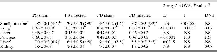
Effects of Heligmosomoides bakeri infection and protein deficiency on maternal serum analyte concentrations at day 20 of lactation 1 , 2

1 Values are means±s.e. (median). PS, protein sufficient; PD, protein deficient.
2 Values that were above or below the linear range of the standard curve were assigned the upper limit (potassium, 14·0 mmol L−1; amylase, 1200 U L−1) or lower limit (BUN, 0·71 mmol L−1) accordingly.
3 Range of sample sizes per experimental group.
4 Two-way ANOVA included diet (D) and infection (I) and their interaction (D×I) with food intake as a covariate. Lower case letters indicate significant differences based on post-hoc Bonferroni comparisons, P<0·05. NS, not significant.
5 Analysis based on log transformed data.
6 Model residuals were not normally distributed even after transformation. P values are based on Friedman's two-way ANOVA of ranks.
Statistical analysis
Analyses were conducted using Proc Mixed models in SAS statistical software (v. 9.3; SAS Institute Inc., Cary, NC, USA) and P values <0·05 were considered statistically significant. Model residuals were checked for normality and data were transformed when necessary. Log values were calculated for total protein, relative lung mass, medulla : cortex ratio and per cent abnormal glomeruli. Square root values were calculated for lobular inflammatory foci number. If transformation did not normalize residuals, non-parametric analyses were used. All values are presented as untransformed means±s.e. and medians are also reported for non-parametric data.
Two-way ANOVAs with main effects of diet (PS vs PD) and infection (sham vs infected) were employed. Maternal relative organ mass as a percentage of total mass was analysed by a two-way ANOVA with litter size at birth as a covariate. Maternal serum analyte concentrations were analysed by two-way ANOVA. Models for relative organ masses and serum analytes were run with and without food intake as a covariate to control for differences in energy intake between PS and PD groups and to differentiate between the effects of protein deficiency and energy restriction. Only relevant differences in analyses are reported. Quantitative histology measurements were analysed by two-way ANOVA. Bonferroni adjustments were used to account for multiple comparisons where appropriate.
When model residuals were not normally distributed even after transformation (serum BUN and potassium concentrations, relative small intestine and liver mass and lymphoid follicle number and width) and when semi-quantitative histology scores were analysed (pancreatitis and portal inflammation scores) Friedman's two-way ANOVA by ranks was employed with covariates as indicated and, where appropriate, Bonferroni post-hoc comparisons were done.
RESULTS
Confirmation of protein deficiency
Not only was body mass lower in dams fed a PD diet, but absolute small intestine, heart and kidney mass were lower as well (data not shown). Dams fed a PD diet had lower concentrations of both total serum protein and albumin than dams fed a PS diet, confirming that the 6% protein diet led to protein deficiency in the dams. In contrast, H. bakeri infected dams had higher total serum protein concentrations than uninfected dams but serum albumin was unaffected by infection (Table 1).
Small intestine
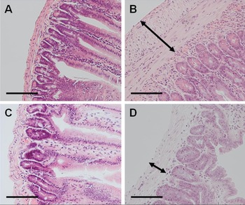
Relative small intestine mass was increased by infection but was not affected by protein deficiency (Table 2). Gross histological examination (Fig. 1) revealed that H. bakeri-infected PS dams had severe transmural inflammation extending from the lamina propria of the duodenum through to the submucosa with many macrophages in the area inhabited by L4 (Fig. 1B). Protein deficiency reduced the infection-induced inflammation (Fig. 1D). This observation was supported by quantification of mucosal mast cells and eosinophils, where a significant interaction between diet and infection was detected. Numbers of both mucosal mast cells (Fig. 2A) and eosinophils (Fig. 2B) were elevated in infected mice and much more so in PS than in PD mice. The number of goblet cells was also higher in infected mice but did not differ between PS and PD dams (Fig. 2C). Infection had no effect on villus height (Fig. 2D) but increased crypt depth (Fig. 2E). In contrast, PD dams had shorter villi than PS dams (Fig. 2D). Furthermore, crypts were shorter in PD infected compared with PS infected dams, suggesting that protein deficiency blunted the infection-induced increase in crypt depth (Fig. 2E).
Cross-sections of duodenum taken on day 20 of lactation from dams fed a protein sufficient (PS; A and B) or protein deficient (PD; C and D) diet and given a sham (A and C) or Heligmosomoides bakeri trickle (B and D) infection, stained with hematoxylin and eosin (H&E). Inflammation extending into the submucosa (arrows) is greatest in PS infected dams (B). Scale bar: 10 μm.

Interaction effect of maternal protein deficiency and Heligmosomoides bakeri infection on (A) mast cell number per villus : crypt unit (VCU) (main effect of diet: P = 0·0082; main effect of infection: P<0·0001; interaction of diet×infection: P = 0·0049); (B) eosinophil number per VCU (main effect of diet: P<0·0001; main effect of infection: P<0·0001; interaction of diet×infection: P<0·0001); (C) goblet cell number per VCU (main effect of diet: P = NS; main effect of infection: P = 0·0387; interaction of diet×infection: P = NS); (D) villus height (main effect of diet: P = 0·0387; main effect of infection: P = NS; interaction of diet×infection: P = NS); and (E) crypt depth (main effect of diet: P = 0·06; main effect of infection: P = 0·0311; interaction of diet×infection: P = NS) in the duodenum of dams on day 20 of lactation. Mast cell numbers were log transformed prior to analysis but values represent untransformed means. PS, protein sufficient; PD, protein deficient; sham infection (solid bars); Heligmosomoides bakeri infection (cross-hatched bars). Different lower case letters represent significant differences among groups based on post hoc tests using Bonferroni adjustments, P<0·05. Values are means±s.e.

Effects of Heligmosomoides bakeri infection and protein deficiency on relative dam organ mass (% body mass) at day 20 of lactation 1 , 2

1 Values are means±s.e. (median). n = 12 per group. PS, protein sufficient; PD, protein deficient.
2 Relative spleen mass was reported previously (Odiere et al. Reference Odiere, Scott, Leroux, Dzierszinski and Koski2013).
3 Two-way ANOVA included diet (D) and infection (I) and their interaction (D×I) with food intake and litter size at birth as covariates. Lower case letters indicate significant differences based on post-hoc Bonferroni comparisons, P<0·05. NS, not significant.
4 Model residuals were not normally distributed even after transformation. P values are based on Friedman's two-way ANOVA of ranks.
5 Analysis based on log transformed data.
Thymus and spleen
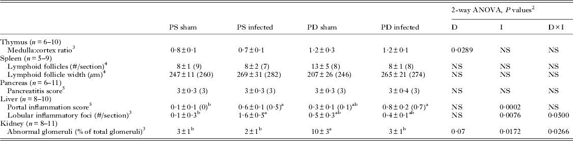
In the thymus, protein deficiency increased the medulla : cortex ratio whereas infection did not (Table 3). To test the hypothesis that infection-induced splenomegaly would be due to altered architecture of the white pulp, we measured lymphocyte follicle number and width (Table 3), but infection had no effect on either, suggesting that infection-induced splenomegaly may be a consequence of changes in the red pulp rather than the white pulp. Protein deficiency had no effect on lymphocyte follicle number or width (Table 3).
Effects of Heligmosomoides bakeri infection and protein deficiency on histopathology of dam extra-intestinal lymphoid and visceral organs at day 20 of lactation 1

1 Values are means±s.e. (median). PS, protein sufficient; PD, protein deficient.
2 Two-way ANOVA included diet (D) and infection (I) and their interaction (D×I). Lower case letters indicate significant differences based on post-hoc Bonferroni comparisons, P<0·05. NS, not significant.
3 Analysis based on log (medulla : cortex ratio and % abnormal glomeruli) or square root (lobular inflammatory foci) transformations.
4 Model residuals were not normally distributed even after transformation. P values are based on Friedman's two-way ANOVA of ranks.
5 Scores assessed by Friedman's two-way ANOVA of ranks.
Heart and lung
Relative heart mass was not affected by either protein deficiency or infection (Table 2). In contrast, both protein deficiency and infection independently increased relative lung mass. Post-hoc analysis based on the borderline significant interaction (P = 0·07) revealed that relative lung mass was highest in dams with both infection and protein deficiency (Table 2).
Pancreas
Dams fed a PD diet had lower relative pancreas mass than dams fed a PS diet (Table 2) but neither diet nor infection influenced the composite pancreatitis score (Table 3), its component measures of oedema, leucocyte infiltration, acinar cell vacuolation and necrosis or fat necrosis (data not shown), or serum amylase concentrations (Table 1). Pancreatitis scores were driven predominantly by leucocyte infiltration in all experimental groups (data not shown) but infected dams tended to have higher leucocyte infiltration scores than uninfected dams (P = 0·07).
Liver
PD dams had lower relative liver mass than PS dams (Table 2). Steatosis was observed in the livers of uninfected dams fed a PD diet (Fig. 3C) but this was less evident in infected PD dams (Fig. 3D). Some perivascular infiltrate was observed in livers of dams fed a PD diet (Fig. 3C and D). Infected mice had higher portal inflammation scores and more lobular inflammation foci than uninfected mice and a significant interaction was detected such that infected PD dams had fewer lobular inflammatory foci than infected PS dams (Table 3). A significant interaction was also detected for serum bilirubin concentrations which were elevated in infected PD dams compared with PS infected dams (Table 1).
Cross-sections of liver taken on day 20 of lactation from dams fed a protein sufficient (PS; A and B) or protein deficient (PD; C and D) diet and given a sham (A and C) or Heligmosomoides bakeri trickle (B and D) infection, stained with hematoxylin and eosin (H&E). Portal inflammation (arrow) is evident in PS infected dams (B). Steatosis is widespread in PD uninfected dams (C). Scale bar: 10 μm.

Kidney
Relative kidney mass was not affected by infection or protein deficiency when controlling for energy intake (Table 2). Histopathological changes were exclusively inside the glomerulus where the cells that are normally arranged evenly were aggregated causing the Bowman's capsule to appear larger (Fig. 4). The percentage of these abnormal glomeruli was reduced by infection, the effect of protein deficiency was borderline (P = 0·07) and a significant interaction was detected such that uninfected PD mice had a higher percentage than any other group (Table 3). Serum creatinine concentrations were elevated independently by infection and protein deficiency, and post-hoc analysis revealed that the highest serum creatinine concentrations were in infected PD dams (Table 1). Neither infection nor diet affected serum electrolyte concentrations (Table 1).
Cross-sections of kidney taken on day 20 of lactation from dams fed a protein sufficient (PS; A and B) or protein deficient (PD; C and D) diet and given a sham (A and C) or Heligmosomoides bakeri trickle (B and D) infection, stained with periodic-acid Schiff (PAS). Abnormal glomeruli (arrows) seen in PD uninfected dams (C) contain aggregated cells causing expansion of the Bowman's capsule. Scale bar: 10 μm.

DISCUSSION
Although it is often assumed that the pathological impact of intestinal nematode infections is predominantly in the intestine, we have shown that, at least during lactation, the negative effects of infection extend beyond the intestine to visceral organs not in direct contact with parasites. In line with our hypothesis that protein deficiency would dampen infection-induced pathology in visceral organs, lobular liver inflammation was reduced in PD dams. Conversely, abnormalities in the glomeruli caused by protein deficiency were absent in PD infected dams. Interestingly, serum analyte concentrations did not reflect these pathologies, as measures of kidney and liver damage were highest when infection and protein deficiency were combined. In contrast to our hypothesis that changes in relative spleen size in response to infection and protein deficiency would be due to changes in the lymphoid compartment of the spleen, we found indirect evidence that the red pulp and not the lymphoid compartment of the spleen was enlarged during infection.
As previously reported (Odiere et al. Reference Odiere, Scott, Weiler and Koski2010b ), dams fed a PD diet beginning on day 14 of pregnancy had decreased food intake throughout lactation, and weighed less at day 20 of lactation compared with dams fed a PS diet. Total serum protein and albumin concentrations were significantly lower in mice fed a PD diet than those fed a PS diet, confirming the deficient status of our PD mice. In contrast, total protein concentrations were higher in infected mice than uninfected mice, likely due to an increase in immunoglobulins (Pritchard et al. Reference Pritchard, Williams, Behnke and Lee1983; Urban et al. Reference Urban, Katona and Finkelman1991) and acute phase response proteins (Stadnyk and Gauldie, Reference Stadnyk and Gauldie1991). As has been consistently reported in non-lactating mice (Boulay et al. Reference Boulay, Scott, Conly, Stevenson and Koski1998; Tu et al. Reference Tu, Koski and Scott2007), worm counts and eggs per gram feces were higher in our PD lactating mice (Odiere et al. Reference Odiere, Scott, Weiler and Koski2010b , Reference Odiere, Scott, Leroux, Dzierszinski and Koski2013).
Gut responses
Infection induced transmural inflammation and infiltration of mast cells, goblet cells and eosinophils, as previously reported (Cywińska et al. Reference Cywińska, Czumińska and Schollenberger2004; Tu et al. Reference Tu, Koski and Scott2007). Interestingly, however, both the mast cell and eosinophil (but not goblet cell) responses to our trickle infection were considerably stronger (approximately 10-fold increase in mast cells and 20-fold increase in eosinophils compared with uninfected PS mice) than previously reported in young adult mice given a single challenge infection with 200 H. bakeri larvae (<50% increase in mast cells and ∼5-fold increase in eosinophils compared with uninfected PS mice; Tu et al. Reference Tu, Koski and Scott2007). Even though protein deficiency reduced the magnitude of infection-associated mastocytosis and eosinophilia as expected (Boulay et al. Reference Boulay, Scott, Conly, Stevenson and Koski1998; Ing et al. Reference Ing, Su, Scott and Koski2000), the magnitudes of both responses were still stronger in lactating PD mice than has been previously reported in non-lactating PS mice. Two possibilities might explain such intense local mastocytosis and eosinophilia in response to H. bakeri infection in this study. First, the trickle infection protocol involved repeated arrival of L3 parasites, accumulation of L4 larvae that are highly immunogenic and recurring tissue damage as the L4 migrated from the serosal musculature into the intestinal lumen, all of which are likely to exaggerate immunological and inflammatory processes in the duodenum (Behnke et al. Reference Behnke, Lowe, Clifford and Wakelin2003) and also reduce the immunosuppressive effects of adult worms (Kamal et al. Reference Kamal, Dehlawi, Rosa Brunet and Wakelin2002), compared with a single infection or drug-abbreviated challenge infection. This ongoing antigenic stimulus may have contributed to the heightened mastocytosis and eosinophilia. Second, lactating dams reallocate resources to expand the absorptive surface of the small intestine during lactation (Hammond, Reference Hammond1997) through a marked increase in relative mass of both mucosal and serosal layers, in villus length and in glucose transport (Casirola and Ferraris, Reference Casirola and Ferraris2003). Consistent with this, the villi of our uninfected PS lactating mice were approximately 50% longer (∼690 μm) than reported previously for uninfected PS non-pregnant mice (∼450 μm; Tu et al. Reference Tetsutani, Ishiwata, Ishida, Tu, Torii, Hamano, Himeno and Hisaeda2007). Thus, local protective defences in the intestine may be amplified along with absorptive surface area as lactating dams maximize nutrient absorption.
Although we expected the villus atrophy typical of H. bakeri infection (Tu et al. Reference Tu, Koski and Scott2007; Hashimoto et al. Reference Hashimoto, Uchikawa, Tegoshi, Takeda, Yamada and Arizono2009), infection had no effect on villus length. Furthermore, our infected lactating mice had deeper crypts, an observation not reported for H. bakeri in non-lactating mice (Tu et al. Reference Tu, Koski and Scott2007; Hashimoto et al. Reference Hashimoto, Uchikawa, Tegoshi, Takeda, Yamada and Arizono2009) but in line with the emerging role of crypt hyperplasia in the inflammation associated with nematode infections (Artis and Grencis, Reference Artis and Grencis2008; Cooper, Reference Cooper2009). Increased cell proliferation in the crypts of infected mice could be explained by cell regeneration in response to mucosal injury by larvae (Umar, Reference Umar2010) and induced by IL-13 (McDermott et al. Reference McDermott, Humphreys, Forman, Donaldson and Grencis2005), which we have previously reported to be elevated by infection in these mice (Odiere et al. Reference Odiere, Scott, Leroux, Dzierszinski and Koski2013). This increased crypt cell proliferation would help to counter any infection-induced villus damage and help the lactating dam to maximize the absorptive surface area. However, our data indicate that these processes were not occurring during protein deficiency where both villi and crypts were shorter, as reported previously (Pond et al. Reference Pond, Ellis, Mersmann, Heath, Krook, Burrin, Dudley and Sheng1996; Tu et al. Reference Tu, Koski and Scott2007). These data indicate that during lactation, infection-induced damage to the absorptive surface of the duodenum is minimized by regenerative responses, but only when dams are fed adequate protein.
Extra-intestinal lymphoid tissues
Thymic atrophy is an established consequence of a low protein diet (Mittal et al. Reference Mittal, Woodward and Chandra1988; Barone et al. Reference Barone, O'Brien and Stevenson1993). Immature CD4+/CD8+ cells in the cortex are most susceptible to protein deficiency but during prolonged deficiency, the number of mature thymocytes decreases as well (Barone et al. Reference Barone, O'Brien and Stevenson1993). Our histological examination of the thymus extends these findings to lactating mice where the medulla : cortex ratio was increased by protein deficiency but unaffected by infection. The absence of an infection effect is consistent with previous reports that thymus mass is not altered by H. bakeri infection (Boulay et al. Reference Boulay, Scott, Conly, Stevenson and Koski1998; Ing et al. Reference Ing, Su, Scott and Koski2000), even in lactating mice (Odiere et al. Reference Odiere, Scott, Leroux, Dzierszinski and Koski2013).
In contrast to the thymus, the spleen responded to both infection and protein deficiency. We previously reported that protein deficiency inhibited infection-induced splenomegaly in these lactating PS mice (Odiere et al. Reference Odiere, Scott, Leroux, Dzierszinski and Koski2013). Infection may increase production and/or storage of lymphocytes in the spleen, especially if mesenteric lymph nodes are unable to process all the parasite antigen (Ali and Behnke, Reference Ali and Behnke1985). To date, however, evidence that H. bakeri alters lymphocyte or dendritic cell populations in the spleen is limited and inconsistent. Although a small increase in splenic CD4+CD25+ T reg cells and sIG+ B cells has been reported (Su and Dobson, Reference Su and Dobson1997; Finney et al. Reference Finney, Taylor, Wilson and Maizels2007), some studies have shown no effect of H. bakeri on numbers of splenic T reg cells (Tetsutani et al. Reference Tetsutani, Ishiwata, Ishida, Tu, Torii, Hamano, Himeno and Hisaeda2009) or dendritic cells (Su et al. Reference Su, Segura, Morgan, Loredo-Osti and Stevenson2005) and other studies have reported that infection decreases both B and T (Thy1+) cell populations (Parker and Inchley, Reference Parker and Inchley1990). Although we had hypothesized that changes in relative spleen size in response to infection and protein deficiency would be due to changes in the lymphoid compartment of the spleen, neither infection nor protein deficiency altered the size or number of lymphoid follicles of the lactating mouse. Thus, we suggest that the increase in relative spleen mass due to infection, and the blunting of this response in PD infected mice, may reflect changes in the red pulp.
The majority of the spleen consists of red pulp that serves as a reservoir for erythrocytes (Corbin et al. Reference Corbin, Vicente, Martin-Hernando, Acevedo, Pérez-Rodriguez and Gortazar2008) and in mice, is a site of haematopoiesis (Tan and O'Neill, Reference Tan and O'Neill2010) and erythrocyte processing that accelerates under pathological conditions leading to splenomegaly (Jiao et al. Reference Jiao, Okumiya, Saibara, Kudo and Sugiura2001). In our trickle infection protocol, the repeated tissue damage and haemorrhaging caused by the penetration of L3 larvae (Sukhdeo and Mettrick, Reference Sukhdeo and Mettrick1984) and migration of L4 larvae from the serosal musculature to the lumen of the intestine (Crandall et al. Reference Crandall, Crandall and Franco1974) may increase turnover of damaged erythrocytes. The resulting elevation in erythropoietin concentration signals increased production of erythrocytes and other myeloid cells in the spleen (Graber and Krantz, Reference Graber and Krantz1978), accounting for moderate hyperplasia seen in reticuloendothelial tissue 3 weeks after H. bakeri infection (Liu, Reference Liu1965). In contrast, protein deficiency has been associated with decreased numbers of haematopoietic progenitor cells, especially in the spleen (Bell et al. Reference Bell, Hazell and Price1976), and impaired responses of murine splenic cells to erythropoietin (Barrio Rendo, Reference Barrio Rendo1995) which may explain the blunted splenomegaly in PD infected mice compared with PS infected mice. A second possibility relates to the impacts of immunization with small peptides and of chronic inflammation on inappropriate trafficking and accumulation of T cells in the red pulp of the spleen (Zhang et al. Reference Zhang, Lizzio, Chen and Kozlowski2005; Ezernitchi et al. Reference Ezernitchi, Vaknin, Cohen-Daniel, Levy, Manaster, Halabi, Pikarsky, Shapira and Baniyash2006). It would be of considerable interest to determine whether the chronic inflammation occurring during H. bakeri infection leads to an accumulation of T cells in the red pulp, and whether this might account for the infection-induced splenomegaly.
Non-lymphoid visceral organs
A similar pattern to small intestine inflammation emerged in the liver, where infection induced moderate inflammation in the portal and lobular areas. As drainage via the portal blood system travels to the liver directly from the small intestine (Crispe et al. Reference Crispe, Dao, Klugewitz, Mehal and Metz2000), focal non-specific hepatitis during H. bakeri infection may result from lytic products of intestinal damage transported to the liver (Liu, Reference Liu1965). In addition, systemic markers of inflammation such as CD4+ Th2 cells (Mohrs et al. Reference Mohrs, Harris, Lund and Mohrs2005), eosinophils and mucosal mast cell protease (Ing et al. Reference Ing, Su, Scott and Koski2000) and monocyte chemoattractant protein 5 (Tu et al. Reference Tu, Koski and Scott2007) are elevated during H. bakeri infection and may signal inflammation in the liver. Protein deficiency blunted the infection-induced lobular inflammation, an observation consistent with the reduction by protein deficiency of disseminated portal fibrosis and hepatic granulomas around trapped eggs in mice infected with Schistosoma mansoni (Coutinho et al. Reference Coutinho, De Souza, Silva, Cavalcanti, De Araujo, Barbosa, Cheever and Andrade1997). Although we expected elevated serum bilirubin concentrations to be associated with liver damage, as swollen hepatocytes block bilirubin elimination causing it to circulate back into the blood (Stogdale, Reference Stogdale1981), bilirubin concentrations were higher in PD than PS infected dams. This suggests that either the combination of protein deficiency and infection impairs liver function despite the absence of histopathology or that increased bilirubin is due to pre- or post-hepatic damage.
In addition to the lower relative liver mass in PD mice, relative pancreas mass was also reduced by protein deficiency, consistent with the well-established sensitivity of the exocrine pancreas to dietary components including protein (Brannon, Reference Brannon1990; Baumler et al. Reference Baumler, Koopmann, Thomas, Ney and Groblewski2010). The composite pancreatitis score in all groups was driven predominantly by leucocyte infiltration, which tended to be higher in infected dams, possibly due to infection-associated systemic inflammatory signals. Cell damage, however, was minimal, suggesting that the pancreas is largely protected from protein deficiency and infection. This was supported by the finding that serum amylase concentrations were not increased in PD or infected dams.
We observed several differences in the effects of protein deficiency and infection on the kidney when energy intake was not controlled for in our analyses. Specifically, relative kidney mass was lower and sodium and potassium concentrations were higher in PD dams and infection had no effect on BUN concentrations, suggesting that the kidney is more susceptible to energy restriction than protein deficiency. However, we did observe interesting interactive effects of diet and infection on kidney histology. Our observation of aggregation of cells in the glomeruli and an increase in Bowman's capsule space in response to protein deficiency is in line with previous reports of glomerular nephrosis and decreased glomerular size as consequences of protein deficiency (Klahr and Alleyne, Reference Klahr and Alleyne1973). Decreased filtering ability of damaged glomeruli may also explain the reduced serum total protein, albumin and BUN that we observed in the PD mice. Why the combination of H. bakeri infection and protein deficiency would cause the percentage of abnormal glomeruli to be lower is not known as, to our knowledge, the only studies to consider the impact of H. bakeri infection on the kidney showed no change in kidney mass (Kristan and Hammond, Reference Kristan and Hammond2004), even in lactating mice (Kristan, Reference Kristan2002). In contrast to the antagonistic effects of protein deficiency and infection on abnormal glomeruli, additive effects were observed for serum creatinine concentrations, with the highest concentrations in infected PD mice. Further research is needed to examine how H. bakeri influences kidney structure and function.
The ∼40% increase in relative lung mass in infected PD dams compared with uninfected PS dams might be explained by increased metabolic demands in response to infection and protein deficiency that could be achieved through recruitment of pulmonary capillaries to increase the surface area for oxygen transport (Hou et al. Reference Hou, Gheorghiu, Huxley and Pfeifer2010) or through elevation of resting metabolic rate (RMR). However, RMR of our dams on day 15 of lactation was not affected by infection and was lowered (not elevated) by protein deficiency (Odiere et al. Reference Odiere, Scott, Weiler and Koski2010b ). Alternatively, increased lung mass may be associated with a systemic inflammatory response. Heligmosomoides bakeri infection induces systemic dissemination of CD4+ Th2 cells, basophils and eosinophils to the lung airways (Mohrs et al. Reference Mohrs, Harris, Lund and Mohrs2005) that may increase lung mass and this response may be exacerbated during our trickle infection protocol.
Although PD dams ate up to 50% less food than PS dams during peak lactation (Odiere et al. Reference Odiere, Scott, Weiler and Koski2010b ), we have calculated the actual energy and nutrient intakes and determined that all mice were at least four times above the National Research Council mouse requirements for all nutrients except energy (NRC, 1995). Although some of the PD mice were below recommended NRC intakes for energy during peak lactation, the average energy intake of PD mice was within the normal range for lactating mice and all mice ingested more calories than the minimum requirement for non-lactating mice (NRC, 1995). The inclusion of food intake as a covariate in our analyses allowed us to differentiate between the effects of reduced energy intake and protein deficiency. This demonstrated that only the kidney was more sensitive to energy than protein intake.
In conclusion, the increased crypt depth caused by H. bakeri infection was only apparent in our lactating dams if they were fed adequate protein, perhaps because increased crypt depth maximizes nutrient absorption to support lactation. A more complex picture emerged with regard to extra-intestinal pathologies. Whereas H. bakeri infection minimized the glomerular histopathology induced by protein deficiency, lobular inflammation of the liver induced by infection was dampened by protein deficiency. The limited inflammation when the two conditions were combined did not extend, however, to the elevation of serum analytes of liver and kidney function which were highest in PD infected dams. Furthermore, our results suggest that the splenomegaly typical of H. bakeri infection may result from altered haematopoiesis and erythrocyte turnover or from inappropriate T cell trafficking to the red pulp, rather than expansion of the lymphoid compartment of the spleen. Further research is needed to explore the pathophysiological processes underlying extra-intestinal consequences of infection and protein deficiency, and also to determine whether similar responses occur in mice under less extreme energetic demands.
ACKNOWLEDGEMENTS
We thank Carly Langlois for assistance with histology.
FINANCIAL SUPPORT
This work was supported by Natural Sciences and Engineering Research Council of Canada (NSERC) Discovery Grants to M. E. S. and K. G. K. and an NSERC Postgraduate Scholarship to L. M. S. Research at the Institute of Parasitology is supported by a Regroupement stratégique grant from Fonds Québécois de la recherche sur la nature et les technologies.