Introduction
The bone-anchored hearing aid (BAHA) (also known as a bone-anchored hearing device) has become established as a mode of hearing support since the first procedures in 1977 by Tjellström and colleagues in Gothenburg, Sweden.Reference Brånemark, Hansson, Adell, Breine, Lindström and Hallén 1 The clinical indications include conditions for which conventional hearing aids cannot be used, such as persistent or recurrent otitis externa infections, or abnormal pinna anatomy.
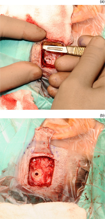
Put simply, the BAHA allows the direct transmission of sound via bone conduction, so that a sound processor on the ‘external’ surface of the bony skull allows sound transmission directly through the bone. The sound processor is attached via a skin-penetrating coupling – the abutment. This is surgically fixed, and requires access to the post-auricular bone. The traditional surgical approach for this is the raising of a skin flap over an area of bone suitable for implanting the abutment (Figure 1).
(a) & (b) Skin flap incision, comprising flap elevation with soft tissue removal.

Complications from the surgical procedure required for BAHA insertion are skin-wound related.Reference Proops 2 – Reference Holgers, Roupe, Tjellström and Bjursten 4 It is on this background that our centre chose to adopt an alternative surgical technique, instead using a linear incision, without soft tissue removal, to access the post-auricular bone. We wanted to identify whether there was an improvement in wound-related complications associated with this change in technique.
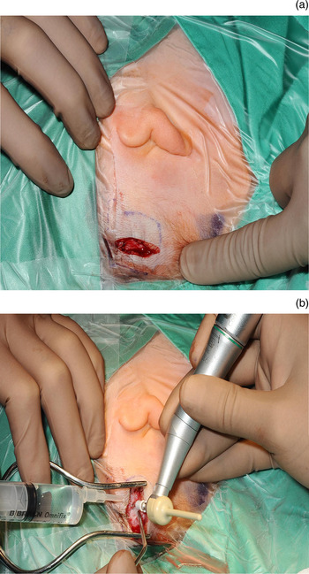
At our centre, the abutment is now placed directly at the level of the linear incision (Figure 2). The abutment is placed at a distance from the main incision, into a separate hole (in the skin flap) created using a biopsy punch. This is a recognised way of performing the procedure. It also avoids skin thinning or removal. All cases in this study involved the direct placement of the abutment at the level of the linear incision. Whilst DermaLock™ hydroxyapatite-coated implants have been used clinically since 2013, 5 they were not involved in the study cases during 2012–2013.
(a) & (b) Linear post-auricular incision, with no removal of soft tissue.

Materials and methods
This paper reports the findings of a retrospective assessment of a single surgeon's (SSMH) outcomes, at a single centre, over two time periods. A total of 117 ears were included. From 1999 to 2011, traditional flap elevation with soft tissue removal was performed (group 1). From 2012 to 2013, a linear incision method was employed (group 2).
The recorded outcome was any complication related to the wound or skin for up to one year post-procedure. The complications recorded included wound infection, skin overgrowth and patient-reported numbness.
Results
There were a total of 86 ears in group 1 (traditional flap elevation with soft tissue removal group) and 31 in group 2 (linear incision access group).
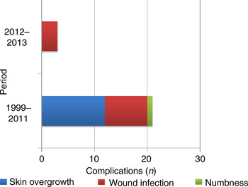
There were 21 cases of complications in group 1; these consisted of 12 cases of skin overgrowth, 8 of wound infection and 1 of numbness (Figure 3). There were three complications in group 2, all of which were wound infections.
Summary of complications in each period (numbers of complications after one year of follow up). The traditional method of skin flap and soft tissue removal was used between 1999 and 2011 (group 1; n = 86), and linear incision without soft tissue removal was used between 2012 and 2013 (group 2; n = 31).

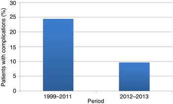
The percentage of patients with complications in group 1 was 24.4 per cent (21 out of 86) and only 9.7 per cent (3 out of 31) in group 2 (Figure 4). Statistical analysis utilising the independent t-test indicated that this difference was significant (p < 0.05; 95 per cent confidence interval = 0.0800–0.4473).
Comparison of complication rates (percentages of patients with complications after one year of follow up) between the two periods (p < 0.05; 95 per cent confidence interval = 0.0800–0.4473). The traditional method of skin flap and soft tissue removal was used between 1999 and 2011 (group 1; n = 86), and linear incision without soft tissue removal was used between 2012 and 2013 (group 2; n = 31).

Discussion
This retrospective analysis of the two groups indicated a statistically significant improvement in complications for those patients undergoing BAHA surgery via the linear incision method.
There is a difference in the case numbers, and the small numbers in group 2 (linear incision access group) must be acknowledged. There is certainly a role for further study utilising a greater number of patients, in order to confirm that the improvement in outcome is a result of the change in surgical technique to the linear incision method.
It is prudent to further comment on the numbers in the groups. In comparing the 2 groups, there were 86 ears in group 1 (traditional flap elevation with soft tissue removal group) over the 12-year time period, suggesting an average of around 7 cases per year for 12 years, compared to 31 cases in 2 years for group 2. This proportion discrepancy is actually a reflection of local trust policy change. For the first seven years of BAHA surgery at this centre, the number of BAHA procedures that could be performed per year was capped at six. There were therefore a larger number of surgical procedures performed in the later years, and this is reflected in the 31 cases performed in 2 years (2012 and 2013) using the linear incision method.
The nature of complications is also important. Notably, in group 1, using the skin flap method with soft tissue removal, 12 patients had the complication of skin overgrowth. This is a very clinically significant issue. If skin grows over the surgically implanted abutment, the hearing aid can no longer be fitted onto the device and it is rendered unusable. This has serious clinical implications. If the abutment is to be used, the patient needs to return to the operating theatre and have the overgrown skin removed. This complication therefore has a significant patient morbidity attached to it, manifesting ultimately as either a return to the operating theatre or a loss of use of the aid. Indeed, all 12 patients with skin overgrowth had to return to the operating theatre.
There was no indication that the complications, including overgrowth, occurred with greater frequency in any particular year. The stable incidence of the skin overgrowth complication seen across the 12-year time period, indicates that the observed improvement in outcomes was not the result of experience or a learning curve of the surgeon. It is suggested that the complications of overgrowth and skin numbness are caused by the soft tissue removal employed in the skin flap method.
-
• Bone-anchored hearing aids (BAHAs) have become established as a mode of hearing support
-
• The clinical indications for BAHAs include conditions for which conventional hearing aids cannot be used
-
• Complications related to BAHA insertion are skin-wound related
-
• At our centre, the surgical method was changed from skin flap elevation to a single linear incision; the latter offers adequate access for BAHA placement
-
• The single linear incision reduced the complication rate and the nature of complications, which are limited and relatively minor
It is important to note that in group 2, in which the linear incision without soft tissue removal was used, skin overgrowth was completely eliminated as a complication. The associated implied morbidity as a result of this complication is therefore also absent in this group. Also of note, the risk of numbness, which was a minor complication using the skin flap method in group 1, has also been entirely removed.
Conclusion
This single centre, single surgeon series offers strong evidence to suggest that the use of a linear incision instead of the traditional skin flap approach is equally suitable, if not surgically superior, for BAHA insertion. This is evidenced not only by the improved complication rate but also by the change in the nature of complications arising.