Introduction
A preauricular sinus is a developmental abnormality which presents as an opening at the anterior margin of the ascending limb of the helix.Reference Tan, Constantinides and Mitchell1 Such sinuses may appear simple and innocuous, but they may have deeper, widespread extensions. Infrequently, these extensions involve the facial nerve branches, thus posing the risk of injury during surgical excision. Preauricular sinus infection can lead to abscess formation, which presents with swelling, tenderness, erythema and/or recurrent discharge.Reference Tan, Constantinides and Mitchell1, Reference Scheinfeld, Silverberg, Weinberg and Nozad2 Isolated, asymptomatic preauricular sinuses require no treatment. However, complete surgical excision of the sinus and its tract is needed for recurrent or persistent preauricular sinus infection.Reference Gur, Yeung, Al-Azzawi and Thomson3 Incomplete excision is the predominant cause of preauricular sinus recurrence.Reference Tan, Constantinides and Mitchell1 The incidence of recurrence has been reported as 0–42 per cent.Reference Prasad, Grundfast and Milmoe4, Reference Kumar, Narayanamurthy, Sumathi and Vijay5
Various techniques have been reported for excision of the preauricular sinus. The standard surgical technique is sinectomy.Reference Leopardi, Chiarella, Conti and Cassandro6 Prasad et al. Reference Prasad, Grundfast and Milmoe4 have described the supra-auricular technique of preauricular sinus excision. Compared with the standard technique, the supra-auricular approach offers better exposure, and hence a better chance of total excision, reduced operating time and lower recurrence rate.Reference Prasad, Grundfast and Milmoe4
In otorhinolaryngology, the role of radiofrequency thermal ablation is well established in the treatment of enlarged tonsils,Reference Back, Paloheimo and Ylikoski7 inferior turbinate hypertrophyReference Back, Hytonen, Malmerz and Yilkoski8 and snoring.Reference Wedman and Miljeteig9
In this study, we explored the use of radiofrequency thermal ablation in the excision of preauricular sinuses, and compared it to ‘cold steel’ surgical excision. We hypothesised that radiofrequency-assisted preauricular sinus excision would result in a reduced operating time, reduced peri-operative bleeding, easier dissection, better healing and, more importantly, a reduced recurrence rate.
Materials and methods
Between October 2007 and March 2009, a prospective study was carried out involving the excision of 60 preauricular sinuses in 50 patients. Patients were randomly divided into two groups of 30 preauricular sinuses each. There were no statistically significant differences between the two groups as regards age or gender distribution. In group one, local, wide, cold steel excision of the preauricular sinus was undertaken, using the supra-auricular approach.Reference Prasad, Grundfast and Milmoe4 In group two, radiofrequency-assisted excision was undertaken, also using the supra-auricular approach. Patients with bilateral preauricular sinuses had both sides operated upon simultaneously; these were considered as two separate procedures. All procedures were performed by the same surgeon (the senior author Dr SK).
Patients underwent detailed clinical history-taking, a complete otolaryngological examination and a systemic examination, to rule out any associated conditions. Routine baseline investigations were undertaken.
Informed, written consent was obtained from all patients.
All procedures were performed under general anaesthesia. First, the sinus was injected with methylene blue dye to delineate the tract course. An elliptical incision was then made around the sinus and extended higher upward to the pre- and supra-auricular region as needed. In group one, dissection was commenced with dissecting scissors until the temporalis fascia was identified. Tissue superficial to this fascia was removed together with the preauricular sinus. A portion of the auricular perichondrium was also excised. The incision was closed in two layers and a compression bandage applied.
The operative procedure in group two was identical except that dissection was performed using radiofrequency probes (Ellman Surgitron; Ellman International, Oceanside, New York State, USA), as shown in Figure 1. A frequency of 3.8 MHz was used.
Intra-operative photograph showing preauricular sinus excision performed with a radiofrequency probe.

After surgery, patients were observed overnight, and were discharged the next day with oral analgesics for 3 days (paracetamol syrup 10–15 mg/kg per dose, 8 hourly for patients younger than 12 years) and antibiotics for 5 days (co-amoxiclav syrup 20–40 mg/kg per day in three divided doses). Patients were recalled after one week for suture removal.
Patients were followed weekly for a month, and monthly thereafter. The minimum follow-up period was six months and the maximum follow-up period 24 months (average, 15 months). At each visit, observations were recorded regarding the incision site and any discharge, abscess, swelling or wound dehiscence. Patients with unhealthy wounds were continued on the same antibiotics for another 5 days. Recurrence was defined as any discharge, swelling or abscess occurring in the follow-up period which did not heal after a course of antibiotics.Reference Currie, King, Vlantis and Li10 One year post-operatively, the surgical scar was observed for hypertrophy or keloid formation or recurrence. All variables were recorded on a proforma.
Post-operatively, we recorded on a proforma the duration of surgery, occurrence of bleeding and ease of dissection. The latter two parameters were graded on a visual analogue scale (VAS).Reference Haghighi11 For bleeding, 0 indicated minimal bleeding and 10 bleeding which completely obscured the surgical field and required ligation. For ease of dissection, 0 indicated minimum ease of dissectionReference Haghighi11 and 10 maximum ease of dissection.
Student's t-test was used to compare operative time, bleeding and ease of dissection in the two groups. Fisher's exact test was used to compare recurrence. A p value of <0.05 was considered as significant.
Observations and results
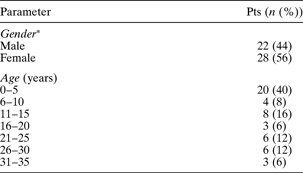
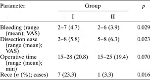
Sixty procedures were performed on 50 patients. Patients' ages varied from two to 34 years, with a mean age of 12.26 years. Table I shows the patients' demographic profile. The average follow-up period was 15 months (range, six to 24 months). The maximum incidence of preauricular sinus presentation was seen in patients aged zero to five years (40 per cent). All patients (n = 60 preauricular sinus cases) had a history of persistent discharge from the sinus; four cases (13.3 per cent) in group one and three (10 per cent) in group two presented with abscess along with persistent discharge. Other peri- and post-operative observations are summarised in Table II.
Patients' demographic profile

*n = 50 patients (pts)
Surgical and clinical observations by group

VAS = visual analogue scale score; min = minutes; recc = recurrence
Discussion
Preauricular sinus is also referred to as preauricular pit, preauricular fistula, preauricular tract and preauricular cyst.Reference Tan, Constantinides and Mitchell1 It can present as an isolated abnormality or be associated with other congenital malformations (3–10 per cent) such as deafness and branchio-oto-renal syndrome.Reference Scheinfeld, Silverberg, Weinberg and Nozad2 The incidence of preauricular sinus is reported as 0.6–22 per 1000 live births, depending on the population.Reference Emery and Salama12 Scheinfeld has suggested that bilateral cases are more likely to be familial.Reference Scheinfeld, Silverberg, Weinberg and Nozad2
The majority of preauricular sinuses are asymptomatic, and no intervention is required. In symptomatic cases, there can be recurrent or persistent discharge, cyst, abscess, dermatitis, ulcerations or scarring which is cosmetically unacceptable.Reference Lau13 In these cases, it is necessary to excise the sinus surgically.Reference Lau13 Patients (mostly young children) generally present for treatment only when the sinus becomes acutely inflamed or infected.
The definitive treatment of preauricular sinus is total extirpation of the sinus tract.Reference Martin-Granzo, Pervez Herrero and Sanchez-Cueller14 There may be intricate arborisation and tortuosity of the sinus tract, with many ramifications upward and medially.Reference Leopardi, Chiarella, Conti and Cassandro6 This causes difficulty in identifying the complete sinus tract, and results in retention of viable squamous epithelium, leading to recurrence. Generally, the sinus terminates as a globular mass, consisting of cystic spaces containing cheese-like sebaceous material, closely adherent to the fibrocartilage of the helix. Surgical treatment comprises excision of the entire sinus and the cyst-like collection.Reference Ahuja, Marshall, Roebuck, King and Metreweli15
The standard operative technique (sinectomy) involves an elliptical incision around the sinus opening and subsequent dissection of the duct tract and its ramifications in the subcutaneous tissue, under direct vision.Reference Leopardi, Chiarella, Conti and Cassandro6 Many additional techniques have been used to correctly identify the sinus tract; the use of a small lacrimal probe,Reference Kumar, Narayanamurthy, Sumathi and Vijay5, Reference Leopardi, Chiarella, Conti and Cassandro6, Reference Martin-Granzo, Pervez Herrero and Sanchez-Cueller14 intra-operative injection of methylene blue dyeReference Leopardi, Chiarella, Conti and Cassandro6, Reference Currie, King, Vlantis and Li10, Reference Martin-Granzo, Pervez Herrero and Sanchez-Cueller14 and pre-operative sinography aimed at grossly evaluating the sinus courseReference Leopardi, Chiarella, Conti and Cassandro6, Reference Ahuja, Marshall, Roebuck, King and Metreweli15 have all been described.
Prasad et al. Reference Prasad, Grundfast and Milmoe4 have described the supra-auricular technique for preauricular sinus excision. Compared with the standard technique, this approach offers better exposure, an increased chance of total excision and a reduced operative time. Lam et al. Reference Lam, Soo, Wormald and Van Hasselt16 have shown that the use of the supra-auricular approach for preauricular sinus excision results in a statistically significantly lower recurrence rate, compared with simple sinectomy.
We undertook surgery on 60 cases of preauricular sinus, using the supra-auricular approach introduced by Prasad. All cases had a history of recurrent discharge, and seven cases (11.6 per cent) also presented with preauricular abscess. In these latter cases, an initial incision and drainage procedure was employed and appropriate antibiotics given, and definitive surgery performed six weeks later.
There was no statistically significant difference in operative time between the two groups. However, there was significantly less intra-operative bleeding in group two. This supports the view that, as radiofrequency thermal ablation both coagulates and cuts during dissection, it reduces bleeding to a minimum.Reference Gupta17 The use of radiofrequency ablation also clarifies the surgical field, thereby facilitating dissection of the sinus and its tract.Reference Gupta18
There were no cases of post-operative incision scar hypertrophy or keloid formation in either group. This suggests that radiofrequency incisions follow the usual course of wound healing, and involve similar pathogenetic mechanisms to standard wound healing.Reference Powell, Riley, Troell, Li, Blumen and Guilleminault19
Dissection was significantly easier in group two. The use of radiofrequency ablation reduced the amount of bleeding, keeping the surgical field clear and improving visualisation of anatomical structures, and thereby facilitated easy, precise dissection.Reference Gupta18
• Various techniques have been described for preauricular sinus excision, the standard being sinectomy
• This study compared the results of radiofrequency-assisted excision versus ‘cold steel’ excision, for supra-auricular excision of preauricular sinus
• Radiofrequency-assisted, wide, local excision appeared suitable for treatment of preauricular sinuses, with a lower recurrence rate, improved visualisation and easier dissection, compared with cold steel excision
The incidence of preauricular sinus recurrence was 23.3 per cent in group one and 3.3 per cent in group two (the mean recurrence time was three weeks). This difference in recurrence rate was statistically significant (p = 0.016). This supports our hypothesis that radiofrequency ablation would result in a reduced incidence of preauricular sinus recurrence, as it involves thermal burning of all the squamous epithelium associated with the sinus tract. Radiofrequency ablation also minimises peri-operative bleeding, thus helping to clarify the surgical field and thereby facilitating sinus tract identification and excision. Thus, the precision and relative ease of dissection enabled by the radiofrequency probe leads to a lower recurrence rate.
Conclusion
Our study observations indicated that radiofrequency-assisted, wide, local excision is suitable for treatment of preauricular sinus, with a reduced recurrence rate (3.3 per cent), improved surgical visualisation, easier dissection and minimal bleeding, compared with cold steel excision.
Acknowledgements
We would like to express our thanks to Dr J K Sahni (MS, MNAMS), Director Professor and Head of Department of ENT, Lady Hardinge Medical College, New Delhi, for his valuable suggestions, advice, support and guidance during this study.