Acute myocarditis in most children is an inflammatory state of the myocardium that is caused by common viral infections. Its clinical presentation is variable, from asymptomatic or mild respiratory symptoms to acute fulminant disease that may require cardiopulmonary mechanical support.Reference Cooper 1 Among children who are admitted to the hospital with myocarditis, the outcomes may also vary from full recovery or dilated cardiomyopathy to cardiac transplantation or death.Reference Abe, Tsuda and Miyazaki 2 , Reference Ghelani, Spaeder and Pastor 3 The outcomes of paediatric acute myocarditis have been reported by some studies.Reference Abe, Tsuda and Miyazaki 2 , Reference Kuhn, Shapiro and Walls 4 – Reference Anderson, Silver and Richmond 6 Studies performed in adults have shown that acute presentation and a small or normal left ventricular chamber at diagnosis are associated with a good prognosis versus a dilated left ventricle with arrhythmia.Reference McCarthy, Boehmer and Hruban 7 , Reference Hare and Baughman 8 In particular, some reports showed that clinical characteristics and outcome in children confined acute fulminant myocarditis;Reference Lee, Tsai and Hsu 9 – Reference Lee, Lee and Kim 12 however, data showing an association between mortality and left ventricular size in children are limited. The aims of this study were to assess the association between left ventricular dilatation and mortality in children with acute myocarditis, to identify factors that can predict mortality among children admitted to hospital with acute myocarditis, and to determine factors that are predictive of incomplete recovery in the survivor group.
Materials and methods
This was a retrospective study of all patients aged⩽18 years who were hospitalised with the diagnosis of acute myocarditis at the Pusan National University Children’s Hospital from October, 2008 to August, 2014. The diagnosis of acute myocarditis included a clinical diagnosis, recent prodromal viral or infectious symptoms before presentation, evidence of ventricular dysfunction, with a left ventricular fractional shortening<28% as assessed by echocardiography at admission, the elevation of at least one of the biomarkers of cardiac injury, and electrocardiographic findings suggestive of cardiac injury. Acute fulminant myocarditis was defined as the need for intravenous inotropic support to maintain cardiac output, the presentation of a distinct onset of heart failure symptoms within 1–2 days, and acute viral illness (<2 weeks).Reference Hare and Baughman 8 The study was approved by the Pusan National University Yangsan Hospital Institutional Review Board.
The histories of 63 patients who were hospitalised with acute myocarditis were reviewed, and 55 patients were enrolled in this study according to inclusion criteria of hospitalised patients with acute myocarditis aged⩽18 years and a follow-up>6 months. Exclusion criteria included patients with a vague history of acute viral illness, CHD, cardiomyopathy, and genetic disease, as well as myocarditis secondary to sepsis, toxins, Kawasaki disease, or arrhythmias.
We classified patients into three groups according to the outcomes at last follow-up: full recovery was classified as group I, incomplete recovery was classified as group II, and death was classified as group III. Full recovery was defined as a left ventricular fractional shortening⩾28%, and incomplete recovery was defined as a left ventricular fractional shortening<28%. Moreover, we investigated demographic factors such as age at admission, duration of admission, ICU stay, time from onset to admission, follow-up duration, initial symptom, laboratory data including initial pH and C-reactive protein, myoglobin, creatine kinase-myoglobin, troponin I, and brain natriuretic peptide levels, and echocardiographic variables; we also reviewed electrocardiographic data, the presence of significant arrhythmia including sustained supraventricular tachycardia and sustained ventricular tachycardia, and high-degree or complete atrioventricular block. We also investigated the history of medication, including intravenous immunoglobulin, steroids, and inotropic agents, and management, including pacemaker, extracorporeal membrane oxygenation, and ventilator or continuous renal replacement therapy. We analysed each variable as a primary outcome for the prediction of mortality including late death. Moreover, we assessed the predictors of incomplete recovery among survivors.
Echocardiography was performed using an Acuson SC 2000 apparatus (Siemens AG, Healthcare Sector, Erlangen, Germany). Using the echocardiographic data, we investigated the left ventricular end-diastolic dimension, left ventricular end-systolic dimension, left ventricular posterior wall thickness dimension, left ventricular ejection fraction, and left ventricular fractional shortening at admission and at the last follow-up. M-mode tracing for left ventricular end-diastolic dimension, left ventricular end-systolic dimension, left ventricular posterior wall thickness dimension, left ventricular ejection fraction, and left ventricular fractional shortening was measured at the level of the posterior mitral leaflet in the parasternal long-axis view. Body surface area-normalised z scores for left ventricular end-diastolic dimension were then calculated.Reference Kampmann, Wiethoff and Wenzel 13 Brain natriuretic peptide values were transformed to log brain natriuretic peptide values. A viral study was performed to identify the causes of the symptoms. Viral pathogens were detected via multiplex polymerase chain reaction of specimens collected by nasopharyngeal swabs, and enterovirus detection was performed via polymerase chain reaction of a specimen collected from patients’ stools. Patients were always treated with intravenous immunoglobulin except for one patient, and six patients with methylprednisolone, according to patient state. In cases where the blood pressure was low according to age, we initially administered dopamine and dobutamine; in the absence of a response, we added epinephrine, and considered extracorporeal membrane oxygenation if cardiogenic shock persisted despite the administration of full medical support.
Statistical analyses were performed using SPSS 21. Continuous variables are presented as medians (range). Categorical variables are presented as counts with percentages. The three groups were compared using one-way analysis of variance for continuous variables and the χ2 test for categorical variables. The correlation between variables and mortality and incomplete recovery was analysed by univariate analysis and by Cox proportional hazard regression. Variables with p values<0.05 in univariate analyses were evaluated together in multivariate analyses. Receiver operating characteristic curves were generated to show the optimal value of the predictor of mortality and incomplete recovery. Time-to-occurrence mortality was used for the creation of a Kaplan–Meier curve. Statistical significance was set at p<0.05.
Results
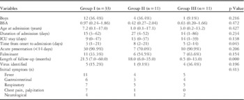
Group I included 33 of the 55 patients, and groups II and III included 11 patients each. The overall mortality was 20%. The clinical characteristics of patients according to their outcomes are presented in Table 1. The median age at admission was 6.2 years (range, 0.1–17.1 years). In total, 17 patients were boys (30%). Sex, body surface area, age at admission, duration of admission, and ICU stay were not statistically different between the three groups. Days from onset to admission were different between the three groups (p=0.043); group II exhibited the longest period from onset to admission (median, 8 days; range, 2–21 days); however, acute presentation (<14 days) was not different between the three groups. The length of follow-up was short in group III, because the outcome of almost all these patients was hospital death, with the exception of one late death (13 months). In all, 24 patients exhibited acute fulminant myocarditis (43%); the frequency of acute fulminant myocarditis was not different between the three groups. The virus detection rate was relatively lower in our data. The initial symptoms were variable, from fever and gastrointestinal, respiratory, and cardiologic manifestations to neurological symptoms.
Comparison of clinical characteristics between the three groups of patients.

BSA=body surface area
Values are the median (range)
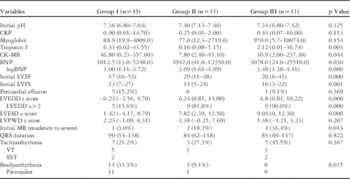
The laboratory data are shown in Table 2. There was no difference in initial pH or C-reactive protein and myoglobin levels. The levels of troponin I and creatine kinase-myoglobin were both higher in group I compared with groups II and III. On the other hand, brain natriuretic peptide exhibited a different pattern, in that its median value was lowest in group I and highest in group II; moreover, groups II and III were more likely than group I to have a high brain natriuretic peptide level (p=0.030), although there was no significant difference after transforming brain natriuretic peptide values to log brain natriuretic peptide values (p=0.080). Regarding the echocardiographic findings, the systolic function of the left ventricle was worse in groups II and III compared with group I, and the initial left ventricular ejection fraction and left ventricular fractional shortening were lowest in group III and highest in group I.
Comparison of laboratory data and echocardiographic data between the three groups of patients.

BNP=brain natriuretic peptide; CK-MB=creatine kinase-myoglobin; CRP=C-reactive protein; LVEDD=left ventricular end-diastolic dimension; LVEF=left ventricular ejection fraction; LVESD=left ventricular end-systolic dimension; LVFS=left ventricular fractional shortening; LVPWD=left ventricular posterior wall thickness dimension; MR=mitral regurgitation; SVT=supraventricular tachycardia; VT=ventricular tachycardia
Values are the median (range)
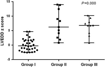
Left ventricle dilation was obvious in groups II and III, as the median value of the left ventricular end-diastolic dimension z score in group I was −0.23 (−2.56–4.70), whereas in group II it was 6.24 (0.81–14.00) and in group III it was 6.8 (0.81–10.22). When we set the standard value of left ventricle dilation at 2, the ratio of >2 of the left ventricular end-diastolic dimension z score was far higher in groups II and III compared with group I, in that nine belonged to group II (81.8%), nine belonged to group III (90.0%), and five belonged to group I (15.6%). The left ventricular end-systolic dimension z score exhibited a similar pattern to the left ventricular end-diastolic dimension z score. The left ventricular posterior wall thickness dimension z score was not different between the three groups. Significant mitral regurgitation, defined as that with a moderate-to-severe degree, was higher in group III (p=0.013).
Of the 55 patients, 15 (27%) had significant tachyarrhythmia, with no differences between the three groups, but bradyarrhythmia was complicated by acute myocarditis in 12 patients, who all received insertion of a temporary pacemaker. Moreover, not only did they all survive, all but one of these patients exhibited a complete recovery (p=0.035).
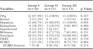
The treatment data are listed in Table 3. The majority of patients (98%) received intravenous immunoglobulin, and six patients (10%) received steroids in addition to intravenous immunoglobulin. Dopamine, isoproterenol, or milrinone infusion was performed according to the haemodynamic state, with no significant differences between the three groups. The patients in group III received significantly more epinephrine infusion (n=9 patients (81.8%); p=0.000). There was no difference regarding the application of a ventilator and continuous renal replacement therapy between the three groups. Of the 55 patients, 16 (29%) received extracorporeal membrane oxygenation, and patients in group III were significantly more likely to receive extracorporeal membrane oxygenation (eight out of 11 patients (72.7%); p=0.004); however, there was no difference in extracorporeal membrane oxygenation duration between the three groups (p=0.274).
Comparison of treatment between the three groups of patients.

CRRT=continuous renal replacement therapy; ECMO=extracorporeal membrane oxygenation; IVIg=intravenous immunoglobulin
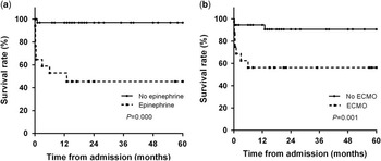
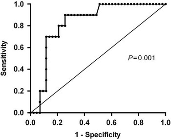
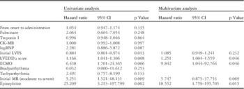
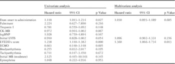
The results of the analysis of the predictors of mortality are presented in Table 4. The univariate analysis showed that the initial left ventricular fractional shortening, left ventricular end-diastolic dimension z score, extracorporeal membrane oxygenation, moderate-to-severe mitral regurgitation, and epinephrine infusion were related to mortality (p<0.05). Subsequently, we included these predictors in a multivariate analysis, which showed that a higher left ventricular end-diastolic dimension z score was associated with an increase in the odds of mortality (odds ratio (OR), 1.251; 95% confidence interval (CI), 1.004–1.559), extracorporeal membrane oxygenation application was associated with an increase in mortality (OR, 9.842; 95% CI, 1.044–92.764), and epinephrine infusion was also associated with an increase in mortality (OR, 18.552; 95% CI, 1.759–195.705). The survival rate in patients treated with epinephrine was significantly lower compared with patients who did not receive epinephrine (p=0.000), whereas the survival rate in patients treated with extracorporeal membrane oxygenation was significantly lower compared with patients who did not receive extracorporeal membrane oxygenation (p=0.001) (Fig 1). The receiver operating characteristic curve of the left ventricular end-diastolic dimension z score at diagnosis showed a cut-off level of 3.01 for predicting mortality, with an area under the curve of 0.831, sensitivity of 90%, and specificity of 74.4% (95% CI, 0.714–0.948) (Fig 2).
Survival rate depending on ( a ) epinephrine use and ( b ) extracorporeal membrane oxygenation (ECMO) application during hospital admission.

The receiver operating characteristic curve of left ventricular end-diastolic dimension (LVEDD) z score at diagnosis, predicting mortality. The cut-off value of the LVEDD z score that predicted mortality was 3.01 (area under the curve=0.831; sensitivity of 90%; specificity of 74.4%).

Predictors of mortality as assessed using univariate and multivariate analyses.

BNP=brain natriuretic peptide; CI=confidence interval; CK-MB=creatine kinase-myoglobin; ECMO=extracorporeal membrane oxygenation; LVEDD=left ventricular end-diastolic dimension; LVFS=left ventricular fractional shortening; MR=mitral regurgitation
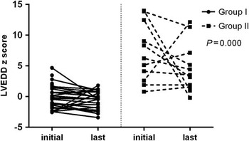
We performed univariate and multivariate analyses in the survivor group to identify predicting factors of incomplete recovery. From onset to admission, a lower left ventricular fractional shortening and a higher left ventricular end-diastolic dimension z score at diagnosis were significant predictors in the univariate analysis. In the multivariate analysis, a higher left ventricular end-diastolic dimension z score was associated with an increase in the odds of incomplete recovery (OR, 1.360; 95% CI, 1.066–1.734; p=0.013) (Table 5). Thus, a higher left ventricular end-diastolic dimension z score at diagnosis was a significant predictor of an adverse outcome including mortality and incomplete recovery. Figure 3 shows the different distribution of initial left ventricular end-diastolic dimension z score according to outcome. The distribution of the left ventricular end-diastolic dimension z score in group I ranged from −2.56 to 4.70; 17 patients had a left ventricular end-diastolic dimension z score<0 – that is, group I was more likely to have a lower initial left ventricular end-diastolic dimension compared with groups II and III. Figure 4 shows the change in the left ventricular end-diastolic dimension z score from diagnosis to the last follow-up according to outcome in survivors. The variation pattern was significantly different between two groups, as group I changed to a recovery of left ventricle dilatation within the normal range (−3.40–1.83), whereas group II changed to a variable value of left ventricular end-diastolic dimension z score from a normal to an extremely increased value (−0.20–12.14).
Distribution of initial left ventricular end-diastolic dimension (LVEDD) z score according to outcome. Group I, complete recovery; group II, incomplete recovery; group III, death. Line, median with range.

Change in left ventricular end-diastolic dimension (LVEDD) z score according to outcomes in survivors. Group I, complete recovery; group II, incomplete recovery.

Predictors of incomplete recovery among survivors as assessed using univariate and multivariate analyses.

BNP=brain natriuretic peptide; CI=confidence interval; CK-MB=creatine kinase-myoglobin; ECMO=extracorporeal membrane oxygenation; LVEDD=left ventricular end-diastolic dimension; LVFS=left ventricular fractional shortening; MR=mitral regurgitation
Discussion
Ventricular enlargement was a major factor that affected mortality in children with acute myocarditis in our study, and extracorporeal membrane oxygenation application and epinephrine infusion were factors that affected mortality significantly. Ventricular enlargement in the acute phase reflects severe myocardial damage, and epinephrine or extracorporeal membrane oxygenation application reflect a haemodynamically compromised state. Moreover, predictors that affect incomplete recovery showed that a higher left ventricular end-diastolic dimension z score was also a major factor that affected incomplete recovery, even though it had a short follow-up. Therefore, we can conclude that ventricular enlargement at diagnosis was the only significant predictor of mortality and morbidity. Moreover, it can be interpreted that a small or normal left ventricle chamber, which is a common feature of fulminant myocarditis, can be a predictor of good outcome in children with acute myocarditis.
To review the outcomes and predictors of mortality in paediatric acute myocarditis in the literature, Lee et alReference Lee, McCrindle and Bohn 5 investigated clinical outcomes in 36 individuals among a paediatric population with histologically confirmed lymphocytic myocarditis; they found high rates of survival (84%), which was consistent with the survival rate observed in our study (80%). Nevertheless, it differed from our result regarding ventricular size, as there was no significant difference in left ventricular end-diastolic dimension between survivors and the death/transplant group, although left ventricular end-diastolic dimension data were only available for four patients in the death group. English et alReference English, Janosky and Ettedgui 14 showed that the outcome of paediatric myocarditis of freedom from death or transplantation at 1 year was 81% and focussed on whether the mode of treatment such as intravenous immunoglobulin or steroid administration influenced the outcomes. Mechanical circulatory support was a risk factor for adverse outcome, which was similar to our results. Ghelani et alReference Ghelani, Spaeder and Pastor 3 also found a good survival rate (heart transplantation/death, 11.5%), and extracorporeal membrane oxygenation, ventricular assist device, and vasoactive medications were independently associated with increased mortality/transplantation. Miyake et alReference Miyake, Teele and Chen 15 showed that in-hospital arrhythmias were associated with worse outcome, defined as the need for mechanical support, orthotopic heart transplant, or death, in children with acute myocarditis. Anderson et alReference Anderson, Silver and Richmond 16 also reported data from a large population that supported the results of Miyake, focussing on in-hospital arrhythmias, which was a significant predictor of outcomes in paediatric patients with acute myocarditis. Regarding arrhythmia, our data showed that bradyarrhythmia with pacemaker was significantly elevated in the complete recovery group and that the death group included no patients with bradyarrhythmia; moreover, tachyarrhythmia was not associated with mortality.
Consistent with our data, Kuhn et alReference Kuhn, Shapiro and Walls 4 showed that left ventricular dilatation was a predictive factor at admission of poor outcome, although poor left ventricular systolic function and moderate-to-severe mitral regurgitation were also significant factors, which differed from our data.
Our data were distinct in that initial left ventricle dilatation, rather than left ventricular systolic function or arrhythmia, was strongly associated with mortality. Severe left ventricular dilation reflects severe myocardial damage in the acute phase,Reference Kindermann, Kindermann and Kandolf 17 as well as ongoing injury with persistent viral infection or immune response at the diagnostic point. Left ventricle dilation is a common feature in acute lymphocytic myocarditis versus fulminant lymphocytic myocarditis, which is associated with a small left ventricle chamber size and increased wall thickness.Reference Felker, Boehmer and Hruban 18 Several studies have reported the outcomes of children with fulminant myocarditis and of those receiving extracorporeal membrane oxygenation, who exhibited relatively good outcomes as the severity decreased.Reference Teele, Allan and Laussen 11 , Reference Wilmot, Morales and Price 19 , Reference Duncan, Bohn and Atz 20 It is known that fulminant myocarditis has a relatively good outcome in adults;Reference McCarthy, Boehmer and Hruban 7 however, it is also known that the fulminant form is more common in adults than in the paediatric group,Reference Cooper 1 and that children have a more variable outcome, from complete recovery to death. Thus, our data are meaningful in that we confirmed that left ventricle dilatation, which is a known common feature in non-fulminant myocarditis, was also a predictor of mortality in acute myocarditis, not only in adults but also in children.
We can see the character of distribution of the left ventricular end-diastolic dimension z score in the complete recovery group, which ranged below −2. This means that a small-to-normal left ventricle is a significant factor in the recovery of the myocardium. We obtained a nearly normal range for the cut-off value of the left ventricular end-diastolic dimension z score for predicting incomplete recovery. A more exact cut-off value for the left ventricular end-diastolic dimension z score for predicting incomplete recovery can be expected if the patients recruited into the incomplete recovery group have a longer follow-up period. When detecting changes in the left ventricular end-diastolic dimension z score in the survivor group, we found that left ventricle normalisation was rare in group II, with the exception of two patients. A study performed in children with idiopathic dilated cardiomyopathy showed that younger age and a lower left ventricular end-diastolic dimension z score at diagnosis independently predicted normalisation of the left ventricle, and that normalisation of the left ventricle occurred at a frequency of only 22% within 2 years of diagnosis, as assessed using serial echocardiographyReference Everitt, Sleeper and Lu 21 – that is, patients with a higher left ventricle dimension had a reduced potential to recover compared with those with a normal dimension.
In total, 17 patients received extracorporeal membrane oxygenation, and the survival rate was 52% in our study, which was lower than that reported by another study (70%).Reference Teele, Allan and Laussen 11 Patients who received extracorporeal membrane oxygenation exhibited increased mortality (by 9.8-fold) in multivariate analyses. Thus, extracorporeal membrane oxygenation application seems to reflect not only disease severity but also complications of extracorporeal membrane oxygenation or a variable outcome according to centre. Epinephrine use also increased mortality (by 18.5-fold) in multivariate analyses. It can be interpreted that sicker patients have higher mortality; however, these factors were less important predictors of mortality, because these treatment modalities are surrogates for a more fulminant presentation and do not represent clinical manifestations or laboratory and echocardiographic findings.
Interestingly, troponin I and creatine kinase-myoglobin were higher in group I, whereas brain natriuretic peptide was higher in groups II and III, although they were not significant predictors of mortality in the univariate analysis. This can be explained by the difference of duration from onset to admission, as group I had a relatively short interval to admission, in that troponin I and creatine kinase-myoglobin reflect acute cardiac injury. The high brain natriuretic peptide levels observed in groups II and III can be explained by a positive correlation between left ventricle dimension and brain natriuretic peptide. Of note, all patients with significant bradyarrhythmia who received a pacemaker survived, which could lead to the interpretation that those patients presented more fulminant and severe symptoms such as seizure; therefore, we were able to make a fast diagnosis and provide appropriate interventions to these patients.
We are aware that the clinical diagnosis of acute myocarditis was a limitation in our study. We did not perform endomyocardial biopsy or cardiac MRI in any of the patients because of the difficulty in performing an endomyocardial biopsyReference Baughman 22 and the age limit for cardiac MRI. Thus, we were not able to prove definite myocarditis histologically for our patient group. To overcome these limitations, that is, the lack of biopsy or cardiac MRI, we attempted to detect the clinical signs of viral infection and to exclude patients with acute heart failure who potentially did not have myocarditis. Nevertheless, we were not able to exclude the possibility that our study included non-symptomatic patients with existing idiopathic dilated cardiomyopathy who were incidentally infected with some virus at the diagnostic point.
In addition, the size of the sample included in our study was small, and the number of patients with a proven virus identified in laboratory examinations was also small. Moreover, we did not perform a volumetric echocardiographic study to evaluate left ventricle systolic function. Although this was a preliminary study aimed at assessing a small sample in a single centre, the results were clinically meaningful: left ventricle dilatation at admission was an important echocardiographic finding for clinicians, because echocardiography is a tool that is most readily available to cardiologists.
In conclusion, we found that extracorporeal membrane oxygenation application, epinephrine infusion, and a high left ventricular end-diastolic dimension z score were significant predictors of mortality in children with acute myocarditis. A high left ventricular end-diastolic dimension z score at admission was a significant predictor of worse outcomes, both regarding mortality and incomplete recovery. A dilated left ventricle at diagnosis reflected severe myocardial damage and widespread involvement in the acute phase; therefore, if children with acute myocarditis present with a dilated left ventricle at diagnosis, clinicians should perform intensive monitoring to avoid aggravation of the state of the patient or consider proper management to avoid poor outcomes and missing the critical time of treatment. The results of our study warrant further investigation and replication.
Acknowledgements
This work was supported by a 2-Year Research Grant of Pusan National University.
Financial Support
This research received no specific grant from any funding agency, commercial, or not-for-profit sectors.
Conflicts of Interest
None.