INTRODUCTION
Pediatric concussion is a prevalent public health burden, with it being estimated that one in every five youth will sustain a concussion before the age of 18 years (Ilie, Boak, Adlaf, Asbridge, & Cusimano, Reference Ilie, Boak, Adlaf, Asbridge and Cusimano2013). Although the majority of youth who sustain a concussion recover from their post-concussion symptoms within 4–6 weeks (Barlow et al., Reference Barlow, Crawford, Stevenson, Sandhu, Belanger and Dewey2010; Davis et al., Reference Davis, Anderson, Babl, Gioia, Giza, Meehan, Moser, Purcell, Schatz, Schneider, Takagi, Yeates and Zemek2017; Zemek et al., Reference Zemek, Barrowman, Freedman, Gravel, Gagnon, McGahern, Aglipay, Sangha, Boutis, Beer, Craig, Burns, Farion, Mikrogianakis, Barlow, Dubrovsky, Meeuwisse, Gioia, Meehan, Beauchamp, Kamil, Grool, Hoshizaki, Anderson, Brooks, Yeates, Vassilyadi, Klassen, Keightley, Richer, De Matteo, Osmond, Xie, Chatfield, Dow, Papadimitropoulos, Levesque, Langford, Tran, McGahern, DiGirolamo, Mazza, Lagace, Cook, Fitzpatrick, MacIntyre and Moore2016), concerns have arisen that these injuries can cause alterations in brain microstructure that may persist long after the injury has occurred (Churchill et al., Reference Churchill, Hutchison, Richards, Leung, Graham and Schweizer2017).
Advanced neuroimaging techniques such as diffusion magnetic resonance imaging (dMRI) have provided a non-invasive means of examining and quantifying white matter microstructure through in vivo measures of water diffusion (Beaulieu, Reference Beaulieu2002; Bigler, Reference Bigler2013). Studies utilizing dMRI in concussion populations have demonstrated that the corpus callosum is a commonly altered white matter pathway acutely following concussion (Aoki, Inokuchi, Gunshin, Yahagi, & Suwa, Reference Aoki, Inokuchi, Gunshin, Yahagi and Suwa2012; Dodd, Epstein, Ling, & Mayer, Reference Dodd, Epstein, Ling and Mayer2014; Schmidt et al., Reference Schmidt, Hayward, Brown, Zwicker, Ponsford, van Donkelaar, Babul and Boyd2018; Virji-Babul et al., Reference Virji-Babul, Borich, Makan, Moore, Frew, Emery and Boyd2013), likely due to its position on the midline axis that increases its risk of axonal shearing (Aoki et al., Reference Aoki, Inokuchi, Gunshin, Yahagi and Suwa2012). Axonal shearing can lead to alterations in diffusion metrics such as fractional anisotropy (FA), mean diffusivity (MD), axial diffusivity (AD), and radial diffusivity (RD), with more severe injuries tending to cause more drastic alterations in diffusivity (Kraus et al., Reference Kraus, Susmaras, Caughlin, Walker, Sweeney and Little2007).
Despite reports of alterations in diffusivity measures following concussion, the direction of these changes has been a source of substantial disagreement (Dodd et al., Reference Dodd, Epstein, Ling and Mayer2014; Eierud et al., Reference Eierud, Craddock, Fletcher, Aulakh, King-Casas, Kuehl and Laconte2014; Honce, Nyberg, Jones, & Nagae, Reference Honce, Nyberg, Jones and Nagae2016; Moore, Kay, & Ellemberg, Reference Moore, Kay and Ellemberg2018; Narayana, Reference Narayana2017; Schmidt et al., Reference Schmidt, Hayward, Brown, Zwicker, Ponsford, van Donkelaar, Babul and Boyd2018). Most dMRI studies have focused on adult or mixed age samples in the acute periods following injury, and very few studies have focused on more chronic time points (i.e., greater than 3 or 6 months post-concussion). Studies that have examined chronic time points have reported decreases (Dean, Sato, Vieira, McNamara, & Sterr, Reference Dean, Sato, Vieira, McNamara and Sterr2015; Grossman et al., Reference Grossman, Ge, Jensen, Babb, Miles, Reaume, Silver, Grossman and Inglese2012; Inglese et al., Reference Inglese, Makani, Johnson, Cohen, Silver, Gonen and Grossman2005; Veeramuthu et al., Reference Veeramuthu, Narayanan, Kuo, Delano-Wood, Chinna, Bondi, Waran, Ganesan and Ramli2015), increases (Henry et al., Reference Henry, Tremblay, Tremblay, Lee, Brun, Lepore, Theoret, Ellemberg and Lassonde2011; Lipton et al., Reference Lipton, Kim, Park, Hulkower, Gardin, Shifteh, Kim, Zimmerman, Lipton and Branch2012; Orr et al., Reference Orr, Albaugh, Watts, Garavan, Andrews, Nickerson, Gonyea, Hipko, Zweber, Logan and Hudziak2016), and no differences (Chamard, Lefebvre, Lassonde, & Theoret, Reference Chamard, Lefebvre, Lassonde and Theoret2016) in corpus callosum FA long after injury. Decreased FA is generally associated with axonal demyelination and degradation and is often expected after injury (Shenton et al., Reference Shenton, Hamoda, Schneiderman, Bouix, Pasternak, Rathi, Vu, Purohit, Helmer, Koerte, Lin, Westin, Kikinis, Kubicki, Stern and Zafonte2012). However, increased FA has also been observed and may be indicative of cytotoxic edema acutely after injury (Wilde et al., Reference Wilde, McCauley, Hunter, Bigler, Chu, Wang, Hanten, Troyanskaya, Yallampalli, Li, Chia and Levin2008) or a loss of crossing fibers chronically after injury (Henry et al., Reference Henry, Tremblay, Tremblay, Lee, Brun, Lepore, Theoret, Ellemberg and Lassonde2011; Orr et al., Reference Orr, Albaugh, Watts, Garavan, Andrews, Nickerson, Gonyea, Hipko, Zweber, Logan and Hudziak2016). Though often reported in terms of FA, similar inconsistent findings for measures of MD, AD, and RD in the corpus callosum have been reported as well (Eierud et al., Reference Eierud, Craddock, Fletcher, Aulakh, King-Casas, Kuehl and Laconte2014; Honce et al., Reference Honce, Nyberg, Jones and Nagae2016; Moore et al., Reference Moore, Kay and Ellemberg2018; Narayana, Reference Narayana2017; Schmidt et al., Reference Schmidt, Hayward, Brown, Zwicker, Ponsford, van Donkelaar, Babul and Boyd2018). Unfortunately, few studies have utilized dMRI in pediatric-only populations with concussion. A recent systematic review on pediatric neuroimaging (Schmidt et al., Reference Schmidt, Hayward, Brown, Zwicker, Ponsford, van Donkelaar, Babul and Boyd2018) found that only one dMRI study thus far has evaluated microstructural changes in the corpus callosum long after pediatric concussion (Van Beek, Vanderauwera, Ghesquière, Lagae, & De Smedt, Reference Van Beek, Vanderauwera, Ghesquière, Lagae and De Smedt2015). This longitudinal study found white matter alterations acutely after injury and these alterations had resolved by 6 months post-injury (Van Beek et al., Reference Van Beek, Vanderauwera, Ghesquière, Lagae and De Smedt2015). Although this study provides the first insight into longer-term dMRI outcomes 6 months following pediatric concussion, further investigation is needed given the large discrepancies found in the dMRI literature and using even longer-term follow-up periods.
Acutely following concussion, clinical or behavioral manifestations of white matter alterations may include worse post-concussion symptom severity and poorer cognitive performance (Geary, Kraus, Pliskin, & Little, Reference Geary, Kraus, Pliskin and Little2010; Hellstrøm et al., Reference Hellstrøm, Westlye, Kaufmann, Trung Doan, Søberg, Sigurdardottir, Nordhøy, Helseth, Andreassen and Andelic2017; Khong, Odenwald, Hashim, & Cusimano, Reference Khong, Odenwald, Hashim and Cusimano2016; Womack et al., Reference Womack, Paliotta, Strain, Ho, Skolnick, Lytton, Turtzo, McColl, Diaz-Arrastia and Bergold2017). A systematic review conducted by Khong and colleagues concluded that decreased FA and increased MD and RD are correlated with greater symptom severity and prolonged post-concussion symptoms (Khong et al., Reference Khong, Odenwald, Hashim and Cusimano2016). These findings were also supported by a study of adults who were at 1-year post-concussion (Hellstrøm et al., Reference Hellstrøm, Westlye, Kaufmann, Trung Doan, Søberg, Sigurdardottir, Nordhøy, Helseth, Andreassen and Andelic2017). Similarly, altered diffusivity acutely after the concussion is associated with decreased verbal memory and executive functioning, and increased reaction time at 1-year post-injury (Geary et al., Reference Geary, Kraus, Pliskin and Little2010; Womack et al., Reference Womack, Paliotta, Strain, Ho, Skolnick, Lytton, Turtzo, McColl, Diaz-Arrastia and Bergold2017). These studies suggest that microstructural alterations may underlie the cognitive and behavioral problems that present following concussion, but further research is needed to determine if these associations still hold true in children and adolescents long after injury.
The present study aimed to explore whether microstructure of the corpus callosum is altered several years after pediatric concussion and if associations exist between white matter microstructure, post-concussion symptoms, and cognitive performance. Based on previous findings (Van Beek et al., Reference Van Beek, Vanderauwera, Ghesquière, Lagae and De Smedt2015), we hypothesized that children and adolescents with concussion would not exhibit significant differences in FA, MD, AD, and RD in the corpus callosum compared to youth with orthopedic injury (OI) several years after injury. Despite no group differences hypothesized in the first aim, we hypothesized that diffusivity metrics of the corpus callosum (i.e., lower FA, higher MD, and higher AD) would be correlated with greater post-concussion symptom severity and lower cognitive performance.
METHODS
Participants
Sixty-one children and adolescents between 8 and 19 years of age with a history of concussion (n = 40) or OI (n = 21) were recruited for this study. Three participants from the concussion group were unable to complete neuroimaging (all under the age of 10 years) and one participant in the concussion group had unusable imaging data due to motion artifact (12 years old). All four participants and their parents completed symptom ratings, with two participants reporting symptom scores slightly above the concussion group average and two of the participants reporting symptom scores below the group average. All OI participants completed the neuroimaging protocol and had usable imaging data. This left a final sample size of N = 57 (concussion, n = 36; OI, n = 21). Participants were identified and invited to participate from existing research databases that included children and adolescents who presented to concussion clinics, sports medicine clinics, or emergency departments in Calgary, Alberta, Canada between September 2014 and November 2016. Children and adolescents with concussion were included if they were diagnosed with a concussion by a physician at the time of injury at one of the recruitment sites. Concussion was defined as including an impact/acceleration of the head coupled with one or more of the following: report of at least one new post-concussive-type symptom at time of injury (e.g., headache, dizziness, or confusion) (Carroll et al., Reference Carroll, Cassidy, Holm, Kraus and Coronado2004), a Glasgow Coma Scale (GCS) score between 13 and 15 (Teasdale & Jennett, Reference Teasdale and Jennett1974), loss of consciousness lasting less than 30 min (or not present), and/or post-traumatic amnesia lasting less than 24 hr (or not present) (Lee, Reference Lee2007). Neuroimaging is not routinely done at these recruitment sites for youth presenting with a concussion. In a few cases, neuroimaging may have been ordered if clinically indicated, but the presence of any finding would have excluded an individual from eligibility for this study.
OI controls were included if they had sustained a minor injury to the thorax, upper extremity, or lower extremity with a score of 3 or less on the Abbreviated Injury Scale (Garthe & States, Reference Garthe and States1999). OI participants must have also presented to a healthcare professional for the management of their OI and must not have had a history of concussion or any traumatic brain injuries. Participants with an OI were excluded if it was suspected that they sustained a concussion or if there was an indication of a head or facial injury. Participants from either group were excluded if they had a history of substance abuse, any psychiatric hospitalizations, neurological disorders (e.g., seizures, hydrocephalus), visual, hearing, motor or language skills that prevented testing, or any contraindication to MRI (e.g., braces, metal in body, pregnancy). Participants for either group were not excluded if they had learning disabilities, attention problems, or mental health issues (e.g., anxiety, depression), as these difficulties are often present in this population, are associated with a risk of sustaining an injury, and are associated with recovery times (Brooks, Iverson, Atkins, Zafonte, & Berkner, Reference Brooks, Iverson, Atkins, Zafonte and Berkner2016; Iverson, Atkins, Zafonte, & Berkner, Reference Iverson, Atkins, Zafonte and Berkner2016; Iverson, Wojtowicz, et al., Reference Iverson, Wojtowicz, Brooks, Maxwell, Atkins, Zafonte and Berkner2016; Zemek et al., Reference Zemek, Barrowman, Freedman, Gravel, Gagnon, McGahern, Aglipay, Sangha, Boutis, Beer, Craig, Burns, Farion, Mikrogianakis, Barlow, Dubrovsky, Meeuwisse, Gioia, Meehan, Beauchamp, Kamil, Grool, Hoshizaki, Anderson, Brooks, Yeates, Vassilyadi, Klassen, Keightley, Richer, De Matteo, Osmond, Xie, Chatfield, Dow, Papadimitropoulos, Levesque, Langford, Tran, McGahern, DiGirolamo, Mazza, Lagace, Cook, Fitzpatrick, MacIntyre and Moore2016; Zemek, Farion, Sampson, & McGahern, Reference Zemek, Farion, Sampson and McGahern2013). The University of Calgary Health Research Ethics Board approved the study, and informed consent and assent were obtained prior to participation.
Measures
Background history
At the beginning of the appointment, parents completed a form that collected information on demographics and background history. This information included date of most recent injury, number of previously diagnosed concussions, details regarding loss of consciousness or post-traumatic amnesia, medication status, and any history of attention concerns or attention deficit hyperactivity disorder, learning concerns or learning disorder, mental health concerns, or headache or migraine.
MRI image acquisition
All participants completed neuroimaging at the Alberta Children’s Hospital using a 3-T MR scanner (GE MR750W, GE Healthcare) with a 32-channel head coil (MRI instruments). The imaging protocol included a 3D anatomical T1 -weighted [fast spoiled gradient-echo brain volume, with repetition time (TR) = 8.2 ms, echo time (TE) = 3.2 ms, flip angle θ = 10°, field of view (FOV) = 240 mm, 230 slices, 0.8 mm thickness, and scan duration = 5.53 min] with 0.8 mm3 isotropic resolution and whole brain coverage. During the diffusion scan, 32 non-collinear diffusion encoding gradient directions (b = 750 s/mm2, voxels = 2.2 mmisotropic) and 3 non-diffusion encoding directions (b = 0 s/mm2) were sampled using 60 contiguous axial slices (TR = 11,500 ms, TE = 70 ms, FOV = 220 mm, scan duration = 6 mins).
Symptom ratings
The 26-item Post-Concussion Symptom Inventory (PCSI) (Rutland-Brown, Langlois, Thomas, & Xi, Reference Rutland-Brown, Langlois, Thomas and Xi2006) was used to measure post-concussion symptoms. Both self-report and parent-report versions were completed. The PCSI takes approximately 5–10 min to complete and allows participants to rank their symptoms on a seven-point Likert scale (ranging from 0 = never to 6 = almost always). PCSI total scores range from 0 to 156, with higher scores indicating increased symptom severity. The PCSI has adequate reliability (Gioia, Schneider, Vaughan, & Isquith, Reference Gioia, Schneider, Vaughan and Isquith2009; Rutland-Brown et al., Reference Rutland-Brown, Langlois, Thomas and Xi2006), validity (Barlow, Crawford, Brooks, Turley, & Mikrogianakis, Reference Barlow, Crawford, Brooks, Turley and Mikrogianakis2015; Barlow et al., Reference Barlow, Crawford, Stevenson, Sandhu, Belanger and Dewey2010), and has been included in the National Institute of Neurological Disorders and Stroke (NINDS) pediatric brain injury common data elements (McCauley et al., Reference McCauley, Wilde, Anderson, Bedell, Beers, Campbell, Chapman, Ewing-Cobbs, Gerring, Gioia, Levin, Michaud, Prasad, Swaine, Turkstra, Wade and Yeates2012).
Cognitive testing
The CNS Vital Signs (CNSVS) (Gualtieri & Johnson, Reference Gualtieri and Johnson2006) computerized neurocognitive test battery was used to measure cognitive performance. CNSVS domain scores generally have medium to large correlations with more conventional neuropsychological measures used in youth populations (Plourde, Hrabok, Sherman, & Brooks, Reference Plourde, Hrabok, Sherman and Brooks2018) and marginal (r = .63) to high (r = .82) test–retest reliabilities (Brooks & Barlow, Reference Brooks and Barlow2011). CNSVS has sensitivity in samples with mixed neurological disorders (Brooks & Sherman, Reference Brooks and Sherman2012), concussion (Brooks, Khan, Daya, Mikrogianakis, & Barlow, Reference Brooks, Khan, Daya, Mikrogianakis and Barlow2014), and moderate to severe traumatic brain injury (TBI) (Plourde & Brooks, Reference Plourde and Brooks2017) and is part of the NINDS pediatric brain injury common data elements (McCauley et al., Reference McCauley, Wilde, Anderson, Bedell, Beers, Campbell, Chapman, Ewing-Cobbs, Gerring, Gioia, Levin, Michaud, Prasad, Swaine, Turkstra, Wade and Yeates2012). CNSVS domain scores have a normalized mean of 100 (SD = 15) based on a standardized sample. In the present study, select domains scores for verbal memory, reaction time, and cognitive flexibility were used. These measures have previously been shown to differentiate youth with concussions from youth with OI in the emergency department (Brooks et al., Reference Brooks, Khan, Daya, Mikrogianakis and Barlow2014).
The Test of Memory Malingering (TOMM) (Tombaugh, Reference Tombaugh1996) was included as a measure of performance validity during cognitive testing. Cutoff scores used for this sample to determine pass or fail were consistent with recommendations from the technical manual (Tombaugh, Reference Tombaugh1996) and prior research with youth (Brooks, Sherman, & Krol, Reference Brooks, Sherman and Krol2012). One participant in the concussion group scored below the acceptable cutoff on the TOMM, therefore, their cognitive scores and symptom ratings were excluded from analyses. Their imaging data were still included.
Analyses
MRI imaging pre-processing and analyses
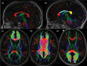
Head motion and eddy current correction were performed on the diffusion tensor volumes using FMRIB Software Library (FSL) (Woolrich et al., Reference Woolrich, Jbabdi, Patenaude, Chappell, Makni, Behrens, Beckmann, Jenkinson and Smith2009). MRTrix3 (Tournier, Calamante, & Connelly, Reference Tournier, Calamante and Connelly2012) was used to calculate the diffusion tensor for each voxel and generate FA maps that were subsequently color coded by the primary diffusion direction. Based on previous literature in pediatric concussion (Maugans, Farley, Altaye, Leach, & Cecil, Reference Maugans, Farley, Altaye, Leach and Cecil2012; Mayer et al., Reference Mayer, Ling, Yang, Pena, Yeo and Klimaj2012) and TBI populations (Rutgers et al., Reference Rutgers, Fillard, Paradot, Tadié, Lasjaunias and Ducreux2008), three regions of interest (ROIs) within the corpus callosum were utilized. Together, these ROIs encompassed the entire corpus callosum, with fibers oriented predominantly in the left–right plane (Figure 1A). These ROIs specifically isolated the genu, body, and splenium of the corpus callosum and were manually outlined on the color-encoded FA map on the central mid-sagittal slice (Figure 1B).
ROI placements and tractography representations. (A) Central mid-sagittal slice of the corpus callosum where fibers are oriented predominantly in the left–right plane (red on color map). (B) Genu (red), body (blue), and splenium (yellow) regions of interest (ROIs), drawn on five mid-sagittal slices. (C) Tractography representation of the genu generated from the red ROI in B. (D) Tractography representation of the body generated from the blue ROI in B. (E) Tractography representation of the splenium generated from the yellow ROI in B.

Tractography
The ROIs above were used as seed regions to reconstruct tracts of interest corresponding to the genu, body, and splenium of the corpus callosum. ROIs were manually expanded laterally in both directions to include five mid-sagittal slices of the corpus callosum (Chamard et al., Reference Chamard, Lefebvre, Lassonde and Theoret2016). Tract reconstructions were created using a probabilistic tractography algorithm within MRTrix3 (FA threshold = 0.20, angle = 90°, iterations = 7500) (Tournier et al., Reference Tournier, Calamante and Connelly2012). Exclusion ROIs were drawn to exclude streamlines from other identifiable fiber bundles (e.g., cingulum) and any spurious streamlines. Resulting tractography representations (illustrated in Figure 1C, D, and E) were saved as binary masks, and FA, MD, AD, and RD values were extracted for each tract of interest giving an average over the entire tract for each diffusion metric. These ROIs were drawn and tracts were generated using a method previously shown by our group to be highly reliable with intraclass correlation coefficients ranging from .97 to .99 (Carlson et al., Reference Carlson, Laliberté, Brooks, Hodge, Kirton, Bello-Espinosa, Hader and Sherman2014).
Along-tract analysis
To more closely investigate differences along the length of the tracts and because variations in diffusivity values tend to exist within white matter tracts (Colby et al., Reference Colby, Soderberg, Lebel, Dinov, Thompson and Sowell2012), both the genu and splenium of participants were subsampled into 21 equally spaced regions along each tract, resulting in 10 samples in each hemisphere and 1 at midline (Groeschel et al., Reference Groeschel, Tournier, Northam, Baldeweg, Wyatt, Vollmer and Connelly2014). Only the genu and splenium ROIs were analyzed using this method due to their increased vulnerability to white matter damage as compared to the body of the corpus callosum after brain injury (Rutgers et al., Reference Rutgers, Fillard, Paradot, Tadié, Lasjaunias and Ducreux2008). The choice of 21 subdivisions is consistent with the methodology used in previous studies (Groeschel et al., Reference Groeschel, Tournier, Northam, Baldeweg, Wyatt, Vollmer and Connelly2014; Grohs, Reynolds, Dewey, & Lebel, Reference Grohs, Reynolds, Dewey and Lebel2018).
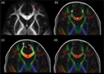
To standardize arc definition for the genu and splenium across participants, three points (Figure 2A) were drawn on the Illinois Institute of Technology Human Brain Atlas V.5.0 template FA map in standard space (Zhang & Arfanakis, Reference Zhang and Arfanakis2018). These arc points were subsequently warped into patient space using inverted linear transformations calculated between the template FA map and each patient’s FA map in native space (FSL FMRIB (Functional MRI of the Brain) Linear Image Registration Tool) (Woolrich et al., Reference Woolrich, Jbabdi, Patenaude, Chappell, Makni, Behrens, Beckmann, Jenkinson and Smith2009). Arc points were visualized overlaid on each participants’ FA map to ensure accurate placement (Figure 2B). Coordinates for the center of each arc point were used to define the arc and resample 21 sections of each tract in each participant’s native space (Figure 2C & D). Streamlines were only sampled if they passed through all three arc points. Because of increased individual variability at the ends of tracts, values for tracts projecting into the very anterior portion of the frontal pole were not sampled (as illustrated in Figure 2D). FA, MD, AD, and RD values were then extracted for each subsample.
An illustration of along-tract methods. (A) Target regions of interest (ROIs) for arc definition were defined using a template fractional anisotropy (FA) map. (B) ROIs were warped into patient space and overlaid on color map to ensure proper placement. (C) Tractography was performed in patient space. (D) Twenty-one subsamples along the genu arc were obtained and FA, mean (MD), axial (AD), and radial (RD) diffusivity were subsequently sampled. This procedure was repeated for the splenium. Images are from an orthopedic injury participant.

Statistical analyses
All statistical analyses were conducted using IBM Statistics SPSS 24 (Version 24.0; IBM corp, Armonk, NY). A mixed model analysis of covariance with age as a covariate was conducted to compare differences in diffusivity values between groups and tract regions. Sex was not associated with any statistical differences (p > 0.05) in diffusion metrics (FA, MD, RD, and AD) in any of the regions (genu, body, and splenium) and was thus not included as a covariate to preserve degrees of freedom. All other group comparisons used ANOVA for continuous data and chi-square (χ 2) for categorical data. Effect sizes are reported using partial eta squared (η p2). PCSI data were square-root transformed due to positive skewness and used for all subsequent analyses. A Shapiro–Wilk test was used to determine normality of the distribution of data, and Spearman’s or Pearson’s correlations were used to determine associations between symptom reports, cognitive data, and diffusivity values of the three subregions of the corpus callosum. Correlations were conducted between diffusion metrics for both the entire sample and separately for each group between PCSI parent proxy and self-reported symptom scores and verbal memory, cognitive flexibility, and reaction time domain scores from the CNSVS. False discovery rate (FDR) correction was used independently for each set of analyses to determine significant p-values and limit type 1 error (Benjamini & Hochberg, Reference Benjamini and Hochberg1995).
RESULTS
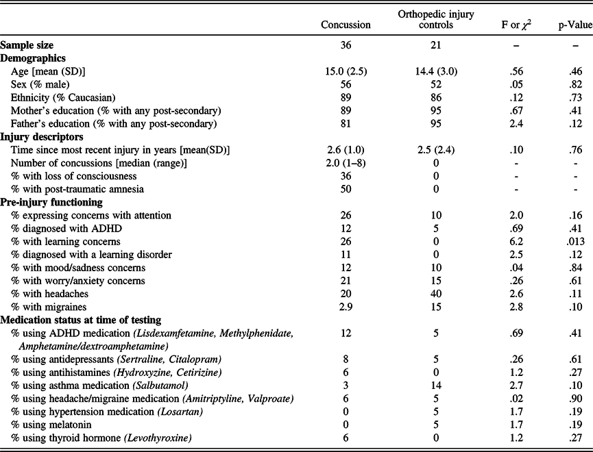
Participant characteristics for both groups are presented in Table 1. The groups did not differ significantly in demographics or average time since most recent injury. In terms of pre-injury functioning, the two groups did not differ significantly except that parents in the concussion group reported concerns about pre-injury learning difficulties more often than parents in the OI group (χ 2(1) = 6.2, p = .013). Both groups consisted primarily of sport-related injuries (67% of injuries in the concussion group and 80% of injuries in the OI group), followed by fall-related injuries (29% of injuries in the concussion group and 20% of injuries in the OI group). The concussion group also included a small proportion of participants who were injured in motor vehicle collisions (4%). The concussion group included 15 individuals with one reported concussion, 9 with two, 9 with three, 1 with six, 1 with seven, and 1 with eight.
Participant characteristics

Notes: SD = standard deviation; % = Percent; ADHD = attention deficit hyperactivity disorder.
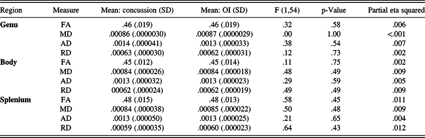
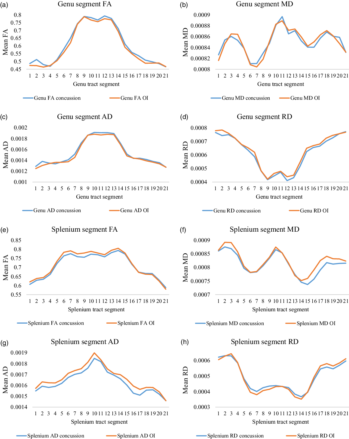
No significant group differences in diffusivity were found in the whole-tract tractography analysis (Table 2). Similarly, after correcting for multiple comparisons, no significant group differences in diffusivity were found in the 21 subdivisions of the genu and splenium (Figure 3).
Group comparisons: white matter tractography

Notes: FA = fractional anisotropy; MD = mean diffusivity; RD = radial diffusivity; AD = axial diffusivity; OI = orthopedic injury control; SD = standard deviation.
Along-tract group comparisons. Graphs representing a comparison between diffusivity values sampled from 21 regions along the genu and splenium of the corpus callosum in youth with a history of concussion (blue) or orthopedic injury (OI; orange). No significant differences were found between groups in any of the 21 regions. FA = fractional anisotropy. MD = mean diffusivity. AD = axial diffusivity. RD = radial diffusivity.

Table 3 summarizes group differences in symptom ratings and cognitive measures. Parents of youth with a history of concussion reported higher levels of post-concussion symptoms than parents of youth with a history of OI (F(1,54) = 10.46, p = .002, η p2 = .162). Similarly, youth with a history of concussion self-reported higher levels of post-concussion symptoms compared to youth with a history of OI (F(1,54) = 4.37, p = .041, η p2 = .074). The groups did not differ significantly on any of the three CNSVS domains. No significant correlations were found between diffusivity metrics, post-concussion symptoms (self- and parent-reported), and cognitive test scores in the whole-tract tractography analysis. This was true when examining correlations for the entire sample as well as within each of the two groups separately. In the along-tract analysis, no significant correlations were found between subdivision diffusivity metrics (FA, MD, AD, and RD), self- and parent-reported post-concussion symptoms, and cognitive test scores after performing FDR correction.
Group differences in symptom and cognitive measures

Notes: PCSI = Post-Concussion Symptom Inventory; CNSVS = CNS Vital Signs; TOMM = Test of Memory Malingering; SD = standard deviation; Square rooted Post-Concussion Symptom Inventory scores were used for analyses.
DISCUSSION
The current dMRI literature in chronic pediatric concussion is sparse. This study aimed to use dMRI to gain a better understanding of the long-term impact of pediatric concussions on white matter microstructure and the related associations with symptoms and cognitive functioning. As hypothesized, the present results do not provide evidence of group differences in corpus callosum diffusivity between children and adolescents with concussion and those with OI who are on average 2.5 years post-injury.
Our findings extend those of the only other study examining axonal health of the corpus callosum long after pediatric concussion. Van Beek and colleagues (Van Beek et al., Reference Van Beek, Vanderauwera, Ghesquière, Lagae and De Smedt2015) found no white matter abnormalities present at 6 months post-concussion, suggesting axonal repair may be complete as early as 6 months post-injury in children and adolescents. However, this finding of rapid repair may potentially have been due to the young age of their sample, which consisted of 16 children with concussion aged 7–13 years. As noted by Lebel (Lebel & Beaulieu, Reference Lebel and Beaulieu2011), large changes in corpus callosum microstructure are observed in young adolescents and then taper off in older adolescence. The current study had a broader age range (8–18 years), with the mean age of the sample being 15 years, and 20 of 36 participants in the concussion group were older than 13 years when their injury occurred. Therefore, youth who suffer a concussion in their teens may also display no overall long-term alterations in corpus callosum microstructure. A recent dMRI study that conducted 22–47 years after injury on former high school football players who sustained two or more concussions found no long-term white matter differences in the corpus callosum compared to a control group further supports this (Terry, Mewborn, & Miller, 2019). However, studies conducted in adults with multiple concussions and persistent cognitive impairment or depression long after injury have suggested the presence of alterations in white matter microstructure (Hart et al., Reference Hart, Kraut, Womack, Strain, Didehbani, Bartz, Conover, Mansinghani, Lu and Cullum2013; Lo, Shifteh, Gold, Bello, & Lipton, Reference Lo, Shifteh, Gold, Bello and Lipton2009; Strain et al., Reference Strain, Didehbani, Cullum, Mansinghani, Conover, Kraut, Hart and Womack2013; Tremblay et al., Reference Tremblay, Henry, Bedetti, Larson-Dupuis, Gagnon, Evans, Théoret, Lassonde and De Beaumont2014). In the current study, only three participants in the concussion group had more than three concussions. These three participants were not outliers in terms of diffusion metrics and symptom scores compared to the rest of the sample. Ultimately, concerns still exist regarding the impact that multiple concussions may have on long-term white matter microstructure. Further longitudinal studies in larger samples of children and adolescents with multiple concussions remain needed.
The current results contrast with dMRI findings in adult populations that have indicated microstructural alterations persisting in the corpus callosum long after injury (Chamard et al., Reference Chamard, Lefebvre, Lassonde and Theoret2016; Dean et al., Reference Dean, Sato, Vieira, McNamara and Sterr2015; Grossman et al., Reference Grossman, Ge, Jensen, Babb, Miles, Reaume, Silver, Grossman and Inglese2012; Henry et al., Reference Henry, Tremblay, Tremblay, Lee, Brun, Lepore, Theoret, Ellemberg and Lassonde2011; Inglese et al., Reference Inglese, Makani, Johnson, Cohen, Silver, Gonen and Grossman2005; Lipton et al., Reference Lipton, Kim, Park, Hulkower, Gardin, Shifteh, Kim, Zimmerman, Lipton and Branch2012; Orr et al., Reference Orr, Albaugh, Watts, Garavan, Andrews, Nickerson, Gonyea, Hipko, Zweber, Logan and Hudziak2016; Veeramuthu et al., Reference Veeramuthu, Narayanan, Kuo, Delano-Wood, Chinna, Bondi, Waran, Ganesan and Ramli2015). The results also contrast with a more severe pediatric TBI sample that found alterations in corpus callosum microstructure many years after injury (Treble et al., Reference Treble, Hasan, Iftikhar, Stuebing, Kramer, Cox, Swank and Ewing-Cobbs2013). Considering that the present study has a pediatric concussion-only sample that averaged 2.5 years post-injury, continued myelination post-injury coupled with ample recovery time may have allowed for axonal repair to occur. As speculated by others (Henry et al., Reference Henry, Tremblay, Tremblay, Lee, Brun, Lepore, Theoret, Ellemberg and Lassonde2011; Orr et al., Reference Orr, Albaugh, Watts, Garavan, Andrews, Nickerson, Gonyea, Hipko, Zweber, Logan and Hudziak2016), these axonal repair capabilities may not be present to the same extent in adult-only samples, who may show alterations in dMRI metrics many years post-injury (Eierud et al., Reference Eierud, Craddock, Fletcher, Aulakh, King-Casas, Kuehl and Laconte2014). Previous work by Lebel (Lebel & Beaulieu, Reference Lebel and Beaulieu2011) has shown that healthy brain development involves large changes in the diffusion parameters of projection and commissural tracts of the corpus callosum in early adolescence. These changes are mostly complete by late adolescence and young adulthood (Lebel & Beaulieu, Reference Lebel and Beaulieu2011). Thus, axonal repair mechanisms may be more efficient in youth whose brains are undergoing more rapid development compared to adults.
While methods of analyzing dMRI across different studies are largely comparable, variations in techniques (i.e., ROI-based vs. whole-brain voxel-based) may also help account for inconsistent findings that are present in the chronic dMRI literature. ROI-based analyses average all voxels in a region together, potentially washing out localized differences (Snook, Plewes, & Beaulieu, Reference Snook, Plewes and Beaulieu2007). Like van Beek and colleagues (Van Beek et al., Reference Van Beek, Vanderauwera, Ghesquière, Lagae and De Smedt2015), the present study conducted a ROI analysis. However, to minimize the risk of washing out localized group differences, the present study also employed a supplementary robust along-tract analysis, which is novel in the concussion dMRI literature. The absence of group differences in the along-tract analysis lends further credence to the assertion that long-term alterations in corpus callosum microstructure are not apparent following pediatric concussion.
Furthermore, measures of diffusivity were not correlated with symptom severity or cognitive test performance despite group differences in both self-reported symptoms and parent-reported symptoms. This suggests that altered white matter in the corpus callosum is not a potential explanation for why the concussion group is still experiencing more severe symptoms at approximately 2.5 years post-injury. Very few previous studies have correlated dMRI metrics with symptom severity or cognitive performance long after injury. The strongest evidence of altered white matter microstructure being associated with increased symptom severity chronically after injury comes from Hellstrøm and colleagues (Hellstrøm et al., Reference Hellstrøm, Westlye, Kaufmann, Trung Doan, Søberg, Sigurdardottir, Nordhøy, Helseth, Andreassen and Andelic2017). However, their study consisted of a primarily adult sample, and all participants in their study were hospitalized at a level-one trauma center and therefore most likely represent a population with a more severe concussion than the present sample (none were admitted to hospital).
The current study does have some limitations to highlight. First, the design is cross-sectional, therefore, we are unable to establish whether the concussive injury altered corpus callosum microstructure acutely or subacutely following injury. Although the present study alludes to potential axonal repair in youth who suffered a concussion, corpus callosum microstructure may not have been altered at all following injury. Although most studies report alterations in diffusivity acutely following injury (Dodd et al., Reference Dodd, Epstein, Ling and Mayer2014), a few studies have found no significant differences (Maugans et al., Reference Maugans, Farley, Altaye, Leach and Cecil2012; Zhang et al., Reference Zhang, Johnson, Pennell, Ray, Sebastianelli and Slobounov2010). Ultimately, comprehensive longitudinal studies are needed in pediatric populations to accurately assess the microstructural changes that occur following concussion. Longitudinal studies in turn will allow us to better characterize recovery trajectories based on dMRI alterations. Second, the present sample could be subject to selection bias (i.e., only including youth who presented to a clinic or hospital following injury) and may not be representative of all children and adolescents who incur a concussion. An additional limitation of this study is that background history was obtained via parent report, which may have been subject to recall bias. Parents were asked to report only previous concussions that were diagnosed by a clinician to minimize parental differences in concussion definition. Therefore, some participants in the study may actually have experienced more concussions than reported, if they sustained concussions not diagnosed by a clinician. Third, the collection of acute injury variables was limited with our study methodology. For example, we did not have exact GCS scores documented for these participants, although we did only include those with GCS between 13 and 15. Finally, the modest sample size of the present study restricted statistical power and as such may have limited our ability to detect significant differences between groups as well as the ability to investigate group differences between those with a different number of concussions (i.e., one concussion vs. two concussion). The present study had 57 participants and thus may have been underpowered. However, other studies utilizing dMRI in concussions populations have had a similar number (Churchill et al., Reference Churchill, Hutchison, Richards, Leung, Graham and Schweizer2017; Inglese et al., Reference Inglese, Makani, Johnson, Cohen, Silver, Gonen and Grossman2005),if not even fewer participants (Chamard et al., Reference Chamard, Lefebvre, Lassonde and Theoret2016; Henry et al., Reference Henry, Tremblay, Tremblay, Lee, Brun, Lepore, Theoret, Ellemberg and Lassonde2011; Kraus et al., Reference Kraus, Susmaras, Caughlin, Walker, Sweeney and Little2007; Terry et al., 2019). Therefore, despite potentially being underpowered, the present sample size is consistent within the existing literature.
Conclusions
The present study is the first to examine a pediatric population more than 2 years post-concussion using ROI-based diffusion tensor imaging. The findings provide evidence that white matter microstructure is not altered in the corpus callosum several years after pediatric concussion, suggesting a lack of persistent structural damage to the corpus callosum. Although the results are promising from the perspective of long-term recovery, further research utilizing longitudinal designs are greatly needed to confirm the long-term effects of pediatric concussion on white matter microstructure.
ACKNOWLEDGMENTS
The authors thank the additional members of the “NEURO-detect” study team (Karen M. Barlow, MBChB, MRCPCH (UK), MSc, Ashley Harris PhD, Frank MacMaster PhD, Michael Esser MD PhD, Kathryn Schneider PhD PT, R. Marc Lebel PhD, and Signe Bray PhD). Thanks to Lonna Mitchell BA and Kalina Slepicka BA for scoring protocols and (alphabetically) Amy Bobyn, Dominique Bonneville BA (Hons), Shauna Bulman, Christianne Laliberté-Durish MSc, Cole Sugden, Trevor Low BSc, and Zeanna Jadavji BSc for assistance with data entry/checking. Thanks to Brenda Turley BA (Hons), Carolyn Emery PhD PT, and Kathryn Schneider PhD PT for assisting with recruitment. Recruitment of some orthopedic control participants was done using the Healthy Infants and Children Clinical Research Program database (HICCUP; http://www.ucalgary.ca/paediatrics_hiccup). Thanks to Elodie Boudes PhD, Aneesh Khetani BSc, and the Child and Adolescent Imaging Research (CAIR) technicians for assistance with the MRI. Thank you to the families who participated in this study and generously donated their time to research.
CONFLICT OF INTEREST
Brian Brooks receives royalties for the sales of the Pediatric Forensic Neuropsychology textbook (2012, Oxford University Press) and three pediatric neuropsychological tests [Child and Adolescent Memory Profile (ChAMP, Sherman and Brooks, 2015, PAR Inc.), Memory Validity Profile (MVP, Sherman and Brooks, 2015, PAR Inc.), and Multidimensional Everyday Memory Ratings for Youth (MEMRY, Sherman and Brooks, 2017, PAR Inc.)]. He has previously received in-kind support (free test credits) from the publisher of the computerized cognitive test (CNS Vital Signs, Chapel Hill, North Carolina) used in this study. Keith Yeates receives royalties for book sales from Guilford Press and Cambridge University Press and occasionally serves as a paid expert in forensic cases. No competing financial interests exist for the other authors.
FUNDING
This study (“NEURO-detect”) was funded by the Shaikh Family Research Award (distributed by the Alberta Children’s Hospital Research Institute) awarded to Brian Brooks (study PI), with additional support from the Ronald and Irene Ward Chair in Pediatric Brain Injury (awarded to Keith Yeates). Maya Sohn acknowledges summer studentship funding from Alberta Innovates-Health Solutions. Shane Virani acknowledges salary funding by the Vi Riddell Children’s Pain and Rehabilitation Program and the Integrated Concussion Research Program. Trevor Low acknowledges summer studentship funding from the Alberta Children’s Hospital Research Institute (University of Calgary), the Integrated Concussion Research Program (University of Calgary), and Alberta Innovates-Health Solutions. Brian Brooks acknowledges salary funding from the Canadian Institutes for Health Research (CIHR) Embedded Clinician Researcher Salary Award.