Introduction
Periorbital cellulitis is seen as both an ENT and an ophthalmological emergency, as it is an orbital complication of sinusitis. There is the potential for serious morbidity and even mortality if the condition remains unrecognised. Delay in diagnosis and appropriate treatment may result in serious complications, including blindness and intracranial infection.Reference Jones, Walker, Bassi, Jones and Punt1 The symptoms and signs at presentation often make it difficult to distinguish pre-septal from post-septal infection, and this can result in inappropriate investigation and management, for example, the overuse of computed tomography (CT).
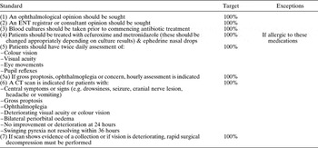
In 2004, interdisciplinary guidelines on how to manage periorbital cellulitis and abscess were published in order to prevent mismanagement and the development of complications.Reference Howe and Jones2 These guidelines provide guidance on the criteria for admission of such cases and emphasises the importance of joint care involving ENT, paediatrics and ophthalmology services. It determines the appropriate investigations required (including which cases warrant CT imaging), the recommended antibiotic therapy, and the circumstances in which surgical intervention is needed (see Table I).
Audit standards derived from 2004 published guidelines

CT = computed tomography
These guidelines provide the basis for management of such cases. At the Queen's Medical Centre in Nottingham (a tertiary referral centre), these guidelines were provided to all ENT and ophthalmology junior doctors during their initial induction to the ENT department. They were also used as a basis for guidance on the management of such cases, provided within the ENT junior doctors' handbook at Kingsmill Hospital, Mansfield (a neighbouring district general hospital).
The aim of this study was to ensure that patients with periorbital cellulitis or abscess were being managed appropriately and promptly, hence preventing complications.
Materials and methods
We performed a retrospective audit of patients seen in the above tertiary referral centre and district general hospital (DGH) since the 2004 publication of the above guidelines. Clinical coding was used to locate the medical notes of patients with orbital disease. This was done using the International Statistical Classification of Disease 10 classification of acute inflammation of the orbit (i.e. H05.0). This coding includes not only periorbital cellulitis and abscess but also osteomyelitis, tendonitis and periostitis of the orbit. The medical notes were then examined to determine whether cases represented periorbital cellulitis or abscess and whether the management criteria had been met.
In the tertiary referral centre, 76 patients were included within the relevant coding. Of these, 22 cases were diagnosed as pre-septal cellulitis after clinical assessment by ENT and/or ophthalmology (with the causes anterior to the orbital septum and not involving the sinuses), and were thus excluded. Nine patients had other diagnoses and were therefore also excluded, and six case notes were unavailable as they were being used in other departments or could not be found. After exclusions, there was thus a total sample size of 39 cases of periorbital cellulitis or abscess seen at the tertiary referral centre after 2004.
In the DGH, 75 patients were included within the relevant coding. Twenty cases diagnosed as pre-septal cellulitis were excluded. Fourteen patients had other diagnoses and were also excluded, and three case notes were unavailable as they could not be located. After exclusions, there was thus a total sample size of 38 cases of periorbital cellulitis or abscess seen at the DGH after 2004.
In order to analyse the data sufficiently, audit standards were created from the published guidance. These standards are shown in Table I.
Results
Having analysed all of the cases at both hospitals, it was established that no patients had come to any harm. It is important to emphasise that this outcome is adequate, as several lapses in management were identified which might have caused problems had any rapid deterioration gone undetected.
Standards one and two
It is important that both a senior ENT surgeon and a member of the ophthalmology team review all patients. In the tertiary referral centre, 37 patients (95 per cent) were seen by an ophthalmologist; however, in the district general hospital (DGH) only 24 patients (63 per cent) were seen thus. In the tertiary referral centre, 32 patients (82 per cent) were seen by a senior ENT surgeon; however, in the DGH only 15 patients (39 per cent) were seen thus.
Standard three
The guidelines recommend taking samples for blood cultures prior to commencing antibiotic treatment.
At the tertiary referral centre, 14 (36 per cent) patients had blood cultures taken prior to commencing treatment; of these patients, 36 per cent were pyrexial. Whether or not a patient was pyrexial determined whether blood cultures were taken (p = 0.006).
At the DGH, 13 (34 per cent) patients had blood cultures taken prior to commencing treatment; of these patients, 45 per cent were pyrexial. The odds ratio of a pyrexial patient having blood cultures taken was 2.7634 (95 per cent confidence interval 0.69–12.00), with p = 0.0778.
Standard four
At the tertiary referral centre, 29 patients (76 per cent) were correctly treated with intravenous cefuroxime and metronidazole, compared with 26 patients (68 per cent) at the DGH. Eight patients (21 per cent) were treated with alternative antibiotics at the tertiary referral centre, e.g. flucloxacillin, gentamicin and co-amoxiclav. Nine patients (24 per cent) were treated with alternative antibiotics at the DGH.
At the tertiary referral centre, 27 patients (69 per cent) were prescribed topical sympathomimetic nasal drops or sprays, compared with 18 patients (60 per cent) at the DGH.
Standard five
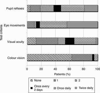
All patients with suspected periorbital cellulitis or abscess should have various ophthalmological criteria monitored at least twice daily. This is important to ensure there is no deterioration which may require urgent surgical intervention. These criteria include eye movements, pupil reflexes, colour vision and visual acuity. In both centres, no patients had twice daily monitoring of all four of these criteria. Figures 1 and 2 indicate how regularly the patients were monitored.
Frequency of ophthalmological criteria monitoring, for patients with periorbital cellulitis at the tertiary referral centre.

Frequency of ophthalmological criteria monitoring, for patients with periorbital cellulitis at the district general hospital.

It is clear from Figures 1 and 2 that patients were monitored more frequently at the tertiary referral centre than the DGH.
It is also clear that colour vision was poorly monitored at both centres. In the tertiary referral centre, 24 patients (63 per cent) did not have their colour vision monitored at all, compared with 30 patients (79 per cent) at the DGH. However, no patient had twice daily monitoring of this test criterion at either centre.
In the tertiary referral centre, five patients (13 per cent) did not have their visual acuity checked at all, while one patient had twice daily monitoring of this test criterion. At the DGH, 14 patients (37 per cent) did not have their visual acuity checked at all, and no patient had twice daily monitoring of this test criterion.
In the tertiary referral centre, 17 patients (44 per cent) had twice daily monitoring of eye movements, compared with only seven patients (18 per cent) at the DGH.
At the tertiary referral centre, nine patients (23 per cent) had twice daily monitoring of pupil reflexes, compared with only two patients (5 per cent) at the DGH.
Standard 5a
Gross proptosis or ophthalmoplegia indicates the development of an abscess which may require urgent surgical intervention. At the tertiary referral centre, 15 patients (39 per cent) had gross proptosis and/or ophthalmoplegia, compared with only four patients (11 per cent) at the DGH.
At the tertiary referral centre, only one patient had hourly assessments to ensure that deterioration of their vision and health did not occur; at the DGH, no patient had hourly assessments.
Standard six
According to the published guidance, a CT scan is indicated in patients presenting with: central symptoms or signs (e.g. drowsiness, seizure, cranial nerve lesion, headache or vomiting); gross proptosis; ophthalmoplegia; deteriorating visual acuity or colour vision; bilateral periorbital oedema; no improvement or deterioration at 24 hours; or swinging pyrexia not resolving within 36 hours.
A CT scan was indicated, as per the above list, for 28 patients at the tertiary referral centre and 17 patients at the DGH. At both centres, one patient who presented with a central symptom (a headache in both cases) did not receive a CT scan.
Standard seven
If a CT scan shows a collection (i.e. an orbital abscess) or if a patient has rapidly deteriorating vision, then they require surgical drainage or decompression. At the tertiary referral centre, 13 patients were found to have a collection on CT scan, all of whom were taken to the operating theatre for surgical decompression. At the DGH, three patients were found to have a collection on their CT scan; one of these patients was taken to the operating theatre for surgical decompression, while the other two were not as they were improving clinically with intravenous antibiotics and nasal ephedrine treatment alone.
Discussion
The interdisciplinary 2004 guidelines for the management of periorbital cellulitis and abscess represent the standard by which patient management is measured.Reference Howe and Jones2 Starkey and Steele's 2001 study reported that medical management successfully treated five out of six cases of periorbital cellulitis, while one required surgical drainage.Reference Starkey and Steele3 However, this report provided no clear guidance as to who should have surgery. Furthermore, the report stated that the investigation of choice was a CT scan, but did not provide clear guidance on when this was indicated.
Without clear guidance on the management of such cases, inadequate treatment may occur.
For example, in children inadequate treatment of periorbital cellulitis can lead to cerebral complications, include cavernous sinus thrombosis (1 per cent), meningitis (2 per cent), carotid occlusion, epidural abscess, subdural abscess, brain abscess (1 per cent) and death.Reference Arun and Rubin4
It is of great concern that, in cases of periorbital cellulitis or abscess, complications may develop due to inadequate monitoring of patients' vision. Previous case reports have shown that patients can experience visual loss secondary to periorbital cellulitis or abscess; therefore, patients' vision should be monitored so that early loss can be detected and therefore treated.Reference Davis and Stearns5
Patients with periorbital cellulitis or abscess may develop raised retro-orbital pressure akin to that occurring in cases of retro-orbital haemorrhage, although the onset of the latter is more acute.Reference Sacks, Lawson, Edelstein and Green6 The orbit can be decompressed either via a lateral canthotomy and inferior cantholysis, or by incising the medial periorbital periosteum after removal of the lamina papyracea. In cases of retro-orbital haemorrhage, orbital decompression should be performed within 60 minutes otherwise visual loss is likely to become permanent (although there are reports of partial recovery following intervention 120 minutes after trauma).Reference Hayreh, Kolder and Weingest7 Because the increase in posterior compartment pressure is less abrupt with a periorbital abscess, twice daily monitoring of visual acuity, colour vision and eye movement is thought to be sufficient, as there are no reports of irreversible vision loss occurring in a shorter period which has not been reversed by decompression. Slavin and Glaser have described cases of irreversible visual loss developing over a period of one day (without regular monitoring) or more.Reference Slavin and Glaser8
Limitations of this audit
In this audit, we used clinical coding to locate the medical records of cases of periorbital cellulitis or abscess. This method of acquisition relies greatly on accurate clinical coding. There may have been cases that were not correctly coded and therefore not included within our data collection process.
There were some cases at the tertiary referral centre (six patients) and at the district general hospital (three patients) for which clinical records could not be found or had been destroyed.
• Periorbital cellulitis is considered both an ENT and an ophthalmological emergency, as it is an orbital complication of sinusitis
• Delay in diagnosis and appropriate treatment may result in serious complications, including blindness and intracranial infection
• This audit found a lack of strict monitoring for deterioration in such patients, regardless of the type of centre the patient attended
• Patient management was better at the tertiary referral centre than the district general hospital
Our method of collecting data from case notes also relied on doctors correctly documenting their consultations. Lack of such documentation may have conveyed the impression that patients were not being monitored as regularly as they were.
Conclusions
ENT surgeons and ophthalmologists did not see all of our patients on admission, and prescribing habits were suboptimal in both centres (although better in the tertiary referral centre than the district general hospital (DGH)).
Twice daily monitoring of ophthalmological criteria was poor at both hospitals, particularly colour vision monitoring.
Interestingly, more patients presented with gross proptosis or ophthalmoplegia at the tertiary referral centre (15 patients) than at the DGH (four patients). These patients should be monitored hourly; however, only one patient at the tertiary referral centre was monitored hourly, and none of the DGH patients were monitored thus.
Both centres showed high compliance with performing CT scans when indicated. Unfortunately, in both centres, one patient in whom a CT scan was indicated did not receive it (both patients presented with headache). Fortunately, both patients recovered well with systemic treatment.
At the tertiary referral centre, all patients found to have a periorbital pus collection on CT were correctly taken to the operating theatre for urgent surgical decompression. At the DGH, one of the two such patients was not taken to theatre, as they were found to be improving clinically with systemic treatment alone.
In summary, it can be seen that patients were not managed in the anticipated manner, given the published guidelines. This was particularly the case in respect to regular monitoring of patients for deterioration. The standard of patient management was clearly better at the tertiary centre, compared with the district general hospital.
Fortunately, no patient suffered untoward morbidity as a result of this. However, visual loss is a well recognised complication of raised pressure in the posterior compartment of the eye.Reference Slavin and Glaser8 Thus, we wish to make the following recommendations.
(1) All suspected cases of periorbital cellulitis or abscess should be reviewed by both ENT and ophthalmology services. It was reassuring to observe that many cases of pre-septal cellulitis were seen by both specialties, particularly at the tertiary referral centre, to exclude any sinus involvement. However, there were still some cases that were not seen by either specialty. Review by both specialties would ensure that the correct treatment was initiated promptly and the severity of the condition assessed. This should also be the case if it is unclear whether the patient has pre-septal cellulitis alone or true periorbital cellulitis.
(2) Guidelines should be discussed regularly and agreed by ENT, ophthalmology and paediatric services.
(3) Guidance should be made available on the ENT intranet site. At the time of audit, there was no such guidance available. Such intranet information is typically the first port of call when seeking guidance on patient management throughout the hospital, particularly amongst junior doctors. The availability of such intranet guidance would ensure that all necessary steps (e.g. blood cultures) are completed regardless of where the patient presents. This would also allow guidance to be accessible when such patients present to different specialties (e.g. the one patient who presented to healthcare services for the elderly).
(4) Regular ophthalmological examination needs to be implemented. These checks should be documented.
(5) A pro forma could be drafted to help manage such patients. When these patients present, this pro forma could be inserted into their notes and followed as step-by-step guidance on correct management.
Acknowledgements
We thank Sonia Gill, clinical audit officer at Kingsmill Hospital, Faye Bradley, clinical audit support worker at Kings Mill Hospital, Andy Carden, clinical audit officer at Queen's Medical Centre, and Barry Feurtado, clinical case notes provider at Queen's Medical Centre.