Introduction
Acquired atresia of the external ear canal has been categorised into several aetiological subclasses: postinflammatory, traumatic, post-operative and neoplastic.Reference Becker and Tos1 We have earlier suggested that idiopathic, inflammatory, medial meatal, fibrotising otitis is a separate disease entity,Reference Hopsu and Pitkäranta2 with a different clinical aetiopathology from that of the other postinflammatory meatal atresias. Idiopathic, inflammatory, medial meatal, fibrotising otitis is a continuous inflammatory process affecting only the glabrous, medial meatal ear canal skin, usually bilaterally.Reference Hopsu and Pitkäranta2
Lichen planus is a chronic, inflammatory epithelial disease of glabrous skin and mucosae, which has manifold clinical and histological presentations, usually with bilateral involvement.Reference Boyd and Neldner3, Reference Sugerman, Savage, Walsh, Zhao, Zhou and Khan4 Its pathogenesis is complex and its aetiology unknown.Reference Sugerman, Savage, Walsh, Zhao, Zhou and Khan4 The lesions are generally self-limiting over a period of several months or years. However, especially in oral forms of lichen planus, some lesions fail to resolve spontaneously.Reference Cribier, Frances and Chosidow5
We report three adult patients with idiopathic, inflammatory, medial meatal, fibrotising otitis who also presented with oral lichen planus.
Patients
All three patients were referred to the otorhinolaryngology department of Helsinki University Hospital because of continuous external ear canal infections. In all patients, the condition was restricted to the medial meatal canals. On presentation, two patients had already developed fibrous atresia of the medial canal in one ear. All three patients also suffered from severe or erosive oral lichen planus, which had been previously diagnosed and histologically confirmed. Clinically, no patient had any ongoing middle-ear or mastoid problems. Histologic specimens from the ear canal process showed no specific pathological condition.
Case one
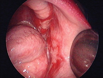
A 78-year-old woman had for over two years experienced a blocked feeling and some discharge in her left ear. This had been treated as an external ear canal infection with several courses of topical antibiotic or antiseptic eardrops. She suffered from polymyalgia rheumatica and rheumatoid arthritis, treated with gold and oral corticosteroid. Several years earlier, she had been diagnosed with ulcerative oral lichen planus (Figure 1) and treated with topical corticosteroid.
Ulcerative, oral lichen planus on the buccal mucosa of patient one.

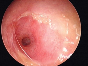
Clinical examination revealed external, medial meatal inflammation and developing atresia in the patient's left ear canal. The skin was defective and eroded and the canal was narrowed but still open up to the tympanic membrane (Figure 2). The right ear was healthy. An audiogram showed a 30 dB conductive hearing loss in the left ear, but the patient had never experienced any noticeable middle-ear problems.
Left ear of patient one; the medial ear canal skin is defective and ulcerative, with epithelial defects. The canal has not yet fully fibrosed and is still open. The conductive hearing level was 30 dB.

Topical corticosteroid (betamethasone) eardrops were commenced, but the ear canal atresia was already so far advanced that the medication was unable to prevent progressive fibrous atresia.
Case two
A 66-year-old woman with insulin-treated diabetes mellitus and bronchial asthma (without regular treatment) presented with a history of continuous, bilateral external ear canal symptoms, for which she had regularly used topical antiseptic or antibiotic-corticosteroid eardrops over the previous four years. Although the ear canal symptoms began in the right ear, the left ear was the first to develop medial meatal atresia. The right ear canal was still symptomatic.
Audiometry showed mixed hearing loss at 45–55 dB, of which bone conduction was 25–30 dB.
The patient had suffered fairly severe oral lichen planus for several years and had used local intra-oral corticosteroids. She also had lichen planus skin involvement in her shoulder region.
Case three
A 70-year-old woman presented with a four-year history of bilateral ear canal symptoms. Prior to this, she had had no ear problems. Oral lichen planus had been diagnosed and histologically confirmed several years earlier, treated with local intra-oral steroids.
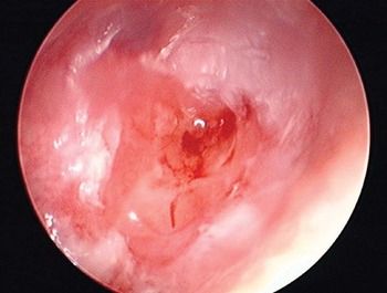
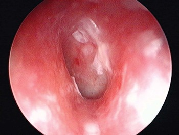
On the patient's first clinic visit, the right ear showed total medial meatal atresia, but the skin was still defective over the atresia (Figure 3). Following cleansing of the epithelial crust over the medial meatal skin, the left ear canal was found to be open. The medial canal skin was inflamed but there was no erosive epithelial destruction or granulation in the left ear (Figure 4).
Right ear of patient three; an almost mature fibrous plug formation is seen in the ear canal. The skin is still defective and ulcerative, but with no clinical infection.

Left ear of patient three; only the medial canal skin is inflamed and chronically symptomatic, without epithelial erosion or destruction.

Local betamethasone treatment was commenced at the first visit. Over subsequent visits, the skin became less inflamed and the condition did not progress; there was no sign of atresia formation in the left ear.
At the time of writing, the patient continued to use corticosteroid drops once or twice weekly in the left ear, and the need to cleanse the ear canal had become minimal.
Discussion
In the present study, we considered the possible association between idiopathic, inflammatory, medial meatal, fibrotising otitis and lichen planus. Both are peculiar epithelial diseases of unknown aetiology. Lichen planus typically presents on the glabrous skin,Reference Boyd and Neldner3 as does idiopathic, inflammatory, medial meatal, fibrotising otitis. In the latter condition, the inflammatory process – including secondary infection – is always restricted to the glabrous medial meatal skin over the bony part of the ear canals and tympanic membranes, whereas the middle ears and lateral or cartilaginous part of the ear canals are unaffected.
An association between bilateral, progressive ear canal stenosis and erosive, multifocal lichen planus has been suggested by Martin et al. Reference Martin, Moriniere, Machet, Robier and Vaillant6 In their reported patient, the tympanic membranes were unaffected, and the stenosis was of the ear canal-obliterating type. In idiopathic, inflammatory, medial meatal, fibrotising otitis, the medial canal-blunting type of atresia occurs, with the condition being characteristically restricted to the medial canal skin over the bony canal wall, but also affecting the tympanic membrane. We found no other reports suggesting a connection between progressive meatal stenosis or atresia and lichen planus.
Our three patients had already suffered from severe oral lichen planus for years before their ear canal symptoms manifested. If the ear canal process in our patients represented an infrequent location of lichen planus, the isomorphic response (Koebner phenomenon)Reference Boyd and Neldner7 may explain the pathogenesis in this secondary location, following possible initiation via minor, epithelial ear canal trauma. The clinical picture of the ear canal process in the presented patients was identical to that in our earlier report of idiopathic, inflammatory, medial meatal, fibrotising otitis.Reference Hopsu and Pitkäranta2 However, none of the patients in that earlier report developed lichen planus. Thus, in most cases of idiopathic, inflammatory, medial meatal, fibrotising otitis, the primary and only site of expression of the epithelial disease is in the medial ear canals.Reference Hopsu and Pitkäranta2 This may also be the situation with postinflammatory atresia of unknown aetiology.Reference Katzke and Pohl8, Reference Slattery and Saadat9
In patients with lichen planus, histopathological analysis of skin from the affected ear canal has revealed no specific diagnostic features regarding the lichen planus (to us or to others). In patients with idiopathic, inflammatory, medial meatal, fibrotising otitis, histological specimens of diseased skin in the active (but non-granular and non-infectious) stage showed no epithelial lining.Reference Hopsu and Pitkäranta2 Instead, Martin et al. found a hyperkeratotic picture in auditory canal skin.Reference Martin, Moriniere, Machet, Robier and Vaillant6
• Within the acquired ear canal atresias, idiopathic, inflammatory, medial meatal, fibrotising otitis is suggested to be a distinct disease entity, for reasons of aetiology
• The authors report three more cases of idiopathic, inflammatory, medial meatal, fibrotising otitis and further consider a possible relationship between this condition and lichen planus
• Early, active treatment of idiopathic, inflammatory, medial meatal, fibrotising otitis with local corticosteroids may prevent total medial meatal atresia
Evidently, the pathophysiology of idiopathic, inflammatory, medial meatal, fibrotising otitis is not principally infectious. With thorough treatment, the recurrent or continuous infections and granulation can be prevented, even when the epithelia has been severely damaged. Also, the inflammatory condition can be controlled in the erosive phase by continuous local corticosteroid treatment, so that an almost ‘steady state’ is achieved. However, the already severely damaged skin epithelia does not have the capability to regenerate,Reference Hopsu and Pitkäranta2 and repair occurs via fibrosis. Secondary infectious involvement of the damaged epithelia seems to accelerate the final fibrosis to a great extent, and often represents the principal clinical sign before final atresia develops. In our present patients, as with our previous patients,Reference Hopsu and Pitkäranta2 the clinical process was one of continuous inflammation, culminating in severe epithelial damage due to the irreversibility of the atretic process. However, it is notable that the process is confined to a strictly limited skin area, and immediately ceases once the skin over the bony ear canal and tympanic membrane has been eliminated and replaced by a fibrous plug. After atresia has fully developed, there appear to be no signs of continuing inflammation or skin involvement in the fibrous plug or the lateral or remaining part of the ear canal.Reference Hopsu and Pitkäranta2
Before atresia development, patients with idiopathic, inflammatory, medial meatal, fibrotising otitis typically use a number of antibiotic and antiseptic eardrops over several months or years, with no noticeable effect on their symptoms. If the condition involved a toxic, allergic or contact dermatitis resulting from prolonged topical medical treatment, it could be anticipated that the lateral ear canal skin would also become symptomatic. Local corticosteroid treatment (betamethasone drops) without antibiotic helps the symptoms and signs to resolve, even when epithelial damage has already occurred.Reference Hopsu and Pitkäranta2 Nevertheless, when atresia has begun to develop, it cannot be prevented by medication or even by irradiation.Reference Eichel and Simonton10 In patient three, who received local corticosteroid treatment, the left ear had only vague signs of inflammation and there was no sign of atresia; the epithelia has since remained undamaged and free of infection.
Conclusion
There are certain peculiar skin diseases of unknown aetiology, such as idiopathic, inflammatory, medial meatal, fibrotising otitis and lichen planus. The latter often affects not only the glabrous skin but also the mucous membranes. Although one patient with erosive, multifocal lichen planus and bilateral, progressive ear canal stenosis has been reported, we found no previous reports of patients with (bilateral) medial canal, fibrotising otitis presenting with lichen planus. These diseases have in common a good response to local corticosteroid treatment, if commenced early enough. In idiopathic, inflammatory, medial meatal, fibrotising otitis, early recognition of the disorder and prompt use of topical corticosteroids may even prevent the erosive skin involvement and thus the development of atresia.
In conclusion, we suggest that there may be an association between idiopathic, inflammatory, medial meatal, fibrotising otitis and lichen planus, due to their pathophysiological mechanisms. To our knowledge, no previous report has histologically confirmed the diagnosis of lichen planus in association with medial ear canal atresia.