Introduction
Heart disease and stroke are major causes of death and morbidity in Western countries.Reference Glass and Witztum1 Most heart attacks and strokes are caused by thrombus formation superimposed on disrupted atherosclerotic plaques.Reference Glass and Witztum1–Reference Trzos, Uznanska and Rechcinski3 Kidney failure and high blood pressure are also associated with atherosclerosis.Reference Tian, Penman, Mawson, Manning and Flessner4, Reference Zoccali and Seck5 Thus, not only does atherosclerosis have a considerable impact on the individual's quality of life, but also causes a significant financial burden on the health care systems.
Epidemiologic studies have identified a number of risk factors associated with atherosclerosis. Obesity, tobacco consumption, elevated levels of low-density lipoprotein and low levels of high-density lipoproteins due to diet, high blood pressure, excessive levels of glucose, physical inactivity and low birth weight are all factors causally linked to atherosclerosis.Reference Hackam and Anand6–Reference Leduc, Levy, Bouity-Voubou and Delvin8
Early atherosclerotic lesions tend to manifest as an accumulation of lipid-laden macrophage foam cell beneath the endothelium known as fatty streaks.Reference Libby, Ridker and Maseri9 These lesions are present in young people and may progress to become advanced plaques or, alternatively, may also disappear and never cause any symptoms at all. The probability that a lesion will develop into a plaque will increase in the presence of the above-mentioned risk factors. Generally, an atherosclerotic plaque is made up of two components: a core region that consists of foam cells (prototypic of atherosclerosis) and extracellular lipid droplets, and a fibrous cap that consists of smooth muscle cells and a collagen-rich matrix.Reference Hansson10, Reference Stary, Blankenhorn and Chandler11
Inflammation significantly drives the formation, progression and rupture of atherosclerotic plaques. Immune cells, particularly macrophages, T cells and mast cells, are present in both early and advanced lesions.Reference Hansson10 Sites of lesion rupture are rich in immune cells that produce proinflammatory cytokines such as interferon (IFN)-γ and tumor necrosis factor (TNF)-α, as well as proteolytic enzymes including matrix metalloproteinases (MMPs) and cysteine proteases that weaken the fibrous cap of the plaque and accelerate plaque rupture (apoptotic cell death is also a major contributor of plaque rupture). Proinflammatory cytokines can also cause endothelial cells to become activated, triggering them to upregulate their expression of adhesion molecules that aid in the recruitment of monocytes. Furthermore, activated endothelial cells can trigger the differentiation of monocytes to become macrophages. Macrophages secrete proinflammatory cytokines causing irreversible tissue damage. Subsequently, tissue damage leads to the recruitment of T cells that further infiltrate the atherosclerotic plaques.Reference Libby, Ridker and Maseri9, Reference Hansson10 CD4+ T cells that infiltrate the atherosclerotic plaques recognize protein antigens presented to them on the major histocompatibility complex (MHC) II molecules, become activated and promote a Th1 inflammatory response. Thus, activated T cells differentiate into Th1 effector cells and produce (IFN)-γ, augmenting the synthesis of (TNF)-α. The presence of both (IFN)-γ and (TNF)-α prompts the production of many inflammatory molecules that advance inflammation and promote atherosclerosis.Reference Hansson10
Fetal programming, also termed ‘developmental origin of health and disease’, is a process where a stimulus or challenge at a critical period of fetal development can result in lifelong effects. In a pioneering study, David BarkerReference Barker12 was able to show in a cohort of over 15,000 men and women that low birth weight as a result of maternal malnutrition led to a higher risk for developing coronary heart disease in later life. From this study, Barker hypothesized that challenges during pregnancy affect fetal development and trigger the onset of disease in later life. More recently, emerging evidence suggests that prenatal challenges, specifically environmental factors such as maternal cigarette smoking, maternal malnutrition and hypercholesterolemia, impair fetal development and program the fetus to become susceptible for diseases such as atherosclerosis, coronary heart disease and cardiovascular disease later in life.Reference McGill, McMahan and Herderick13–Reference Goharkhay, Sbrana and Gamble15 Nevertheless, emerging research advocates that additional prenatal challenges, that is, the psychological status of women during pregnancy, can affect fetal development. Here, high maternal stress perception at critical periods of fetal development may program the fetus to become vulnerable, thereby contributing to the development of immune diseases such as atherosclerosis, asthma and allergies later in life.Reference Knackstedt, Hamelmann and Arck16–Reference Andersson, Jiang and Davidge18
As the origin of inflammation-induced atherosclerosis is postulated to occur in utero, and maternal stress challenge during pregnancy is known to affect fetal development and increase the risk to develop chronic immune diseases later in life,Reference Knackstedt, Hamelmann and Arck16, Reference Andersson, Jiang and Davidge18 the present study aimed to investigate whether prenatal stress challenge perpetuates the development of atherosclerosis and inflammation in apolipoprotein E (apoE−/−)-deficient mice, a well-established mouse model of hyperlipidemic-induced atherosclerosis. Our findings indicate that prenatal stress challenge accelerates atherosclerotic lesion development, which correlates with enhanced systemic inflammation.
These mice have a target deletion of the gene for apoE, which is essential for the catabolism of lipid lipoprotein constituents, such as cholesterol.Reference Andersson, Jiang and Davidge18 When fed a normal chow diet, apoE−/− mice display total cholesterol levels that are five times higher than those observed in wild-type animals, corroborating that deficiency in apoE results in the accumulation of lipids in the endothelium and contributes to the development of atherosclerosis, beginning at an age of ∼10 weeks. Not only is it well established that apoE−/− mice are at a higher risk of developing atherosclerosis, but the plaques that they develop are similar to those seen in humans, making them a common and valuable animal model in atherosclerosis research.
Materials and methods
Mice and dietary conditions
ApoE−/− mice on a C57BL/6J background were purchased from Jackson Laboratories (Bar Harbor, ME, USA) and bred in the specific pathogen-free facilities at St. Joseph's Healthcare, Hamilton Ontario, Canada. Animal care and experimental procedures were conducted in accordance with the animal ethics research board of McMaster University.
Maternal stress exposure and experimental set-up of the offspring
Six- to eight-week-old C57Bl/6 apoE−/− females were mated with age-matched C57Bl/6 apoE−/− males. Two females to every male were placed together in a cage overnight and successful mating was assumed upon the appearance of a vaginal plug the next day. In total, n = 10 females were used for mating. Once a mating plug as a sign for successful mating could be identified, females were randomly assigned to two different groups: one group of pregnant females was left undisturbed during pregnancy in order to provide control offspring (n = 5 females). The study group was exposed to two 24-h stress periods commencing on gestation days 12 and 14 (n = 5). Stress was provided by a rodent repellent, which produced a beeping sound of 70 db at a frequency of 300 Hz in intervals of 15 s.
Delivery dates of the litters were documented and females with their respective litter were kept in individual cages and left undisturbed for a period of 21 days. No differences in terms of gestation length have been observed between groups. In addition, no increase in the fetal loss rate in response to the prenatal stress challenge can be deduced, as the litter sizes between groups did not significantly differ. This absence of fetal loss was anticipated and desired, as we wanted to test the effect of prenatal stress challenge on fetal growth and development rather than fetal rejection. Hence, when initially establishing the experimental approach of prenatal stress challenge in the context of fetal programming of asthma in a different mouse strain,Reference Pincus-Knackstedt, Joachim and Blois17 we had conducted extensive kinetic studies identifying the time point at which prenatal stress challenge would not increase fetal loss, but impair fetal development, which turned out to be if dams where challenged on gestation days 12 and 14 for 24 h, respectively. Further, as apoE−/− dams are known to show aggressive maternal behavior and cannibalism (which we could confirm in pilot experiments independent of the data presented here), BALB/c foster dams were used to replace the aggressive apoE−/− mothers. This was carried out by ensuring that there was a pregnant BALB/c dam that was about to deliver on the same day or at most 3 days before the delivery date of the apoE−/− dam. Hence, within 24 h upon delivery, apoE−/− pups were transferred and added to the litter of the BALB/c dam and left undisturbed until weaning. The total number of apoE−/− offspring per group was 9 male and 13 female pups from nonstressed group, and 8 male and 9 female pups from prenatally stressed females. At the age of 15 weeks, the offspring were killed in order to collect the heart, blood and spleen for further analyses. On the basis of the variations with regard to the age based on the onset of birth and delivery, individual offspring were euthanized once they reached the age of 15 weeks. Hence, individual offspring were killed over a period of 6 weeks. Subsequently, data arising from respective individual mice were merged according to group and gender.
Tissue collection and preparation
Fifteen-week old offspring were anesthetized using isoflurane; blood was collected from the right ventricle using a 1 ml syringe and put into 15 ml falcon tubes containing 50 μl of heparin. Following blood collection, 3 ml of phosphate buffered saline (PBS) was administered into the left ventricle for flushing. Mice were then killed by cervical dislocation and spleens were collected and placed into PBS. Finally, hearts were transversely removed to include the aortic root and perfusion fixed with 10% neutral buffered formalin for a minimum of 24 h. After fixation, hearts were embedded in paraffin in preparation for sectioning.
The blood collected was processed for fluorescence-activated cell sorting (FACS) and flow cytometry. Processing of blood required treatment with 1× red blood cell (RBC) lysis buffer followed by centrifugation at 1500 rpm for 5 min at 4°C. Supernatants were then decanted and resuspended in PBS. This was repeated three times and cells were resuspended in FACS buffer (0.5% bovine serum albumin (BSA)/PBS) in the final treatment. A cell count was carried out on the blood sample by staining with trypan blue and using a bright field microscope.
In vitro stimulation of splenocytes
Splenocytes were harvested by gently teasing apart the spleen using the frosted ends of sterile glass microscope slides. Cells collected were filtered through a sterile nylon filter into a 50 ml sterile falcon tube containing complete media (Roswell Park Memorial Institute medium, 10% fetal calf serum, 1% glutamine and 1% penicillin streptomycin) and were pelleted by centrifugation at 1400 rpm for 5 min at 4°C. Following centrifugation, supernatants were decanted and cells were disrupted by ‘racking’ the tubes. RBCs were then lysed in 2 ml of 1× RBC lysis buffer for 12 min at room temperature. Following lysis, cells were washed with PBS to stop the reaction and filtered through a nylon filter into a new 50 ml falcon tube. Cells were again centrifuged followed by the decanting of supernatant and resuspension of cells in 2 ml of complete media. The number of splenocytes were counted. Cells were then resuspended in complete media to produce a cell concentration of 0.5 × 10⁶/200 μl into a 96-well plate. Plates were centrifuged for 3 min at 1500 rpm at 4°C and subsequently collected and stored at −20°C until further analysis.
Hematoxylin and eosin staining (H&E) and histomorphological analysis
A microtome was used to produce 4 μm serial sections of the aortic root, sections were collected on superfrost glass microscope slides until the valves were no longer visible.Reference Paigen, Morrow, Holmes, Mitchell and Williams19, Reference Dickhout, Lhoták and Hilditch20 Sections were then stained with H&E to evaluate plaque size and cellular morphology. Plaque sizes were examined using a Zeiss Axiophoto light microscope and grouped as follows: very small plaques = 1–7999 μm2; small plaques = 8000–22,999 μm2; moderate plaques = 23,000–51,999 μm2; and large plaques =52,000–110,000 μm2. Photomicrographs were taken at 10× magnification using the Zeiss AxioVision software.
FACS
FACS was conducted for the analysis of cell surface marker expression. The cell surface expression of CD11 allows identification of leukocytes involved in the innate immune system, including monocytes, granulocytes, macrophages and natural killer cells, whereas the expression of (MHC) II molecules is found on specialized cell types, including macrophages, dendritic cells and B cells.Reference Solovjov, Pluskota and Plow21 F4/80 is a transmembrane protein present on the cell surface of myeloid mouse macrophages.Reference Leenen, de Bruijn, Voerman, Campbell and van Ewijk22 Lymphocyte function-associated antigen 1, also known as LFA-1, is found on all T cells and also on B cells, macrophages and neutrophils, and is involved in the migration of cells to the site of infection.Reference Davignon, Martz, Reynolds, Kurzinger and Springer23
Hence, to evaluate the frequency, phenotype and migratory potential of macrophages, the following antibodies were used: FitC-anti-F480, PE-anti-LFA-1, PE-Cy5-anti-CD11b, PE-Cy7-anti-LFA-1 and APC-anti-(MHC) II. For the analyses of CD4 and Treg cell frequencies, PE-Cy5-anti-FoxP3, PE-Cy7-anti-CD4, APC-anti-CD25 and APC-Cy7-anti-CD3 were used. Antibodies were purchased from eBioscience or BD Biosciences. In brief, cells were washed and blocked with CD16/CD32 in FACS buffer for 15 min on ice and then surface-stained for 30 min with the antibodies mentioned above. Stained cells were analyzed using a FACS-Canto flow cytometer (BD Biosciences), where 100,000 to 250,000 events per sample were collected. The data were then analyzed using the FlowJo software version 6.3.4 (Tree Star).
Cytometric bead array (CBA)
TNF-α and IFN-γ are proinflammatory cytokines that are secreted by activated CD4 T cells. Their levels were analyzed from supernatants of stimulated splenocytes using the CBA mouse inflammation kit (BD Pharmingen). TNF-α and IFN-γ levels were acquired using FACSCalibur cytometer (BD Biosciences) and data were analyzed using CBA software (BD Biosciences). A standard curve was generated using the cytokine standard provided by the manufacturer. The concentration of the two cytokines in cell supernatants was determined by interpolation to the corresponding standard curve.
Statistical analysis
All statistics were carried out with SPSS version 18.0. A two-tailed independent t-test was used to test for significant differences between groups. For all analyses, P < 0.05 was considered statistically significant.
Results
Prenatal stress induces a skew toward increases in atherosclerotic plaque size
Prenatal stress of apoE−/− offspring mice resulted in a higher frequency of moderate and large plaques at the level of the aortic root in both male and female offspring (Fig. 1a). Particularly in female offspring, a skew towards large lesions was observed in response to prenatal stress challenge. In addition, female offspring without stress challenge had higher frequency (more than double) of large plaques than that of male offspring without any stress. Overall, the frequency of atherosclerotic plaques was higher in female offspring, compared with male offspring. This different pattern of plaque formation in female apoE−/− mice irrespective of prenatal stress challenge has already previously been described and is due to a developmentally induced (∼3–5 weeks) earlier onset of atherosclerosis in females.Reference Dickhout, Lhoták and Hilditch20, Reference Solovjov, Pluskota and Plow21 Therefore, in the context of the findings presented here, a comparison of the effect of prenatal stress on male v. female offspring was neglected. Further, we did not perform statistical analysis. Although the body weight did not show significant differences between prenatally stressed and control offspring, male offspring showed significantly higher body weight compared with females (Fig. 1b). Figure 1c shows representative photomicrographs of very small, moderate and large atherosclerotic plaques.
Atherosclerotic plaque sizes and body weight in prenatally stressed apolipoprotein E-deficient offspring and nonstressed control offspring. (a) Atherosclerotic plaques were morphologically defined by the presence of a necrotic core evaluated at the aortic root in H&E-stained sections. The plaque size, measured in μm2, was quantified using the Carl Zeiss digital imaging processing software, AxioVision 4.7. One slide per mouse was evaluated to obtain the area of the plaque. If more than one plaque was detectable per slide, then the average area was used as the plaque size for that mouse. Plaque sizes were grouped as follows: very small plaques = 1–7999 μm2; small plaques = 8000–22,999 μm2; moderate plaques = 23,000–51,999 μm2; and large plaques = 52,000–110,000 μm2. (b) Mice were weighed using a digital scale before being killed at the age of 15 weeks. Bars represent the mean body weight per group and gender + s.e.m. (c) (top panel, left) Representative photomicrograph (magnification × 50) of H&E-stained heart tissue from a nonstressed offspring with no visible plaques. (Right) area inside the rectangle of top panel at 200× magnification. (Middle and lower panel, left) Representative photomicrograph of prenatally stressed female offspring with moderate (middle panel) and large plaques (lower panel). (Right) area inside the rectangles of middle and lower panel at 200× magnification. In atherosclerotic plaques, the intima layer of the aorta is lifted from the media because of infiltrating immune cells. The necrotic core, present in mice with visible plaques, is situated between the media and the intima and is marked as ‘NC’. The aortic valve, used as a reliable site for the evaluation of atherosclerotic plaques, is marked as ‘AV’. The lumen is marked as ‘L’ and the cardiac muscle is marked as ‘CM’. The intima consists of a single layer of flattened and elongated endothelial cells and is marked as ‘I’ with an arrow. The media, consisting of smooth muscle cells (SMC) is marked ‘M’. Adipoctyes, seen only in one photo, are marked as ‘Ad’.

Prenatal stress reduces frequency of Treg cells in blood
Flow cytometric analyses of CD4 cells revealed no significant effect of prenatal stress challenge on the overall frequency of CD4 cells among blood lymphocytes, although prenatally stressed female mice revealed a significantly higher frequency of CD4+ lymphocytes compared with male mice (Fig. 2a). Further, the frequency of Treg cells among blood lymphocytes, defined as CD4+CD25+++FOXP3+, was significantly reduced in prenatally stressed male and female offspring. Strikingly, higher levels of Treg cells were detectable in female offspring compared with male offspring, irrespective of prenatal stress challenge (Fig. 2b). Figure 2c reveals representative dot plots of female offspring.
Flow cytometric analyses of peripheral blood derived T cells. (a) Frequency of CD4+ T lymphocytes in the blood of prenatally stressed and control male and female offspring. The analyses were performed by gating on the lymphocyte population on the basis of size and granularity. Bars represent the mean frequency of CD4+ cells per group ± s.e.m. *P < 0.05. (b) Frequency of CD4+CD25+++FOXP3+ T lymphocytes in peripheral blood in prenatally stressed and control male and female offspring. The analyses were performed by gating on the CD25+++FOXP3+ cells within the CD4+ lymphocyte population. Bars represent mean frequency of CD4+CD25+++FOXP3+ cells per group ± s.e.m. *P < 0.05 and **P < 0.01. (c) (top panel) Representative flow cytometric dot plots displaying the CD4+ T lymphocyte population in the blood of stressed and nonstressed offspring (bottom panel). Representative flow cytometry dot plots displaying CD4+CD25+++FOXP3+ cells in the blood of stressed and nonstressed offspring.
Prenatal stress alters macrophage populations in blood
Next, the phenotype and frequency of macrophages and monocytes were evaluated within the leukocyte population. Frequencies of CD11b+ cells were significantly lower in both male and female prenatally stressed offspring as compared with the control offspring (Fig. 3a, representative histograms are shown in Fig. 3e), whereas neither prenatal stress challenge nor gender altered the frequency of (MHC) II (Fig. 3b). Prenatal stress also resulted in lower levels of CD11b+ leukocytes that coexpressed LFA-1, although levels of significance were not reached (Fig. 3c). We further evaluated CD11b+ leukocyte subsets, such as F4/80+, and could show a significant increase of CD11b+ F4/80+ leukocyte subsets in prenatally stressed female offspring. In male offspring, the levels of significance were not reached with regard to the frequency of CD11b+ F4/80+ leukocyte subsets in prenatally stressed offspring compared with controls (Fig. 3d). Figure 3e shows representative histograms or dot plots for the results depicted in Fig. 3a–d.
Macrophage and monocyte populations in the blood of stressed and nonstressed apolipoprotein E-deficient (apoE−/−) offspring mice. (a) Frequency of CD11b+ leukocytes in the blood of prenatally stressed and control male and female offspring. The analyses were performed by gating on the leukocyte population, based on size and granularity. Bars represent the mean frequency of CD11b+ cells per group ± s.e.m. *P < 0.05. (b) Frequency of [major histocompatibility complex (MHC)] II+ leukocytes in prenatally stressed and control male and female offspring ± s.e.m. The analyses were performed by gating on the (MHC) II+ cells within the CD11b+ leukocyte population. (c) Frequency of CD11b+LFA-1+ leukocytes in prenatally stressed and control male and female offspring ± s.e.m. The analyses were performed by gating on the CD11b+LFA-1+ cells within the CD11b+ leukocyte population. (d) Frequency of F4/80+ cells in peripheral blood in prenatally stressed and control male and female offspring. The analyses were performed by gating on the F4/80+ cells within the CD11b+ leukocyte population. Bars represent mean frequency of F4/80+ cells per group ± s.e.m. *P < 0.05, **P < 0.01. (e) Shows representative flow cytometric histograms and dot plots for the data presented in a–d.
Prenatal stress increases secretion of inflammatory cytokines in peripheral blood lymphocytes
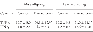
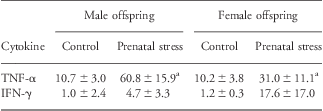
As atherosclerosis is considered an inflammatory disease, we then aimed to analyze cytokine production by CBA. Both TNF-α and IFN-γ levels were higher in cells derived from prenatally stressed offspring (Table 1).
Proinflammatory cytokine secretion in vitro by peripheral blood lymphocytes of prenatally stressed (‘stress’) and control male and female offspring
TNF-α, tumor necrosis factor; IFN, interferon.
Data are presented as mean level ± s.e.m. (pg/ml) of TNF-α and IFN-γ.
aMeans P < 0.05 compared with control offspring of same sex.
Discussion
In the present study, we aimed to identify whether maternal stress challenge during pregnancy in apoE−/− mice would contribute to atherosclerosis and atherosclerosis-related inflammation in the offspring. Hence, a key finding of the present study was that prenatal stress indeed resulted in atherosclerotic plaques that were larger in size at the level of the aortic root, which was particularly profound in female apoE−/− offspring. Second, we provided evidence that prenatal stress challenge skews the apoE−/− adult offspring's immune system toward inflammation, mirrored by a decrease of Treg cells and alterations of the innate immune response, such as an increase in CD11b+ F4/80+ macrophages and a decrease of CD11b+ macrophages/monocytes with migratory potential, such as LFA-1. Further, both prenatally stressed male and female offspring were rendered vulnerable to inflammation, resulting in an increase in TNF-α and IFN-γ concentrations.
In the present study, we used prenatal exposure to sound as a stress challenge. In independent studies, this stress model has been shown to also increase anxiety in the offspring as well as increase in the severity of airway hyperresponsiveness. The latter can be observed in mice upon the induction of experimental asthma (upon sensitization with ovalbumin).Reference Pincus-Knackstedt, Joachim and Blois17 We then used apoE−/− mice and exposed them to prenatal sound stress, as these mice spontaneously develop atherosclerotic plaques without experimental induction. More importantly, apoE−/− mice develop plaques that are very similar to those seen in humans.Reference Whitman24 On the basis of our present findings, we propose that the prenatal stress challenge of apoE−/− dams provides a convincing model to understand the developmental origin of atherosclerosis.
The present study was designed specifically to determine how maternal stress impacts the development of atherosclerotic plaques in the aortic root of offspring apoE−/− mice. Previous work has suggested that stress can alter both human and animal physiology. In humans, stress has been shown to alter immune responses.Reference Kiecolt-Glaser, Glaser, Gravenstein, Malarkey and Sheridan25 Low socioeconomic status (SES) promotes the early onset of atherosclerosis,Reference Kop26 and stress because of high workplace demands influences the atherosclerotic process and contributes to the development or progression of plaques in the arteries.Reference Everson, Lynch and Chesney27 In addition to laboratory mice, studies in cynomolgus monkeys have revealed significantly greater atherosclerosis as a result of stress due to low SES.Reference Kaplan, Manuck, Clarkson, Lusso and Taub28 Overall, stress perception is emerging as a critical threat to health and well-being,Reference Knackstedt, Hamelmann and Arck16–Reference Andersson, Jiang and Davidge18, Reference Kiecolt-Glaser, Glaser, Gravenstein, Malarkey and Sheridan25–Reference Chen, Hanson and Paterson29 particularly in the context of atherosclerosis, as it may alter the immune response.
It is widely accepted that changes to the fetal environment affect patterns of development in the fetus. Barker has proposed that particularly in humans, malnutrition of the fetus influences its development and has lifelong consequences, predisposing it to a number of chronic immune diseases including atherosclerosis or other immune disorders, such as allergies.Reference Pincus-Knackstedt, Joachim and Blois17, Reference Andersson, Jiang and Davidge18 Fetuses with low birth weight are thought to experience a postnatal ‘catch-up’ period where growth is accelerated and inclines the child toward obesity, increasing its risk for atherosclerosis.Reference Barker12 In addition to this, Barker was also able to show that low birth weight, as a result of under nutrition, also leads to a higher risk for coronary heart disease.Reference Barker12 It would have been very interesting to see data on birth weight of the fetuses in the present study. However, we refrained from generating these data because of the tendency of neonate cannibalism in the apoE−/− mothers. Interestingly, data on birth weight using the same stress paradigm are available from prenatally stressed BALB/c offspring, which revealed that offspring of prenatally stressed dams had lower birth weight than those of the nonstressed control group.Reference Pincus-Knackstedt, Joachim and Blois17 These data may be extrapolated to the experimental setting used in the present study, albeit, lower birth weight as a consequence of prenatal stress would have to be confirmed in future studies for the apoE−/− mouse model.
The adaptive immune response, for example, increased proliferation/frequency of CD4 T cells that we described here in prenatally stressed female offspring, may be of central importance with regard to the severity of atherosclerosis. Treg cells are known to play a key role in the suppression of inflammation, for example, Treg cells suppress inflammation by releasing anti-inflammatory cytokines, such as TGF-β and IL-10, which in turn suppress proliferation of T effector cells or macrophages.Reference Hansson and Hermansson30 Thus, the loss of Treg cells in the prenatally stress offspring provides a plausible explanation for the increased inflammatory response and cell proliferation.
Previous studies have suggested that females are more prone to developing atherosclerosis. Interestingly, the plaques of the female apoE−/− offspring were larger in size than plaques from the male offspring, even though the female offspring had significantly higher frequency of Treg cells than male offspring. However, we also show that female offspring have higher basal frequencies of CD4 T cells and CD11b+ F4/80+ macrophages. This suggests a certain ‘disadvantage’ because of higher inflammation in female offspring, suggesting that prenatally stressed females are more susceptible to developing atherosclerosis.
Adhesion molecules such as intracellular adhesion molecule 1 (ICAM-1) also play an important role in inflammation and it has been shown that atherosclerosis causes upregulation of ICAM-1.Reference Nakashima, Raines, Plump, Breslow and Ross31 In this study, we observed upregulated LFA-1 expression of leukocytes in prenatally stressed offspring compared with control offspring. It can be assumed that endothelial ICAM-1 expression may also be upregulated in response to stress, as an upregulation of ICAM-1 has been shown in response to stress challenges in adult mice.Reference Blois, Tometten and Kandil32 Future studies are needed to test whether prenatal stress has a similar effect on the expression of ICAM-1.
Cross-talk between LFA-1+ cell subsets and the endothelial expression of ICAM-1 allows cell infiltration into the artery wall Reference Nakashima, Raines, Plump, Breslow and Ross31 and into tissue. This is especially crucial for the infiltrating T cells and macrophages as they make up the core region of the atherosclerotic plaque.Reference Libby, Ridker and Maseri9, Reference Hansson10, Reference Hansson and Hermansson30 Considering the decreased frequency of LFA-1+ CD11b+ cells in blood, we propose that this cell subset already migrated into the arterial wall and triggered or aggravated the atherosclerotic lesions seen in these mice.
Another characteristic of inflammation is the release of proinflammatory cytokines, particularly TNF-α.Reference Libby, Ridker and Maseri9 Our data reveal significantly higher levels of TNF-α as a result of prenatal stress. Upon verifying the inflammatory response of atherosclerosis, it was sensible to inquire whether there is a relationship between the inflammation noted and the severity of atherosclerosis. In support of this concept, the levels of TNF-α positively correlated with the increased plaque size measured from the aortic root. This correlation, along with the observed decrease in the Treg cell population of prenatally stressed offspring, suggests that inflammation is a key factor to the progression of atherosclerosis in prenatally stressed apoE−/− mice.
Taken together, prenatal stress challenge may trigger the onset of atherosclerosis by enhancing a proinflammatory response that contributes to the progression and severity of the disease.
Acknowledgments
The authors wish to thank Evelin Hagen for her excellent technical support.