I. INTRODUCTION
An area of considerable current interest in nanoparticle research is the assembly of inorganic nanoparticles to form ordered structures that can display novel collective behavior. This includes distinctive mechanical, magnetic, electronic, and optical properties.Reference Glotzer and Solomon1 Magnetic nanoparticles are of considerable interest both because of fundamental scientific interest and for applications in diverse areas such as high-density magnetic recording, spintronics, catalysis, separation, biomedicine, and biotechnology.Reference Lu, Salabas and Schüth2–Reference Gao, Gu and Xu7 As the size of the nanoparticle decreases below a critical value (typically around 10–30 nm), which is dependent upon the material properties, the individual nanoparticles behave as a single magnetic domain and can exhibit superparamagnetic behavior. They will rapidly respond to an applied magnetic field but exhibit negligible residual magnetism and coercivity. These features make superparamagnetic nanoparticles very attractive for a wide range of applications since agglomeration resulting from strong magnetic interaction can be avoided. However, a low magnetization per particle caused by their small size limits their usage since they cannot be effectively separated and controlled by means of moderate magnetic fields. Increasing the particle size increases the magnetic moment, but at the expense of inducing a superparamagnetic-ferromagnetic transition, and thus the nanoparticles are not readily dispersible in solution.Reference Jeong, Teng, Wang, Yang and Xia5, Reference Ge and Yin6 Much effort has therefore been focused on the preparation of large-size superparamagnetic composite particles, such as core-shell structures, nanoparticle-embedded polymer micelles, and others, by encapsulating superparamagnetic nanoparticles in a nonmagnetic matrix.Reference Jeong, Teng, Wang, Yang and Xia5, Reference Ge and Yin6
Self-assembly for forming large complex superparamagnetic (SP) nanoparticle (NP) structures provides an attractive strategy to controllably increase magnetization while retaining SP characteristics. In recent years, several very interesting properties have been discovered for SP NP assemblies that can potentially result in novel practical applications.Reference Jeong, Teng, Wang, Yang and Xia5, Reference Ge and Yin6 The basic principles and concepts regarding the properties and applications of magnetic nanomaterials have been discussed in detail in some recent review papersReference Jeong, Teng, Wang, Yang and Xia5, Reference Ge and Yin6 and book chapters.Reference O’Handley8, Reference Cullity9 In this review, we primarily focus on recent advances in the rapidly evolving field of SP NP assemblies, with an emphasis on the strategies for chemical fabrication of the assemblies, as well as on their novel properties and prospective applications.
II. SUPERPARAMAGNETIC PROPERTY OF NANOPARTICLES
We begin by briefly explaining the superparamagnetic properties of materials. The term magnetism is generally used to describe how a material responds to an applied magnetic field. For example, a ferromagnetic material, the most well known form of magnetism, produces its own persistent magnetic field. In contrast, a majority of materials are magnetized only in the presence of an externally applied magnetic field, a property termed paramagnetism. Generally, a ferromagnetic material undergoes a transition to a paramagnetic state above its Curie temperature. However, superparamagnetism is different from this standard transition since it usually occurs below the Curie temperature of the material.
Superparamagnetism is defined as the property of an assembly of giant magnetic moments that fluctuate when the thermal energy is larger than the anisotropy energy. Depending on the nature of the magnetic material, the size of superparamagnetic particles is normally below 30 nm. At low temperatures, the remanent magnetization of the individual nanoparticles is fixed by the energy minimum provided by the anisotropy of the system. With increasing temperature, the thermal energy can overcome the anisotropy barrier above the so-called blocking temperature and the total magnetization of the particle will thermally fluctuate, such as can be seen by a single spin in a classical paramagnetic material. When an external magnetic field is applied to SP NPs, they tend to align along the magnetic field, leading to a net magnetization. However, the magnetization of SP NPs vanishes rapidly on removal of the magnetic field.
The superparamagnetic properties can be determined by measuring the magnetization curve of a material. Superparamagnetic particles exhibit zero coercivity, and thus no hysteresis is observed in the magnetization curve. The blocking temperature is commonly used to describe the onset of superparamagnetic behavior. The blocking temperature is defined as the temperature below which the magnetization becomes stable. This is due to the fact that the thermal energy is comparable to the magnetic anisotropy energy or the energy barrier for spin reorientation. The blocking temperature depends on the material characteristics, namely the effective anisotropy constant, particle size, and also the applied magnetic field. One can experimentally determine the blocking temperature by measuring the magnetization–temperature curve, as well as the zero-field cooling (ZFC)–field cooling (FC) curve, using a magnetometer. The temperature corresponding to the maximum point on the ZFC curve is the blocking temperature.
Superparamagnetic nanoparticles with a variety of compositions, phases, and sizes have been synthesized. A number of methods, including coprecipitation, micelle synthesis, hydrothermal and solvothermal synthesis, thermal decomposition, and so on, have been developed to render the synthesized nanoparticles to be monodisperse, highly stable, and of controlled size, shape, and structure. Instead of reviewing the work on synthesis of SP NPs, we discuss synthetic strategies, properties, and potential application of ordered assemblies of SP NPs.
III. SYNTHETIC STRATEGIES FOR SUPERPARAMAGNETIC NANOPARTICLE ASSEMBLIES
Improved control of the properties and performance of SP NPs relies strongly on their self-assembled structures. While a number of methods have been developed for the synthesis of SP NPs, their successful assembly into larger self-organized conformations and geometrical architectures remains a major technological challenge.
A. Evaporation-induced self-assembly
In general, uniform size nanoparticles can form close-packed arrays on different substrates through slow evaporation of the solvent from the particle dispersion. This self-assembly process is driven by the balance of van der Waals interaction forces between the particles and the steric repulsion of the attached ligands. At room temperature, magnetic interaction between superparamagnetic particles is isotropic, and thus superparamagnetic particles behave essentially similar to nonmagnetic nanoparticles. The resulting films are composed of a self-assembled nanoparticle superlattice with a long-range ordered structure, such as one-dimensional lines,Reference Nakata, Hu, Uzun, Bakr and Stellacci10 two-dimensional monolayers,Reference Siffalovic, Majkova, Chitu, Jergel, Luby, Capek, Satka, Timmann and Roth11, Reference Hou, Xu and Sun12 and three-dimensional films and superstructures.Reference Zeng, Li, Liu, Wang and Sun13, Reference Lacroix, Lachaize, Falqui, Respaud and Chaudret14
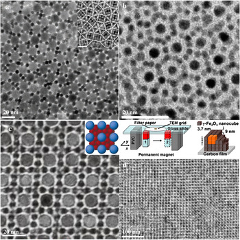
Crystals and superlattices of nanocrystal assemblies exhibit a host of unusual physical properties.Reference Shechtman, Blech, Gratias and Cahn15, Reference Levine and Steinhardt16 These intermediate structures between amorphous solids and regular crystalline materials can be made to self-assemble from binary colloidal nanoparticle systems through evaporation-induced self-assembly. As seen in Fig. 1, by adjusting the particle size ratio, nanoparticles with two different sizes can rearrange themselves into quasicrystals with highly ordered periodic structures [Fig. 1(a)].Reference Talapin, Shevchenko, Bodnarchuk, Ye, Chen and Murray17 With selected size ratios, other binary AB, AB2, AB5, and AB13 colloidal crystals have been prepared using a similar method. For example, an ordered monolayer of a CoPt3 nanoparticle array can display the AB, AB2, and AB5 structures [Fig. 1(b)].Reference Shevchenko, Talapin, Rogach, Kornowski, Haase and Weller18 Mixing two sizes of 11 nm Fe2O3 and 6 nm PbSe nanoparticles results in binary superlattices with AB13 and AB2 structure [Fig. 1(c)].Reference Redl, Cho, Murray and O’Brien19 The evaporation-induced self-assembly method can be combined with the application of an external magnetic field to assemble nanoparticle superlattices. Further, combination with a physical patterning method can produce patterned nanoparticle arrays. As seen in Fig. 1(d), with the use of a modulated magnetic field, long-range order of magnetic nanoparticle arrays with macroscopic dimensions can be obtained.Reference Ahniyaz, Sakamoto and Bergstrom20 Inducing dipolar attraction during the initial stage of the drying-mediated self-assembly process results in assembling the superparamagnetic nanocubes into large and defect-free superstructures with both translational and orientational order. The characteristic dimensions of the superlattice are controlled both by the particle concentration as well as the duration of the externally applied magnetic field. Micrometer-sized three-dimensional mesocrystals have also been obtained when the drying dispersion is subjected to an external magnetic field of moderate strength for the entire duration of the self-assembly process.Reference Ahniyaz, Sakamoto and Bergstrom20 As compared to a stable crystal structure in which the individual elemental constituents are strongly chemically bonded, these nanoparticle assemblies are usually weakly interacting since the nanoparticle subunits are linked by weak hydrogen bond, van der Waals, or electric/magnetic dipole interactions.
(a) Binary colloidal quasicrystal, reprinted by permission from Macmillan Publishers Ltd: Nature, Talapin et al., Reference Talapin, Shevchenko, Bodnarchuk, Ye, Chen and Murray17 copyright 2009. (b) Binary CoPt3 superlattices with AB5 structures, reprinted with permission from Shevchenko et al.,Reference Shevchenko, Talapin, Rogach, Kornowski, Haase and Weller18 copyright 2002 ACS. (c) Binary Fe2O3–PbSe superlattices with AB13 structures, reprinted by permission from Macmillan Publishers Ltd: Nature, Redl et al.,Reference Redl, Cho, Murray and O’Brien19 copyright 2003. (d) Magnetic field-induced self-assembly of oriented superlattices from maghemite nanocubes, reprinted by permission from PNAS, Ahniyaz et al., Reference Ahniyaz, Sakamoto and Bergstrom20 copyright 2007.

B. Template-assisted self-assembly
Template-assisted self-assembly approaches, including organic molecule-assisted self-assembly, porous templates, and nanoparticle templates, have been developed to prepare SP NP assemblies. As compared to the nanoparticle assemblies prepared by the evaporation-induced process, templated nanoparticle assemblies are much more stable since the template acts as a support for stabilizing the nanoparticle assembly. Moreover, the template serves as a structure-directing agent, and depending upon the shape and structure of the template, the shape and structure of the resulting nanoparticle assembly is tunable.
1. Organic molecule templates
Organic molecule-assisted self-assembly methods have been widely used to prepare nanoparticle assemblies.Reference Medintz, Uyeda, Goldman and Mattoussi21, Reference Shenhar, Norsten and Rotello22 The primary approaches include: (i) assembling nanoparticles into clusters and (ii) inducing ordering and orientation of the nanoparticles. Organic molecule templates can be charged surfactant molecules, amphiphilic block copolymers, and so on.Reference Nikolic, Olsson, Salcher, Kornowski, Rank, Schubert, Frömsdorf, Weller and Förster23, Reference Soler-Illia, Sanchez, Lebeau and Patarin24 Ionic surfactants are composed of a hydrophilic head group and a long hydrophobic tail group within the same molecular moiety. Protonation of amphiphilic block copolymers depends upon the solution pH. In an organic molecule templating self-assembly process, both organic molecule and inorganic soluble species direct the synthesis of nanoparticle assemblies, which are strongly dependent on the interaction between the organic molecule and its inorganic precursors.Reference Soler-Illia, Sanchez, Lebeau and Patarin24 As for the case of a single-molecule chain of organic molecule template in the case of ionic surfactants, the formation of the nanoparticle assemblies is mostly governed by electrostatic interactions. Other assembly routes rely on nonionic surfactants, where the primary interaction between the template and the inorganic species is H-bonding or dipolar.
The shape and structure of the nanoparticle assemblies can be further tuned by controlling the structure and molecular weight of the organic molecule micelles in the solvent. Upon progressive increase of surfactant concentration in an aqueous solution, micellar aggregates organize to form different shapes, but always follow the order: direct spheres, direct cylinders, lamellae, inverse cylinders, and inverse spheres.Reference Alexandridis, Holzwarth and Hatton25, Reference Evans and Wennerström26 Other arrangements, such as sponge-like and sheet-like structures, are also observed in a number of reported organic molecule-nanoparticle assemblies.Reference Li, Li, He, Zhu and Zhao27
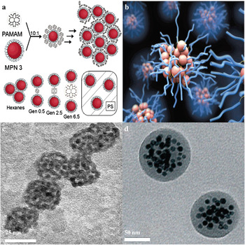
A series of anionic polyamidoamine dendrimers have been used to control the interparticle spacing of SP NP assemblies.Reference Frankamp, Boal, Tuominen and Rotello28 As seen in Fig. 2(a), the resulting assemblies display a systematic increase in the average interparticle spacing over a range of 2.4 nm. The particles can be completely coated by careful control of the ratio and concentration of the nanoparticles and the dendrimer. Further, the amount of dendrimer embedded between adjacent particles enables control of the interparticle spacing. The increase in the spacing between the nanoparticles modulates the collective magnetic behavior by effectively lowering the dipolar coupling between the particles. With the use of polymer micelles as templates, clusters of assembled iron oxide nanoparticles can be encapsulated in the polymer micelles, as seen in Figs. 2(b)–2(d).Reference Euliss, Grancharov, O’Brien, Deming, Stucky, Murray and Held29, Reference Kim, Qiu, Wang and Taton30 The size of uniform composite nanospheres is also tunable, ranging from 10–100 nm.Reference Niu, Li, Ma, Diao, Gu, Chen, Zhao, Ruan, Zhang and Shi31 The magnetization of the assemblies is determined by the number of SP NPs embedded in a single assembly. Although the number of nanoparticles formed in one micelle can be tuned by simply changing the concentration of nanoparticles, the maximum number of nanoparticles is still relatively small, thus resulting in a low magnetization of the final assembly.
(a) Schematic depiction of dendrimer-mediated nanoparticle assembly and control of interparticle spacing, reprinted with permission from Frankamp et al.,Reference Frankamp, Boal, Tuominen and Rotello28 copyright 2005 ACS. (b) Schematic illustration. (c and d) TEM images of the nanoparticle-micellar assemblies, reprinted with permission from (c) Euliss et al.,Reference Euliss, Grancharov, O’Brien, Deming, Stucky, Murray and Held29 copyright 2003 ACS, and (d) Kim et al.,Reference Kim, Qiu, Wang and Taton30 copyright 2005 ACS.

2. Nanopore templates
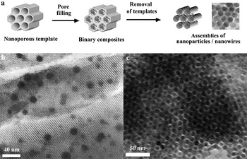
In contrast to organic molecules that are soft templates, porous materials, such as mesoporous materials,Reference Soler-Illia, Sanchez, Lebeau and Patarin24 porous anodic aluminum oxide,Reference Teng, Liang, Rahman and Yang32, Reference Hulteen and Martin33 and so on, can serve as hard templates to grow ordered nanoparticles within the confined space of the nanopore channels. Mesoporous materials provide some advantages, especially in their specific topological stability, highly ordered porous structures, and controllability.Reference Davis34 In the hard template route, the voids of a preformed mesoporous solid are utilized and are impregnated with the desired precursors. The subsequent mineralization/solidification of these precursors and removal of the solid template leads to ordered nanoparticle assemblies with different compositions. Since the inorganic nanoporous templates are stable at relatively high temperatures, post-annealing can be carried out to improve the crystallinity and to form the desired magnetic phase, while maintaining the structure of the nanoparticle assembly. The synthetic procedure is schematically illustrated in Fig. 3(a). It should be noted that in many reported cases there is only partial filling of the pore channels. To prepare highly ordered nanoparticle assemblies by the hard template route, the following three characteristics are generally necessary: (i) the templates should possess a high degree of order and robust thermal stability; (ii) a full impregnation of the desired precursors within the voids of templates is necessary; (iii) the original template should be completely removable to achieve a faithful replica.
(a) Schematic illustration of nanopore-templated assembly and control of nanoparticle assemblies. (b) Assemblies of SP NPs inside mesoporous silica. Reprinted with permission from MacLachlan et al.,Reference MacLachlan, Ginzburg, Coombs, Raju, Greedan, Ozin and Manners35 copyright 2000 ACS. (c) Ordered mesoporous iron oxide with crystalline walls. Reprinted with permission from Jiao et al.,Reference Jiao, Harrison, Jumas, Chadwick, Kockelmann and Bruce40 copyright 2006 ACS.

MacLachlan et al. reported on the synthesis of superparamagnetic ceramic nanocomposites.Reference MacLachlan, Ginzburg, Coombs, Raju, Greedan, Ozin and Manners35 With the use of silaferrocenophanes as the iron precursor, SP NPs are uniformly formed in the pore channels of mesopoeous silica [Fig. 3(b)]. Since the organometallic complexes used in the synthesis are hydrophobic, attachment of product nuclei to the pore walls is induced only after the formation of the Si-O bridges through hydrolysis of the silacyclobutane ring. With the use of less hydrophilic precursors, the nanopore filling ratio is low since the pore surface is hydrophilic. Several papers have also reported the synthesis of magnetic mesoporous silica nanocomposites.Reference Zhao, Gu, Zhang, Chen and Shi36–Reference Giri, Trewyn, Stellmaker and Lin39 However, these materials display poor magnetic response due to the difficulty in increasing the Fe3O4 mass fraction.Reference Kim, Lee, Lee, Yu, Kim, An, Hwang, Shin, Park, Kim and Hyeon37 Moreover, they possess irregular core-shell structuresReference Lin, Wu, Hung, Chou, Chang, Lin, Tsai and Mou38 and poor dispersity in water, which limits their applicability. To completely fill the pore channels, new synthetic strategies of using inorganic salts as precursors have been developed for the preparation of a variety of magnetic nanoparticle/nanowire arrays.Reference Jiao, Harrison, Jumas, Chadwick, Kockelmann and Bruce40–Reference Imperor-Clerc, Bazin, Appay, Beaunier and Davidson42 As seen in Fig. 3(c), highly ordered iron oxide nanoparticle arrays with very large surface area of 210 m2/g have been prepared using iron nitride as the precursor and mesoporous silica as the template. After removing the template, stable and ordered SP NP arrays are obtained.Reference Jiao, Harrison, Jumas, Chadwick, Kockelmann and Bruce40
3. Nanoparticle templates
Forming core-shell composite assemblies by directly growing SP NPs on the surface of presynthesized nanoparticle templates, or by converting the nanoparticle templates to superparamagnetic phase, is an alternate strategy to prepare SP NP assemblies. The nanoparticle templates can be metallic, semiconducting, or even insulating (e.g., silica).
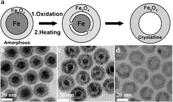
Sun’s group at Brown University has reported the synthesis of unusual core-shell-void Fe-Fe3O4 nanoparticles and hollow Fe3O4 nanoparticles by controlled oxidation of amorphous core-shell Fe–Fe3O4 nanoparticles.Reference Peng and Sun43 Fig. 4(a) provides a schematic illustration of the synthetic procedure. By proper choice of the heating condition, the Fe3O4 shell [Fig. 4(b)] is controlled to be either amorphous or polycrystalline. Isolated core-shell-void Fe–Fe3O4 nanoparticles [Fig. 4(c)] are also obtained under similar reaction conditions. Additional oxidation of core-shell-void Fe–Fe3O4 nanoparticles results in the formation of hollow Fe3O4 nanoparticles and their self-assembled monodisperse superlattice arrays [Fig. 4(d)]. Controlled surface oxidation of metal nanoparticles based on the nanoscale Kirkendall effect provides a simple and elegant approach for fabricating SP NPs with unique structures. The synthetic strategy can be extended to other types of hollow or core-shell-void transition-metal oxide nanoparticles. Moreover, metallic nanoparticles, such as Fe,Reference Peng, Wang, Xie and Sun44 and Co,Reference Chen, Liu, Li and Zeng45 among others, have also been used to prepare monodisperse superparamagnetic core-shell composite nanoparticles and hollow nanostructures based on the Kirkendall effect.Reference Yin, Rioux, Erdonmez, Hughes, Somorjai and Alivisatos46
(a) Schematic illustration of the synthesis of core-shell-void Fe–Fe3O4 and hollow Fe3O4 nanoparticles from Fe–Fe3O4 nanoparticle seeds. TEM images of (b) Fe–Fe3O4 nanoparticle seeds, (c) core-shell-void Fe–Fe3O4, and (d) hollow Fe3O4 nanoparticles. Reprinted by permission from Wiley, Peng and Sun,Reference Peng and Sun43 copyright 2007.

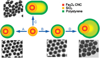
Semiconducting CdSe, PbSe, and PbS nanoparticles and insulating silica nanoparticles have also been utilized as templates to grow SP NP assemblies in the form of core-shell structures.Reference Tartaj and Serna47 Nucleation of SP NPs usually occurs on the surface of the nanoparticle templates. As compared to metallic nanoparticles that can serve as sacrificial reactants for growing superparamagnetic oxide nanoparticle shells, chalcogenide and silica nanoparticles are usually retained in the final product. Thus, the most commonly formed assemblies are core-shell bicomponent nanostructures. Considering the complexity of the assembly procedure, it is not surprising that there are only a few reports in the literature on the synthesis of tricomponent multi-shell core-shell superparamagnetic nanostructures. Yin’s group has reported the synthesis of interesting superparamagnetic composite colloids containing three components.Reference Ge, Hu, Zhang and Yin48 The composite colloids are produced through one-step emulsion polymerization of styrene using Fe3O4/SiO2 as the seed. As seen in Fig. 5, the composite colloids have anisotropic structures such as ellipsoids formed by one-step emulsion polymerization [Fig. 5(c)] and doublets produced with distinct steps [Fig. 5(d)]. Spherical colloids can also be produced in one-step emulsion polymerization, both without [Fig. 5(a)] and with [Fig. 5(b)] using the crosslinker divinylbenzene. All types of colloid particles have superparamagnetic cores that are concentrically or eccentrically located in the polystyrene shells. The formation of such novel shapes and structures is due to the interfacial tension between the hydrophilic seed particle and the hydrophobic monomer.
Synthesis scheme for Fe3O4/SiO2/PS composite colloids through seeded emulsion polymerization. The morphologies of the final products are controlled by: (a) interfacial tension, (b) cross-linking, and (c) single and (d) separated steps of swelling and phase separation. All scale bars represent 400 nm. Reprinted with permission from Ge et al.,Reference Ge, Hu, Zhang and Yin48 copyright 2007 ACS.

C. Free-Template, Direct Self-assembly
Many applications require SP NP assemblies with small size, large magnetization, and special geometrical architectures, such as hollow structures, to achieve facile manipulation of the particles, improved sensitivity for magnetic detection, increased porosity and inner surface area for drug delivery, and so on. However, to directly prepare such SP NP assemblies in a controlled manner is challenging since the uniformity of nucleation, growth, and subsequent assembly of nanoparticles is difficult to achieve simultaneously in a single reaction.
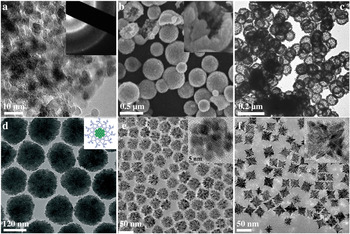
Long et al. have developed a crystalline superparamagnetic nanoarchitecture of iron oxide while still retaining the desirable bicontinuous pore-solid network, significant porosity, and large surface area expected from an aerogel [Fig. 6(a)].Reference Long, Logan, Rhodes, Carpenter, Stroud and Rolison49 The iron oxide aerogels are prepared by minor modification of a previously reported procedure using epoxide-based proton scavengers to initiate the sol-gel reactions for hydrolysis and polycondensation.Reference Gash, Tillotson, Satcher, Poco, Hrubesh and Simpson50 The pore size has a wide distribution, ranging from 7 to 80 nm. The iron oxidation state and related vacancy content of the Fe3O4-Fe2O3 phases can be readily tuned while preserving the nanoscale particle size and pore structure of the aerogel.
TEM images of (a) aerogel-templated mesoporous magnetic architectures, reprinted with permission from Long et al.,Reference Long, Logan, Rhodes, Carpenter, Stroud and Rolison49 copyright 2004 ACS, (b) superparamagnetic ferrite hollow submicrospheres, reprinted with permission from Li et al.,Reference Li, Zhang and Chen51 copyright 2006 ACS, (c) superparamagnetic Fe3O4/ZnS hollow nanospheres, reprinted with permission from Wang et al.,Reference Wang, Wu, Chen and Zhou52 copyright 2009 ACS, (d) superparamagnetic magnetite colloidal Fe3O4 nanocrystal clusters, reprinted with permission from Wiley, Ge et al.,Reference Ge, Hu and Yin53 copyright 2007, and (e–f) self-supported superparamagnetic nanostructures of spherical and rodlike CoFe2O4 nanocrystals, reprinted with permission from Bao et al.,Reference Bao, Shen, Wang, Ma, Mazumdar and Gupta54 copyright 2009 ACS.

The synthesis of hollow structures usually requires sacrificial templates through a successive multistep process involving preparation of a template, coating of the template with desired materials, and removal of the template to leave a hollow center. Li et al. reported a direct self-assembly strategy for preparation of hollow spheres [Fig. 6(b)].Reference Li, Zhang and Chen51 The approach involves the formation of ferrite nanoparticles and the simultaneous self-assembly of nanoparticles and block copolymer into hollow spheres under solvothermal conditions. Furthermore, this approach is general for the preparation of a variety of different ferrite hollow spheres with a composition of M1–xFe2+xO4 (M = Fe, Co, and Mn; 0 < x < 1). Unlike conventional hollow spheres, which are either hydrophobic or hydrophilic, the products prepared using this method exhibit excellent dispersibility in both polar and nonpolar solvents, and are promising for a broad range of applications.
In addition to single phase structures, bicomponent Fe3O4/ZnS superparamagnetic hollow nanospheres [Fig. 6(c)] have been synthesized through reaction of monodisperse FeS particles in a mixture containing zinc acetylacetonate, poly(vinylpyrrolidone), ammonium nitrate, glycol, and water at 150 °C for 10 h.Reference Wang, Wu, Chen and Zhou52 The formation of such hollow structures does not obey the conventional Kirkendall effect since the initial formation of a layer of Fe3O4 and ZnS on the surface of the FeS particles completely hinders any further direct chemical reaction between FeS and zinc acetylacetonate. The authors consider that the FeS and ZnS particles are positively charged, while Fe3O4 is negatively charged. Thus, the position of the Fe3O4 and ZnS particles being determined by the electrostatic interaction, with the Fe3O4 layer located between the positively charged FeS core and the outer ZnS layer. With a decreasing concentration of reactants, Ostwald ripening sets in. The small Fe3O4 and ZnS particles formed around the surface of the FeS continually grow into larger particles and then coalesce to form a shell. Such reactions continue until the FeS core disappears, finally leaving behind the bicomponent Fe3O4/ZnS hollow structures.
SP NPs with dimensions in the range of 10 nm have a low magnetization per particle, so it is difficult to effectively manipulate them using moderate magnetic fields. The strategy of forming clusters of magnetite nanocrystals has the advantage of controllably increasing the magnetization while retaining the superparamagnetic characteristics. Recently, Yin’s group developed a high-temperature hydrolysis process for the preparation of polyacrylate-capped superparamagnetic magnetite (Fe3O4) colloidal nanocrystal clusters with tunable sizes from 30 to 180 nm [Fig. 6(d)].Reference Ge, Hu and Yin53 The clusters are composed of a number of small primary crystals of size 6–8 nm. Since each cluster is composed of many magnetite crystallites that are magnetically decoupled, they retain their superparamagnetic characteristics.
Recently, our group also reported on novel shape- and structure-controlled superparamagnetic nanostructures composed of self-supported spherical and rodlike CoFe2O4 colloidal nanocrystals.Reference Bao, Shen, Wang, Ma, Mazumdar and Gupta54 They are prepared by thermolysis of a stoichiometric Co2+Fe23+-oleate complex in organic solution with periodic injection of hexane to control the nucleation, assembly, and growth of the nuclei. These large self-supported superparamagnetic nanocrystal structures have the advantage of increasing the magnetic moment while retaining the superparamagnetic characteristics. By merely changing the heating rate, the product shape and size can be varied from being spherical [40.5 ± 4.5 nm, Fig. 6(e)] to cubic [36.5 ± 2.5 nm, Fig. 6(f)]. These nanostructures are composed of individual spherical nanocrystals of <5 nm in size, or nanorods with dimensions of ~3.6 nm in diameter and <25 nm in length, respectively. They can form highly ordered two-dimensional assemblies over a large area. The formation of such superparamagnetic nanocrystal assemblies is likely because the burst boiling accompanied by a temperature drop slows down the growth of the nuclei and promotes their assembly to form clusters by particle coalescence (from interaction between the nuclei) or oriented attachment to decrease the surface energy.Reference Zheng, Smith, Jun, Kisielowski, Dahmen and Alivisatos55
IV. PROPERTIES AND POTENTIAL APPLICATIONS
A. Magnetically responsive colloidal photonic crystals
Functional photonic materials and devices can be created by assembling monodisperse colloidal particles into periodically ordered structures. The optical band gap of photonic crystals is determined by the lattice constants and symmetry of the colloidal arrays. An external magnetic field provides an effective means for controlling the lattice spacing of magnetic colloidal photonic crystals and thus the photonic properties. Calderon et al. initially observed that emulsion droplets containing γ-Fe2O3 nanoparticles can diffract light in the visible range by applying an external magnetic field.Reference Calderon, Stora, Monval, Poulin and Bibette56 In later work, Xu et al. developed monodisperse polymer-SP NP colloidal particles for photonic applications. They embedded superparamagnetic iron oxide nanoparticles in a polymer matrix through an emulsion polymerization process.Reference Xu, Friedman, Humfeld, Majetich and Asher57, Reference Xu, Friedman, Humfeld, Majetich and Asher58 By introducing high density charges to the colloid surface, they successfully constructed three-dimensional photonic crystals by taking advantage of the strong repulsive electrostatic interactions. However, because of the small loading of magnetic materials in the polymer, the field-responsive tuning range of these photonic crystals is limited, and the response time is rather slow.
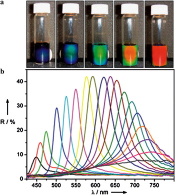
Photonic crystals fabricated using pure colloidal magnetic particles are expected to offer a faster response to an external field. If the colloids possess uniform shape, controllable size, and narrow size distribution, then the lattice constant of photonic crystals can also be precisely tuned. Further, with particle sizes in the range of 100–200 nm, the assembled structures will have periodicities comparable to the wavelength in the visible or near infrared range. Yin’s group has reported the synthesis of superparamagnetic clusters that satisfy all these conditions quite well.Reference Yin, Rioux, Erdonmez, Hughes, Somorjai and Alivisatos46 They successfully prepared polyacrylate-capped superparamagnetic clusters of Fe3O4 nanocrystal assemblies with tunable sizes from 30 to 180 nm, and a narrow size distribution.Reference Ge, Hu and Yin53, Reference Ge, Hu, Biasini, Beyermann and Yin59 Colloidal photonic crystals fabricated using these superparamagnetic clusters [Fig. 6(d)] exhibit highly tunable stop bands across the entire visible spectral region [Fig. 7] because of highly charged polyacrylate-capped surfaces and strong interaction of the magnetite clusters with an external magnetic field. The optical response to changes in the magnetic field strength is quite fast and fully reversible. The superparamagnetic clusters have been used to fabricate field-responsive polymer composite films for potential applications, such as color displays and sensors.
(a) Photograph of colloidal crystals formed in response to an external magnetic field; the magnet–sample distance decreases gradually from right to left. (b) Dependence of the reflection spectra at normal incidence of the colloidal crystals on the distance of the sample from the magnet. Diffraction peaks blue-shift as the distance decreases from 3.7 to 2.0 cm with step sizes of 0.1 cm. Reprinted with permission from Wiley, Ge et al.,Reference Ge, Hu and Yin53 copyright 2007.

B. Biomedical and biological applications
Gold nanoparticles are important reagents for many highly sensitive and selective DNA and protein detection schemes.Reference Rosi and Mirkin60 Magnetic particles are also attractive and have been widely exploited in biology for diagnostic and therapeutic applications. There is a need in the field of biomedicine for the development of composite materials that combin gold for its stability, surface chemistry, and optical properties and superparamagnetic particles for easy manipulation. Stoeva et al. have developed three-layer composite magnetic nanoparticles for detecting DNA.Reference Stoeva, Huo, Lee and Mirkin61 The composite particles are composed of a gold surface, silica core, and superparamagnetic inner layer. The DNA-modified three layer magnetic nanoparticles can be repeatedly centrifuged and redispersed in a buffer solution without aggregation. Moreover, the DNA-modified three-layer magnetic nanoparticles assemble into macroscopic aggregates upon hybridization with complementary linking oligonucleotides.
Superparamagnetic colloids are also ideal systems for bioseparation of specific biological molecules (e.g., DNAs, proteins, and cells) from solution because the external control of magnetization enables the ready separation of adsorbed biomolecules with a magnetic field. In a typical separation procedure, the surface of magnetic particles is first modified with biocompatible molecules, providing conjugation between the magnetic particles and the targeted biological molecules. The biological molecules labeled by superparamagnetic colloids are then separated from the solution by application of an external magnetic field. Magnetic microspheres consisting of an iron oxide core and silica shell have recently attracted attention for bioseparation.Reference Xu, Deng, Gao, Yu, Yang and Zhang62 Zhao’s group has reported on novel sandwich-structured mesoporous silica microspheres composed of a silica-coated magnetite core and ordered mesoporous silica shell with perpendicularly oriented channels.Reference Deng, Qi, Deng, Zhang and Zhao63 Three steps are involved in the synthesis procedure: (i) uniform magnetite microspheres are first coated with a thin silica layer using a sol-gel approach; (ii) a mesostructured silica composite is then deposited on the Fe3O4/nSiO2 microspheres through a surfactant-templating approach using cetyltrimethylammonium bromide as a template; (iii) final removal of surfactant templates by acetone extraction to form a mesoporous SiO2 shell results in well-dispersed tricomponent Fe3O4-nonporous SiO2-mesoporous SiO2 microspheres. The resulting microspheres are superparamagnetic, with a high magnetization of 53.3 emu/g, uniform accessible 2.3 nm mesochannels, high surface area of 365 m2/g, and a large pore volume of 0.29 cm3/g. They exhibit enhanced adsorption efficiency for fast removal of large microcystins (1000 Da) from solution.
Magnetic resonance imaging (MRI) is routinely used for clinical diagnostic imaging. The working principle of MRI is to measure the change in magnetization of protons in a magnetic field after being exposed to a radio frequency pulse. MRI contrast agents are often used to decrease the relaxation time so that the magnetic moment of a proton relaxes back faster to its initial value, thus producing better contrast.Reference Browne and Semelka64 SP NPs are magnetically saturated at the magnetic field strength used in an MRI scanner so they generate local variations in the magnetic susceptibility of the system under a magnetic field. For example, Berret et al. reported on the structural and magnetic properties of such complexes made from 6.3 nm diameter superparamagnetic iron oxide nanoparticles and cationic-neutral copolymers.Reference Berret, Schonbeck, Gazeau, El Kharrat, Sandre, Vacher and Airiau65 The copolymers investigated include poly(trimethylammonium ethylacrylate methyl sulfate)-b-poly-(acrylamide), with molecular weights 5000-b-30,000 g/mol and 110,000-b-30,000 g/mol. The mixed copolymer-nanoparticle aggregates have been characterized using a combination of light scattering and cryotransmission electron microscopy. Their hydrodynamic diameters have been determined to be in the range of 70–150 nm, and the number of nanoparticles per aggregate varies from tens to the hundreds. In addition, magnetic resonance spin-echo measurements show that the complexes have a better contrast in MRI than single nanoparticles and that these complexes are suitable for biomedical applications.
V. OUTLOOK
In this paper we have reviewed the recent progress in the field of SP NP assemblies. Depending on the spacing and alignment of the individual nanoparticles, the SP NP assemblies exhibit novel properties that are attractive for a variety of practical applications. A number of synthetic strategies that have been reported for the fabrication of crystals, films, nanostructures, and patterned superstructures of nanoparticle assemblies are discussed. However, the precise control of the assembly of nanoparticles into ordered nanostructures still remains a challenge.
Research on self-assembly of SP NPs is developing in several directions.
(1) Principles and rules of self-assembly processes, such as thermodynamics and kinetics, need to be better established to design and reproducibly synthesize complex hierarchical assemblies with high yield, well-defined sizes, shapes, aspect ratios, and chemical heterogeneity. This will also help in the development of self-assembly processes that are scalable, low cost, and environmentally friendly for future industrial applications.
(2) The surface functionalization and modification of magnetic nanoparticles to introduce additional functionality is generally a difficult task and will receive increasing attention in coming years. Multifunctional magnetic nanoparticle assemblies with designed active sites and amphiphilic surface appear promising for a variety of applications, including in biology, catalysis, separation, and composites.
(3) Nature provides many examples of remarkably complex, yet highly organized, self-assembled hierarchical structures, and even limited exploitation of some of the forms of self-assembly found in nature can lead to much better control over the types of nanoparticle assemblies that can be produced in the laboratory and to improvements in their quality. It would thus be desirable to adopt some of the bioinspired synthetic approaches.
(4) Exploring novel properties and applications of SP NP assemblies.
ACKNOWLEDGMENTS
This work was supported by NSF under Grant No. ECS-0621850.