Introduction
Primordial germ cells (PGCs) segregate early in the developing mammalian embryo from the proximal epiblast, and migrate and colonize the undifferentiated gonadal ridges. Once in the primordial gonad, PGCs proliferate through successive mitotic rounds and differentiate. Depending on the chromosomal sex of the embryo, mitotic proliferation of PGCs will be followed by its entrance to meiosis I in females, or will enter in close contact with the somatic stratum giving rise to testicular morphogenesis (Tam & Snow, Reference Tam and Snow1981; McLaren, Reference McLaren2001; Zhao & Garbers, Reference Zhao and Garbers2002). Primordial Sertoli cells join each other and encompass PGCs, and seminiferous cords morphogenesis starts (Angelopoulou et al., Reference Angelopoulou, Magre, Patsavoudi and Jost1984, Reference Angelopoulou, Balla, Lavranos, Chalikias, Kitsos, Baka and Kittas2008). Within the sex cords, PGCs will differentiate morphologically and become the so-called prospermatogonia or gonocytes (Angelopoulou et al., Reference Angelopoulou, Balla, Lavranos, Chalikias, Kitsos, Baka and Kittas2008). By that time, the developing mammalian testes are organized in sex cords composed of germ cells, supporting cells, precursors of Sertoli cells that surround germ cells, and the Leydig cells in the interstitial tissue between sex cords. Until postnatal life, the morphology of the testes will remain unchanged but mitotic activity will persist throughout fetal development (Orth, Reference Orth1984; Van Vorstenbosch et al., Reference Van Vorstenbosch, Colenbrader and Wensing1984). Cell proliferation and its counterpart, apoptosis-driven cell demise, are essential processes in the physiology of the developing testis. Moreover, these processes strongly influence the normal adult spermatogenesis (Angelopoulou et al., Reference Angelopoulou, Balla, Lavranos, Chalikias, Kitsos, Baka and Kittas2008). Nevertheless, germ cell proliferation and morphometry of the developing mammalian testis have been scarcely investigated.
The South American plains viscacha, Lagostomus maximus, is a seasonal histricognathe rodent inhabiting the southern area of the Neotropical region. In females, several unique and exceptional reproductive characters have been described (Weir, Reference Weir1970, Reference Weir1971a, Reference Weirb). Interestingly, these females are able to ovulate up to 800 eggs or more per reproductive cycle, representing the highest ovulation rate so far recorded in mammals (Weir, Reference Weir1971a). This massive polyovulation arises from the highly convoluted ovarian anatomy that increases the surface area for ovulation (Weir, Reference Weir1971a), the small size of the ovulatory follicle (Weir, Reference Weir1971a), and the suppression of apoptosis-dependent follicular atresia through the overexpression of anti-apoptotic BCL2 gene and little or no detectable proapoptotic BAX (Jensen et al., Reference Jensen, Willis, Albamonte, Espinosa and Vitullo2006). Suppression of apoptosis is also known to occur in the developing ovary, avoiding massive fetal germ cell demise (Leopardo et al., Reference Leopardo, Jensen, Espinosa and Vitullo2006), and involves all the cell compartments in the adult gonad (Jensen et al., Reference Jensen, Willis, Leopardo, Espinosa and Vitullo2008). This peculiar behaviour of female germ line highlights the need to explore how male germ line develops in this species, particularly during fetal life, as primordial germ cells share a common developmental pathway until they reach the genital ridges and genetic differences dictated through embryo sex chromosomes are put in motion.
Male reproductive physiology in L. maximus has been scarcely investigated. As this rodent is a seasonal breeder, most of the published information focuses on adult testicular changes related to the photo-period, and shows that endocrine variation caused by light/dark cycle fluctuation induces changes in the morphology of Leydig cells, Sertoli cells and spermatogonia (Calvo et al., Reference Calvo, Sagripanti, Peltzer, Guzman and Charreau1986; Sinha Hikim et al., Reference Sinha Hikim, Amador, Klemcke, Bartke and Russel1989a, Reference Sinha Hikim, Amador, Bartke and Russelb; Fuentes et al., Reference Fuentes, Caravaca, Pelzer, Scardapane, Piezzi and Guzman1991; Muñoz et al., Reference Muñoz, Fogal, Dominguez, Scardapane and Piezzi2001). However, there is no information about the development and physiology of the fetal testis in Lagostomus maximus.
The aim of the present study was to analyse the testicular morphometry, to establish the pattern of expression of the proliferation cell nuclear antigen (PCNA) and to evaluate its correlation with the expression of germ cell markers, OCT-4 and VASA, to estimate the percentage of proliferating germ cells in different stages of the embryonic testis throughout development in Lagostomus maximus.
Materials and methods
Animals and tissue collection
The protocol of this study was reviewed and approved by the Ethics and Research Committee of Universidad Maimónides, Argentina. Handling and killing of captured animals were performed in accordance with the CCAC Guide for the Care and Use of Laboratory Animals (CCAA, 2002). Plains viscachas, Lagostomus maximus, were trapped from a natural population at the Estación de Cría de Animales Silvestres (E.C.A.S), Ministry of Agriculture, Villa Elisa, Buenos Aires province, Argentina. Animals were captured at three different time-points during the main breeding season which extends from March–April to August–September, in order to include early-, mid-, and late-gestating females within the 5-month long gestation period. Animals were anesthetized with xylacine/ketamine, bled by intracardiac puncture and immediately killed through the administration of 0.2 ml/kg body weight Euthanyl (sodium pentobarbital, sodium diphenyl hydantoinate; Brouwer S.A) by trained technical staff from E.C.A.S. Blood samples were centrifuged, and sera separated and stored at –20°C until used for hormone measurements. Fetuses were removed, weighted, and crown-head length recorded to estimate gestational age. Embryonic testes were dissected out under a stereo-microscope, fixed in 4% paraformaldehyde and classified in three groups: early- (n = 5), mid- (n = 5) and late-developing (n = 5) testis, according to capture time, embryonic size and previous description on embryo development (Roberts & Weir, Reference Roberts and Weir1973).
Testis histology and morphometry
Fixed testes were embedded in paraffin and serially cut in 5-μm-thick sections, mounted onto cleaned coated-slides, dewaxed in xylenes, rehydrated in graded alcohols, washed in tap water, and processed for routine haematoxylin–eosin staining. For each specimen, at least three to five slides were stained for general histology inspection. The volume densities of the testicular tissue components were determined by light microscopy using a 441-intersection grid placed in the ×10 ocular of a light microscope according to Leal & França (Reference Leal and França2006). Briefly, 15 fields chosen randomly (6615 points) were scored for each animal at a ×400 magnification. Technical artifacts were rarely seen and were not considered in the total number of points used to obtain volume densities. Points were classified as one of the following: seminiferous cords, comprising tunica propria, epithelium and lumen; Leydig cells; blood vessels and lymphatic spaces and connective tissue. The volume of each component of the testis was determined as the product of the volume density and testis volume. The results of the testicular proportions were expressed as percentages.
Immunohistochemistry
Mounted paraffin sections (5 μm) were dewaxed in xylenes, rehydrated in graded alcohols and washed in tap water. Endogenous peroxidase activity was inhibited in tissue sections using 0.5% v/v H2O2/methanol for 20 min at room temperature. Then, sections were blocked for 1 h with 15% normal goat (PCNA) or rabbit (OCT-4) serum in phosphate-buffered saline (PBS) and then incubated overnight at 4°C with primary antibody (1:200 diluted rabbit anti-PCNA, Abcam, UK; 1:500 diluted goat anti-OCT-3/4, Santa Cruz Biotechnology, Inc.). After three rinses in PBS, sections were incubated for 1 h at room temperature with appropriate 1:200 diluted biotinylated secondary antibodies (Vector Labs, UK). After further washing in PBS, sections were incubated for 30 min with 1:100 diluted streptavidin–peroxidase complex (ABC kit, Vector Labs, UK). Sections were washed twice with PBS and development of peroxidase activity was performed with 0.05% w/v 3,3′-diaminobenzidine and 0.1% v/v H2O2 in Tris–HCl. Sections were finally washed with distilled water. Negative controls were processed simultaneously by omitting the primary antibodies or pre absorbing the primary antibody with synthetic peptides.
For double immunohistochemistry, sections were first stained with rabbit anti-VASA (Abcam, UK) using 1:100 diluted streptavidin–peroxidase complex (ABC kit, Vector Labs) for 30 min and visualized with blue Vectastain (Vector Labs). After five rinses in PBS, anti-PCNA primary antibody was applied and subsequent steps were as described above for single immunostaining.
Positively stained cells for PCNA, VASA, and OCT-4 were counted in single sections using an Olympus BX40 microscope at ×1000 magnification. Sections were counted independently by two observers. Approximately, 300 Leydig cells, peritubular cells, and Sertoli cells or 200 in the case of germ cells, were counted per slide. Leydig cells and peritubular cells were identified according to their localization in the interstitial compartment of the testis. Sertoli cells were easily identified because of the small nuclei and localization in the seminiferous cords. Germ cells were identified within the cords, according to their large round nuclei (larger than those from Sertoli cells) and distinct cytoplasm.
Immunofluorescence
Dewaxed and rehydrated tissue sections were blocked for 1 h with 15% normal goat (PCNA) or rabbit (OCT-4) serum in PBS, washed with PBS and then incubated overnight at 4°C with primary antibody (1:200 diluted rabbit anti-PCNA, Abcam, UK; 1:500 diluted goat anti-OCT-4, Santa Cruz Biotechnology, Inc.). After five rinses in PBS, sections were incubated in darkness for 1 h at room temperature with 1:300 diluted FITC–goat anti-rabbit IgG (H+L) Conjugate (Zymed Laboratories) or 1:250 diluted Cy3–rabbit anti-goat IgG (H+L) Conjugate (Zymed Laboratories) secondary antibodies for anti-PCNA or anti-OCT-4 treated sections, respectively. Slides were mounted with DAKO fluorescence mounting medium (Dako) and analysed by using an Olympus BX40 microscope with conventional epifluorescence. Negative controls were processed simultaneously by omitting the primary antibodies or preabsorbing the primary antibody with synthetic peptides.
TUNEL assay
Apoptosis-dependent DNA fragmentation was detected in paraffin-embedded sections by terminal deoxynucleotidyl transferase-mediated deoxyuridine triphosphate nick end labelling (TUNEL) technique, using the In Situ Cell Death Detection Kit (Roche Diagnostics GmbH), with fluorescein-tagged nucleotides, according to the manufacturer's protocol. Treated sections were examined in an Olympus BX40 microscope with conventional epifluorescence with ultraviolet illumination. In order to ascertain negative results, TUNEL-processed sections were incubated with 10IU/ml DNase II (Sigma) in 50 mM Tris–HCl, pH 7.5, 10 mM MgCl2, and 1 mg/ml BSA for 10 min at room temperature. After incubation, slides were thoroughly rinsed and treated again according to the TUNEL protocol. Images were captured with an Olympus Camedia C-5060 camera.
Hormone measurements
Sera from early- (n = 5), mid- (n = 5), and late-gestating females (n = 5) were tested for follicule stimulating hormone (FSH) and luteinizing hormone (LH) by ELISA (EIA) using FSH IEMA well (Radim) and LH IEMA well (Radim), respectively. The hormone values were obtained with a microplate spectrophotometer (μQuant™, BioTek). The sensitivity of the FSH and LH assay was 0.25 mIU/ml.
Statistical analysis
Mean and standard error (SEM) were calculated. The GraphPad Prism Software (Windows GraphPad Software version 5.0) was used for one-way analysis of variance. Newman–Keuls test was used when differences between more than two groups were compared. A p-value of less than 0.05 was considered significant.
Results
Testicular histology and morphometry
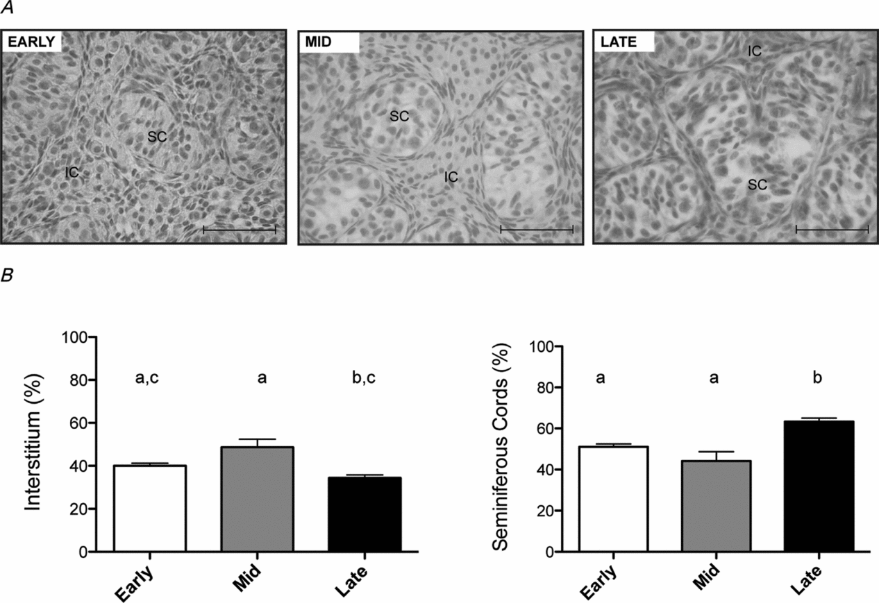
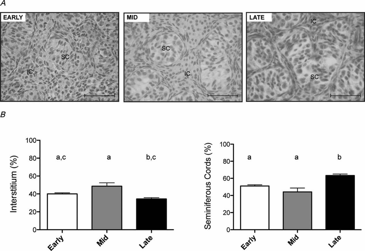
Early-, mid-, and late-developing testes contained well defined seminiferous cords surrounded by interstitial tissue. The testicular morphometry of the three developmental stages studied revealed that seminiferous cords significantly increased in late-developing testis (63.2 ± 0.6%, p < 0.05) when compared with early-developing (51.0 ± 1.4%) and mid-developing testis (44.1 ± 9.1%) (Fig. 1). The density of the interstitium remained almost stable with a significant increase in mid-development (Fig. 1).
General histology (A) and morphometry (B) in early-, mid- and late-developing testes in L. maximus embryos. Note the clear increase in the size of seminiferous cords from early- to late-development (A) mirrored in the significant quantitative decrease of the interstitium and increase of seminiferous cords in late-developing testis (B). Data are plotted as the mean ± SEM (n = 5)a,b,c. Different letters in bars indicate significant differences between groups (p < 0.05). IC: interstitial cells; SC: seminiferous cords. Scale bar: 50 μm.
Cell proliferation
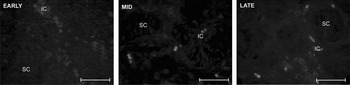
PCNA immunoreaction was detectable in Leydig cells, peritubular cells, Sertoli cells and germ cells at early-, mid- and late-developing testis (Fig. 2). However, each cell type displayed a different pattern of proliferation. The percentage of positive Leydig cells was significantly higher in mid-developing testis (77.35 ± 5.58%, p < 0.05) in comparison with early- (60.10 ± 14.14%) and late-developing testis (55.81 ± 5.40%) (Table 1). A stable rate of proliferation characterized the peritubular compartment and no significant differences were found in the percentage of PCNA-positive peritubular cells between groups (Table 1). In the tubular compartment, there was a significant increase (p < 0.05) in the percentage of PCNA positive germ cells from mid-development (early: 79.10 ± 2.82%; mid: 88.25 ± 1.70%; late: 87.10 ± 5.32%) (Table 1). The percentage of PCNA-positive Sertoli cell was stable throughout the developmental stages analysed with a density around 79% (Table 1).
Mean distribution of PCNA-positive cells in the developing testis of L. maximus throughout gestation

Data are plotted as the mean ± SEM (n = 5).
a,bDifferent letters in columns indicate significant differences between groups (p < 0.05).
Fluorescence immunolocalization of proliferating cell nuclear antigen (PCNA) in the developing testis of L. maximus. PCNA activity was detected throughout development in the interstitial cells (IC) and seminiferous cords (SC). Scale bar: 50 μm.

Testicular apoptosis
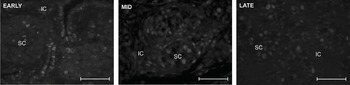
TUNEL analysis showed to be conspicuously negative in testis from early-, mid-, and late-developing embryos (Fig. 3). Only a few positive interstitial cells for TUNEL were detected in the testis from the three time-points studied. It is worth to note that no apoptotic cells were detected in seminiferous cords. In order to confirm that TUNEL-processed sections were truly negative, treated sections were incubated with DNase and reprocessed by TUNEL protocol. All nuclei from all cell types become positive confirming that DNA was originally not fragmented.
DNA fragmentation/integrity analysis in the developing testis of L. maximus by TUNEL assay. Sections in early-, mid-, and late-developing testis were conspicuously negative. Except for occasional positive cells in the interstitium, no positive signal was detected in the nuclei of gonocytes. IC: interstitial cells; SC: seminiferous cords. Scale bar: 50 μm.

Germ cell identity, totipotentiality and proliferation
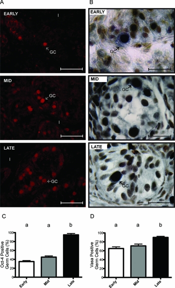
Expression of the totipotentiality marker OCT-4 was detected in gonocytes at all developmental stages analysed (Fig. 4A). In line with the per cent increase in seminiferous cords (see Fig. 1) and the number of PCNA positive germ cells that also increased in mid- and late-developing testis, the totipotentiality marker OCT-4 displayed a conspicuous and significant increase at late-development (Fig. 4). From a constant 40% detectable OCT-4-positive germ cell until mid-gestation, this figure rose up to 90% in late-developing testis (Fig. 4C). The identity of germ cells was confirmed by the co-localization of VASA protein in the cytoplasm of PCNA immunoreactive cells (Fig. 4B). The number of VASA-positive germ cells also showed a significant increase that correlated to the increasing immunodetection of OCT-4-positive germ cells (Fig. 4D).
Immunolocalization of pluripotency marker OCT-4 and co-localization of germ cell marker VASA and proliferating cell nuclear antigen, PCNA in the developing testis of the plains viscacha, L. maximus. (A) OCT-4 positive germ cells (arrows) clearly increase from early- to late-development. (B) Co-localisation of germ cell marker VASA (blue cytoplasmic staining) and PCNA (brown nuclear staining). (C,D) Percentage of positive OCT-4 and VASA germ cells, respectively. Note the increasing proportion of OCT-4 and VASA-expressing germ cells with increasing developmental age. Data are plotted as the mean ± SEM (n = 5). Different letters indicate significant differences between groups (p < 0.05). I: interstitium; GC: germ cell. Scale bar: (A) 50 μm, (B): 20 μm.

LH and FSH levels
Circulating LH levels were undetectable in early-, mid- and late-gestating females. The levels of FSH were lower in early-gestating females (0.7 ± 0.2 mUI/ml), showed a four-fold significant increase by mid-gestation (2.75 ± 0.1 mUI/ml, p < 0.05) and attained a seven times significant higher value (4.9 ± 1.5 mUI/ml, p < 0.05) at late gestation.
Discussion
Normal masculinisation of the fetus, and fertility in the adult life, depend on differentiation and development of Leydig cells, Sertoli cells and germ cells during foetal and neonatal life. Studies in rodents have provided some insight into the maturation and differentiation of the male germ cell lineage in foetal and neonatal life but little is known about the concerted proliferation of germinal and somatic compartments during fetal life (Wilhem et al., Reference Wilhelm, Palmer and Koopman2007), and the balance between proliferation and cell death is still a matter of discussion (Koji & Hishikawa, Reference Koji and Hishikawa2003).
In this study, proliferation activity of male germ cells, and accompanying somatic cells, in the developing testes of the South American plains viscacha was examined from early to late development, presenting the first description of fetal testis morphometry in a mammal whose counterpart, female germ line, is known to develop through abolished apoptosis and unrestricted germ cell proliferation (Jensen et al., Reference Jensen, Willis, Albamonte, Espinosa and Vitullo2006; Leopardo et al., Reference Leopardo, Jensen, Espinosa and Vitullo2006).
The testicular morphometry of early-, mid- and late-developing testis of L. maximus revealed a distinctive pattern of proliferation in Leydig cells, peritubular cells, Sertoli cells and gonocytes. The interstitial compartment showed a significant increase at mid-development. This increase could be associated with the proliferation of Leydig cells as they showed high levels of PCNA expression at this developmental stage. In the fetal rat testis, proliferation of Leydig cells has been associated to the influence of LH/hCG as a single dose of hCG during gestation induces a rapid proliferative response of Leydig cells (Kuopi et al., 1984). However, this does not seem to be the case in L. maximus; LH was not detected throughout gestation. This situation is similar to the fetal mouse development of testis in which Leydig cell number is gonadotrophin-independent and a requirement for gonadotrophin is not established until late in fetal life (Baker & Shaughnessy, Reference Baker and O'Shaughnessy2001).
Histological analysis revealed a clear increase in the size of the seminiferous cords at late development. This increment seems to be linked to an increase in Sertoli cell number (data not shown) although part of it is also associated with the proliferation of germ cells. A high percentage of PCNA positive germ cells was detected in the early-, mid-, and late-developing testis, but mid and late embryos showed significantly more positive PCNA germ cells compared with early embryos. This pattern of proliferation of germ cells in Lagostomus maximus is in contrast with that found in other species. Mitchell et al. (Reference Mitchell, Cowan, Morris, Anderson, Fraser, Mckenzie, Wallace, Kelnar, Saunders and Sharpe2008) reported that the immunoexpression of Ki67, another proliferation marker that follows a pattern of expression close to PCNA (Angelopoulou et al., Reference Angelopoulou, Balla, Lavranos, Chalikias, Kitsos, Baka and Kittas2008), was detected in germ cells of the fetal testis in the marmoset, rat and human. In the three species, a reduction in the number of Ki67-positive germ cells at the end of gestation was detected. Previous studies have also demonstrated Ki67 expression in human germ cells in the third trimester and into early postnatal life (Honecker et al., Reference Honecker, Stoop, de Krijger, Chris Lau, Bokemeyer and Looijenga2004). In the developing testis of the rat, however, germ cell labelling index with PCNA and Ki67 antibodies increases from mid (~14.5 days post coitum) to late gestation, entering then a sharp decrease from birth onwards followed by a new transient increase between 3 and 5 days post partum (Angelopoulou et al., Reference Angelopoulou, Balla, Lavranos, Chalikias, Kitsos, Baka and Kittas2008).
Male germ cell proliferation kinetics in the developing rat testis is comparable to that found in L. maximus, and pinpoints the necessity of further analysis from birth onwards in the viscacha's testis, especially considering that, in contrast to the rat, after a 5-month gestation period viscacha pups are born fully mature, eyes open and fur coated (Roberts & Weir, Reference Roberts and Weir1973; Weir, Reference Weir1970).
The transcriptional factor OCT-4 was immunodetected in early-, mid- and late-developing testis displaying a slight increase from early to mid development, and rising to almost 90% of germ cells in late-developing testis, most likely associated to the increase registered at that time in germ cell proliferation. This pattern of OCT-4 expression is in contrast to that found in other mammals. Several authors detected OCT-4 in a decreasing proportion of the total germ cell population during progression through fetal life in marmosets and humans (Gaskell et al., Reference Gaskell, Esnal, Robinson, Anderson and Saunders2004; Honecker et al., Reference Honecker, Stoop, de Krijger, Chris Lau, Bokemeyer and Looijenga2004; Pauls et al., Reference Pauls, Schorle, Jeske, Brehm, Steger, Wernert, Buttner and Zhou2006; Kerr et al., Reference Kerr, Hill, Blumenthal and Gearhart2008; Mitchell et al., Reference Mitchell, Cowan, Morris, Anderson, Fraser, Mckenzie, Wallace, Kelnar, Saunders and Sharpe2008). Besides its association with proliferation, the rise in OCT-4-expressing germ cells could be associated with the conspicuous increase in levels of circulating FSH we found in mid- and late-gestating females. FSH is recognised as a central player in the regulation of spermatogenesis in mammals whose effects are mediated directly or depend on indirect alteration of the levels of androgens via Sertoli cells (Ruwanpura et al., Reference Ruwanpura, McLachlan and Meachem2010). In rodents, O'Shaughnessy et al. (2010) have recently shown that FSH directly stimulates the increase of spermatogonial number and its entrance to meiosis with no direct effect on meiosis completion. Moreover, the quantitative maintenance of spermatogenesis by optimising germ cell number is a primary effect of FSH (O'Shaughnessy et al., 2010). It is worth to mention that embryo development in L. maximus proceeds in an unusual endocrine ambiance as gestating females ovulate at mid-gestation, display continuous folliculogenesis, and at least two transient rises in FSH take place from mid- to late- gestation (Fraunhoffer et al., Reference Fraunhoffer, Jensen, Willis, Freysselinard, Inserra and Vitullo2008; Jensen et al., Reference Jensen, Willis, Leopardo, Espinosa and Vitullo2008). The addition of further developmental ages at smaller intervals in future analysis will be useful to clarify the influence of FSH in the continuous proliferation of gonocytes.
The increase in proliferation in the somatic compartment and germ cells from early- to late-developing testis was in line with a remarkably negative TUNEL analysis. Moreover, we failed to detect apoptotic germ cells at any developmental time analysed. In mouse, TUNEL positive germ cells are found until 17 days of gestation. Thereafter, no detectable TUNEL staining is found (Koji & Hishikawa, Reference Koji and Hishikawa2003). However, the possibility that germ cells in the developing testis of L. maximus are being lost through a pathway not detected by TUNEL assay cannot be ruled out. Controversial results on the nature of mechanism of male germ cell demise exist. By the end of gestation and early after birth, it has been shown in mouse that germ cells degenerate by a process necrotic in nature rather than apoptotic, although typical features of necrosis were not identified (Wang et al., Reference Wang, Nakane and Koji1998) and TUNEL staining revealed a lack of positive nuclei (Koji & Hishikawa, Reference Koji and Hishikawa2003).
The identity of proliferating germ cells was assessed by co-localization of VASA protein in early-, mid- and late-developing testis. The pattern of VASA expression and its widespread cytoplasmic localisation is similar to that previously described in the rat (Mitchell et al., Reference Mitchell, Cowan, Morris, Anderson, Fraser, Mckenzie, Wallace, Kelnar, Saunders and Sharpe2008) and mouse (Toyooka et al., Reference Toyooka, Tsunekawa, Takahashi, Matsui, Satoh and Noce2000) in which all germ cells express VASA from colonization of the genital ridges. However, it must be emphasized that the continuous rise in VASA-expressing germ cells was accompanied by a conspicuous OCT-4 expression, specially at late gestation, that clearly contrasts with decreasing proportion of OCT-4-expressing gonocytes during progression of foetal life in other mammals.
In conclusion, the morphometric analysis of the South American plains viscacha shows a distinctive pattern of testis development characterized by a sustained proliferation of germ cells most likely driven through the high levels of circulating maternal FSH which directly influence germ cell number, a little or no apoptosis-mediated germ cell demise, and a continuous and conspicuous rise in the number of OCT-4-expressing gonocytes which add novel aspects to the overall understanding of mammalian testis development.
Acknowledgements
We are especially grateful to the personnel of E.C.A.S for their invaluable help in trapping and handling the animals and Ms Clara Ippólito for technical assistance in tissue processing. This study was supported by a PICTO-CRUP (grant No. 30972) from the Agencia Nacional de Promoción Científica y Técnica (ANPCyT) granted to ADV, and intramural funding from Universidad Maimónides.