Introduction
Bilingualism could be defined as the ability to communicate in two or more languages and use them on a regular basis. It is not easy, however, to categorize people as bilinguals or monolinguals, since there are many aspects that need to be considered beyond proficiency in the first and the second acquired languages (L1 and L2 respectively). These include age of acquisition, acquisition methodology, frequency of use, etc. (Bak, Nissan, Allerhand & Deary, Reference Bak, Nissan, Allerhand and Deary2014)
Different authors have studied the potential advantages that bilingualism confers to cognitive performance and brain health. For example, bilingualism has been claimed to provide an advantage in the performance of several cognitive tasks, mainly in the domain of executive functioning (Adesope, Lavin, Thompson & Ungerleider, Reference Adesope, Lavin, Thompson and Ungerleider2010; Costa, Hernández & Sebastián-Gallés, Reference Costa, Hernández and Sebastián-Gallés2008; Stocco & Prat, Reference Stocco and Prat2014), although some studies have failed to identify such benefits among the bilingual population (Cox, Bak, Allerhand, Redmond, Starr, Deary & MacPherson, Reference Cox, Bak, Allerhand, Redmond, Starr, Deary and MacPherson2016; Hernández, Martin, Barceló & Costa, Reference Hernández, Martin, Barceló and Costa2013; Crane, Gruhl, Erosheva, Gibbons, McCurry, Rhoads, Rhoads, Nguyen, Arani, Masaki & White, Reference Crane, Gruhl, Erosheva, Gibbons, McCurry, Rhoads, Rhoads, Nguyen, Arani, Masaki and White2010; Zatorre, Belin, Crane, Gruhl, Erosheva, Gibbons, McCurry, Rhoads, Nguyen, Arani, Masaki & White, Reference Zatorre, Belin, Crane, Gruhl, Erosheva, Gibbons, McCurry, Rhoads, Nguyen, Arani, Masaki and White2001), even when the specific outcomes of age of acquisition and multilingualism were taken into consideration (Paap, Johnson & Sawi, Reference Paap, Johnson and Sawi2014). Bialystok (Reference Bialystok2017) provides an in-depth review of the influence of bilingualism on cognition in children, younger adults and older adults, and proposes executive attention as the key component that accounts for such effects. Beyond the impact of bilingualism on cognition, several authors have delved into the study of how speaking two languages shapes brain structure and function. Bilinguals have been found to present greater grey matter volumes, especially in the basal ganglia (for extensive reviews on this topic, see Grundy, Anderson & Bialystok, Reference Grundy, Anderson and Bialystok2017; Wong, Yin & Brien, Reference Wong, Yin and O'Brien2016). As for diffusion tensor imaging, there is some controversy in the literature regarding whether bilingualism truly contributes to better preserved white matter tracts in older adults in terms of higher fractional anisotropy values (Gold, Kim, Johnson, Kryscio & Smith, Reference Gold, Kim, Johnson, Kryscio and Smith2013; Luk, Bialystok, Craik & Grady, Reference Luk, Bialystok, Craik and Grady2011), although better controlled studies point in that direction both in young (Rossi, Cheng, Kroll, Diaz & Newman, Reference Rossi, Cheng, Kroll, Diaz and Newman2017) and older adults (Anderson, Grundy, De Frutos, Barker, Grady & Bialystok, Reference Anderson, Grundy, De Frutos, Barker, Grady and Bialystok2018). Overall, bilingualism has been shown to produce an advantage in cognition and brain structure integrity, although results concerning structural connectivity are less consistent, leaving a gap about the effect of bilingualism in network organization.
Bilingualism has also been studied as a proxy of cognitive reserve (CR). CR is a construct that emerged to explain inter-individual differences in how the brain copes with underlying pathology. More specifically, bilingualism has been extensively studied as a protective factor against cognitive decline and Alzheimer's disease (AD; Abutalebi, Canini, Della Rosa, Sheung, Green & Weekes, Reference Abutalebi, Canini, Della Rosa, Sheung, Green and Weekes2014; Bialystok, Craik & Freedman, Reference Bialystok, Craik and Freedman2007; Estanga, Ecay-Torres, Ibañez, Izagirre, Villanua, Garcia-Sebastian, Iglesias Gaspar, Otaegui Arrazola, Iriondo, Clerigue & Martinez-Lage, Reference Estanga, Ecay-Torres, Ibañez, Izagirre, Villanua, Garcia-Sebastian, Iglesias Gaspar, Otaegui Arrazola, Iriondo, Clerigue and Martinez-Lage2017; Gold et al., Reference Gold, Kim, Johnson, Kryscio and Smith2013). Nevertheless, several studies have failed to identify such benefits among the bilingual population (Cox et al., Reference Cox, Bak, Allerhand, Redmond, Starr, Deary and MacPherson2016; Zahodne, Schofield, Farrell & Manly, Reference Zahodne, Schofield, Farrell, Stern and Manly2014; Zatorre et al., Reference Zatorre, Belin, Crane, Gruhl, Erosheva, Gibbons, McCurry, Rhoads, Nguyen, Arani, Masaki and White2001). In this regard, Klimova, Valis and Kuca (Reference Klimova, Valis and Kuca2017) concluded that, while prospective studies, in general, do not replicate the finding that bilinguals develop AD later in life, most retrospective studies come to that conclusion. Bilingualism is a complex skill, and differences in its conceptualization and measurement could be behind these discrepancies (Bialystok, Reference Bialystok2017). For instance, immigration, educational attainment or socio-economic status have been pointed to as potential confounding factors (Calvo & Bialystok, Reference Calvo and Bialystok2014; Fuller-Thomson, Reference Fuller-Thomson2015; Guzmán-Vélez & Tranel, Reference Guzmán-Vélez and Tranel2015; Schweizer, Ware, Fischer, Craik & Bialystok, Reference Schweizer, Ware, Fischer, Craik and Bialystok2012). In general, despite some inconsistent results, bilingualism seems to behave as a protective factor in the aging process, delaying the onset of pathological conditions, thus making it relevant to study the potential brain mechanisms underlying such effects.
As has been shown above, most neuroimaging studies about bilingualism in the older population have focused on the potential of bilingualism to delay dementia. However, not as much is known about the neurophysiological changes that bilingual healthy older adults might exhibit. Functional connectivity (FC) is a powerful approach that provides a measure of how the activity of different regions in the brain is temporarily synchronized, which is thought to reflect communication between several key brain regions supporting cognitive functioning (Mesulam, Reference Mesulam1990, Reference Mesulam1998; Varela, Lachaux, Rodriguez & Martinerie, Reference Varela, Lachaux, Rodriguez and Martinerie2001). In different functional magnetic resonance imaging (fMRI) studies, FC has already been applied to the study of bilingual differences in the brain of younger adults (Berken, Chai, Chen, Gracco & Klein, Reference Berken, Chai, Chen, Gracco and Klein2016; Chai, Berken, Barbeau, Soles, Callahan, Chen & Klein, Reference Chai, Berken, Barbeau, Soles, Callahan, Chen and Klein2016; Hsu, Jacobs & Conrad, Reference Hsu, Jacobs and Conrad2015; Perani, Farsad, Ballarini, Lubian, Malpetti, Fracchetti, Magnani, March & Abutalebi, Reference Perani, Farsad, Ballarini, Lubian, Malpetti, Fracchetti, Magnani, March and Abutalebi2017; Zou, Abutalebi, Zinszer, Yan, Shu, Peng & Ding, Reference Zou, Abutalebi, Zinszer, Yan, Shu, Peng and Ding2012). For example, Berken et al. (Reference Berken, Chai, Chen, Gracco and Klein2016) found that bilinguals exhibited greater FC at rest between the inferior frontal gyrus (an area that has been demonstrated to be susceptible to structural and functional changes as a result of second language acquisition) and other brain regions. This increase in FC negatively correlated with age of acquisition. Also, Kousaie, Chai, Sander and Klein (Reference Kousaie, Chai, Sander and Klein2017) showed that simultaneous bilinguals presented stronger anti-correlation between the default mode network (DMN) and the task-positive attention network than sequential bilinguals, and that the former outperformed the latter in cognitive control as well.
The most relevant publication including healthy older adults found that bilinguals presented higher FC at rest than their monolingual counterparts in the DMN, the salience network and the frontoparietal control network, which was interpreted as a potential future advantage (Grady, Luk, Craik & Bialystok, 2015). Similarly, another study employing a slightly different metric reported that bilingual AD patients presented higher metabolic connectivity in the executive control network and the DMN (Perani et al., Reference Perani, Farsad, Ballarini, Lubian, Malpetti, Fracchetti, Magnani, March and Abutalebi2017), which was again interpreted as a sign of greater cognitive reserve, expressed as a better preserved network functioning. Interestingly, different studies have linked normal aging with a decrease in FC, particularly over posterior brain regions, an effect that was even more pronounced in AD patients and at-risk populations (Jones, Machulda, Vemuri, McDade, Zeng, Senjem, Gunter, Przybelski, Avula, Knopman, Boeve, Petersen & Jack, Reference Jones, Machulda, Vemuri, McDade, Zeng, Senjem, Gunter, Przybelski, Avula, Knopman, Boeve, Petersen and Jack2011; López-Sanz, Bruña, Garcés, Martín-Buro, Walter, Delgado, Montenegro, Lopez-Higes, Marcos & Maestú, Reference López-Sanz, Bruña, Garcés, Martín-Buro, Walter, Delgado, Montenegro, Lopez-Higes, Marcos and Maestú2017a). FC decreases have been found to correlate with a decline in cognitive performance in healthy older adults (Andrews-hanna, Snyder, Vincent, Lustig, Head, Raichle & Buckner, Reference Andrews-hanna, Snyder, Vincent, Lustig, Head, Raichle and Buckner2009). Moreover, healthy older adults who are carriers of the most relevant genetic risk factor for AD (namely the apolipoprotein E -APOE- ε4 allele) have been found to present altered FC patterns (Cuesta, Garcés, Castellanos, López, Aurtenetxe, Bajo, Pineda-Pardo, Bruña, Marín, Delgado, Barabash, Cabranes, Fernandez, Del Pozo, Sancho, Marcos, Nakamura & Maestú, Reference Cuesta, Garcés, Castellanos, López, Aurtenetxe, Bajo, Pineda-Pardo, Bruña, Marín, Delgado, Barabash, I, Cabranes, Fernandez, Del Pozo, Sancho, Marcos, Nakamura and Maestú2015). All the above-mentioned studies highlight the relevance of FC in the context of aging and bilingualism. While FC increases have mainly been reported for bilinguals, particularly in regions related to language or task-relevant attention, normal and pathological aging have been repeatedly associated to decreases in FC, particularly over posterior brain regions. Thus, understanding how bilingualism modifies brain networks could be crucial to unravelling the mechanisms through which bilingualism may act as a protective factor during aging.
In this study, we seek to unveil whether late bilingualism results in a different organization of the brain functional networks measured by means of electrophysiological recordings. To rule out the possibility that educational attainment is driving the differences between both groups, we will exclusively focus on highly educated older adults. We will employ magnetoencephalography (MEG), a very useful non-invasive technique to study the brain functional networks. MEG measures brain activity by mapping the magnetic fields that arise perpendicularly to the tiny electrical currents that emerge in the process of neural communication. In this sense, one of the great advantages of neurophysiological techniques is that they measure neuronal activity, thus providing a more direct measure of brain activity compared to other techniques, such as fMRI that relies on BOLD signal. Moreover, it offers an excellent temporal resolution with an adequate spatial resolution. Some studies have already been carried out to address the effect of bilingualism on brain oscillations, mainly accounting for linguistic effects such as foreign language encoding (Pérez, Carreiras, Gillon Dowens & Duñabeitia, Reference Pérez, Carreiras, Gillon Dowens and Duñabeitia2015), decoding (Correia, Jansma, Hausfeld, Kikkert & Bonte, Reference Correia, Jansma, Hausfeld, Kikkert and Bonte2015) or translation (Grabner, Brunner, Leeb, Neuper & Pfurtscheller, Reference Grabner, Brunner, Leeb, Neuper and Pfurtscheller2007). Nevertheless, to the best of our knowledge, this is the first time that MEG has been applied to the study of bilingual differences in FC among the healthy older population. In light of the previous work, we expect bilingualism to act as a protective factor, fostering brain preservation against normal aging and/or the manifestation of different potentially underlying pathological conditions. Thus, we hypothesized that healthy older bilingual adults would show more preserved functional networks, resulting in higher FC compared to their monolingual counterparts.
Materials and Methods
Participants
38 healthy older adults participated in this study. The sample was recruited from three different services: Neurology Department in “Hospital Universitario San Carlos”, “Center for Prevention of Cognitive Impairment” and “Seniors Center of Chamartin District”, all of them in Madrid (Spain).
Healthy status was attributed according to the participants’ scores in a set of neuropsychological tests that included the Mini Mental State Examination (MMSE; Lobo, Ezquerra, Gomez Burgada, Sala & Seva Díaz, Reference Lobo, Ezquerra, Gomez Burgada, Sala and Seva Dıaz1979), the Geriatric Depression Scale – Short Form (GDS-SF, Yesavage, Brink, Rose, Lum, Huang, Adey & Leirer, Reference Yesavage, Brink, Rose, Lum, Huang, Adey and Leirer1983), the Hachinski Ischemic Score (HIS; Rosen, Terry, Fuld, Katzman & Peck, Reference Rosen, Terry, Fuld, Katzman and Peck1980) and the Functional Assessment Questionnaire (FAQ; Pfeffer, Kurosaki, Harrah, Chance & Filos, Reference Pfeffer, Kurosaki, Harrah, Chance and Filos1982).
Subjects were subsequently classified as bilinguals or monolinguals after filling out a questionnaire in which they were asked if they spoke any other language besides Spanish and, if so, about their proficiency in that second language (both in the past and at present); the age of acquisition, if they had lived in a region where a language other than their mother tongue was spoken; as well as about the frequency of use of their second language in the present. More precisely, participants who qualified their L2 linguistic skills as being good or very good were considered bilinguals, while those who described them as poor or fair were not included in the final sample. Only those participants who reported not having acquired a second language or had not lived in a region where a language other than Spanish was socially or formally used were classified as monolinguals. This is important because certain regions of Spain have two official languages. However, all monolingual participants in our sample related having always resided in areas where Spanish was the only official language. In such areas of Spain, it is rare to be exposed to a second language unless the individual actively seeks out such experience. In this regard, we also collected information with respect to whether bilingual older adults still used their L2 in their everyday lives (for example to watch movies, read books or in social interactions), since most of them acquired L2 proficiency due to job requirements but are presently retired.
In Spain, it was not until 1970 that learning a second language became mandatory in the educational system (Ley 14/1970, de 4 de agosto, General de Educación y Financiamiento de la Reforma Educativa). Before that, certain schools incorporated a second language in their academic curriculum (most commonly French and/or English), usually at the age of 11-12. In our specific sample, the age of L2 acquisition ranged between 6 and 57 years. The final sample was composed of 22 bilinguals and 16 monolinguals. The age range of the participants was 65–78. More relevant information can be found in Table 1. It is important to highlight that no significant differences in cognitive status (MMSE) were observed, with all the subjects scoring high (above 27), which reflects a preserved cognitive performance. Furthermore, other relevant factors such as age, formal education and depression (as measured by the GDS) were comparable across groups, as shown in the table.
Sample Characterization
Table 1 includes mean values ± standard deviation for age, years of education, Mini Mental State Examination (MMSE), Geriatric Depression Scale (GDS) and L2 age of acquisition (AoA) for the whole sample (N = 38), bilinguals (n = 22) and monolinguals (n = 16) where appropriate. The size of the subsamples of men (M) / women (W) and APOE4+ / APOE4- are specified. Lastly, the proportion of bilinguals currently using L2 is also reported. P-values (p) for the MannWhitney or Fisher tests (bilinguals vs monolinguals) are shown.

For the purpose of this study, we only included participants who held a postsecondary educational qualification. This was necessary because both in the literature and in our sample there was a close relationship between bilingualism and educational attainment, so in order to isolate the effects of the former variable we homogenised the latter across the whole sample (bilinguals and monolinguals). At the same time, in order to carefully delimit the bilingual subpopulation that was being addressed, we focused on late-bilinguals who were born and raised in Spain, and who were exposed to their L2 for the first time in a formal context. We focused on that particular bilingual subpopulation since it is the most representative of the region where the study took place. Also, it seems to make sense that if interventional studies seeking to improve cognition by means of learning a second language were to be conducted, the target population would be late-learners.
Other exclusion criteria that were applied in this study included: (1) history of psychiatric or neurological disorders or drug consumption that could affect MEG activity such as cholinesterase inhibitors; (2) evidence of infection, infarction or focal lesions in a T2-weighted scan within 2 months before MEG acquisition; (3) alcoholism or chronic use of anxiolytics, neuroleptics, narcotics, anticonvulsants or sedative hypnotics. Before they joined the study, all participants signed an informed consent. The Hospital Universitario San Carlos Ethics Committee approved this study and the procedure was performed in accordance with approved guidelines and regulations.
APOE genotyping
Genomic DNA from each participant was obtained from 10 ml blood samples in ethylenediaminetetraacetic acid (EDTA). Single nucleotide polymorphisms (SNPs) rs7412 and rs429358 genotypes were obtained using TaqMan assays in an Applied Biosystems 7900 HT Fast Real Time PCR machine (Applied Biosystems, Foster City, CA) and APOE haplotype was determined accordingly.
MEG recordings and preprocessing
Each subject underwent four minutes of eyes-closed resting state recording while comfortably sitting in dim light. MEG recordings were done with a Vectorview Elekta system including 102 magnetometers and 204 planar gradiometers inside a magnetically shielded room.
Before recording, each subject's headshape was digitalized and the position of three fiducials (left and right preauricular and nasion) was stored. Additionally, 4 head position information coils were attached and digitalized over bilateral mastoids and forehead using a Fastrak digitalizer (Polhemus, Colchester, Vermont). The recording set up also included 2 vertical electrooculogram electrodes to detect and correct eye blinks and movements at preprocessing.
During the recording, an online anti-aliasing bandpass filter between 0.1 Hz and 330 Hz was applied and continuous information about head position was also acquired. Subsequently, an offline filter was applied using a spatiotemporal signal space separation algorithm with movement compensation (Taulu & Simola, Reference Taulu and Simola2006) with a correlation window of 0.9 and a time window of 10 seconds.
For artifact detection, an automatic algorithm included in the open source Fieldtrip package (Oostenveld, Fries, Maris & Schoffelen, Reference Oostenveld, Fries, Maris and Schoffelen2011) was used. Afterwards, artifacts were visually confirmed by a MEG expert. The remaining artifact-free signal was segmented into 4 second epochs and an ICA-based procedure was employed to remove the electrocardiographic component of the signal. Magnetometers data were employed for further analyses due to the redundancy of the information contained by both types of sensors after spatiotemporal filtering (Garcés, López-Sanz, Maestú & Pereda, Reference Garcés, López-Sanz, Maestú and Pereda2017).
MRI acquisition
Each subject included in the study had a T1-weighted MRI image available, acquired in a General Electric 1.5 T system. A high-resolution antenna was employed and a homogenization PURE filter (Fast Spoiled Gradient Echo sequence, TR/TE/TI = 11.2/4.2/450 ms; flip angle 12°; 1 mm slice thickness, 256 × 256 matrix and FOV 25 cm). MRI images were processed with Freesurfer software (version 5.1.0) and its specialized tool for automated cortical and subcortical segmentation (Fischl, Salat, Busa, Albert, Dieterich, Haselgrove, van der Kouwe, Killiany, Kennedy, Klaveness, Montillo, Makris, Rosen & Dale, Reference Fischl, Salat, Busa, Albert, Dieterich, Haselgrove, van der Kouwe, Killiany, Kennedy, Klaveness, Montillo, Makris, Rosen and Dale2002) in order to obtain the volume of several brain areas that were included in subsequent analyses.
Source reconstruction
We employed a regular grid of 1 cm spacing in the Montreal Neurological Institute (MNI) template, ending up with a 2459 source model homogeneously distributed across the brain. These sources were linearly transformed to subjects’ space between the native T1 image (whose coordinate system was previously converted to match the MEG coordinate system using in-house scripts) and a standard T1 image in the MNI stereotactic space using Fieldtrip and SPM8 (Litvak, Mattout, Kiebel, Phillips, Henson, Kilner, Barnes, Oostenveld, Daunizeau, Flandin, Penny & Friston, Reference Litvak, Mattout, Kiebel, Phillips, Henson, Kilner, Barnes, Oostenveld, Daunizeau, Flandin, Penny and Friston2011). In order to calculate the leadfield, we used a three-shell boundary element method creating three interfaces (brain-skull, skull-scalp and scalp-air) from each subjects’ T1-weighted image. We used OpenMEEG software for the leadfield calculation (Gramfort, Papadopoulo, Olivi & Clerc, Reference Gramfort, Papadopoulo, Olivi and Clerc2010).
Artifact-free activity was then bandpass filtered into 4 different bands: theta (4-8 Hz), alpha (8-12 Hz), beta1 (12-20 Hz) and beta2 (20-30 Hz) with an 1800 order finite impulse response (FIR) filter using Hanning window. The analyses focused on these bands due to increased reliability, most likely linked to higher SNR in this frequency range (Deuker, Bullmore, Smith, Christensen, Nathan, Rockstroh & Bassett, Reference Deuker, Bullmore, Smith, Christensen, Nathan, Rockstroh and Bassett2009; Hardmeier, Hatz, Bousleiman, Schindler, Stam & Fuhr, Reference Hardmeier, Hatz, Bousleiman, Schindler, Stam and Fuhr2014; Jin, Seol, Kim & Chung, Reference Jin, Seol, Kim and Chung2011). Data was filtered in a two-pass procedure as implemented in Matlab's filtfilt function to avoid phase distortion. A padding segment of real data consisting of 2000 samples was kept at each side of clean epochs to avoid edge effects while filtering. To calculate source time-series and solve the inverse problem we employed a Linearly Constrained Minimum Variance beamformer (Van Veen, van Drongelen, Yuchtman & Suzuki, Reference Van Veen, van Drongelen, Yuchtman and Suzuki1997).
Connectivity calculation
FC between all pairs of sources was estimated using phase-locking value (PLV) algorithm. PLV is based on the assumption that measuring the degree of non-uniformity of phase differences between two time series should be a good estimator of their coupling (Lachaux, Rodriguez, Martinerie & Varela, Reference Lachaux, Rodriguez, Martinerie and Varela1999).
 $$PLV_{k,l} = \left \vert {\displaystyle{1 \over T}\mathop \sum \limits_t e^{-j\lpar {\varphi_k\lpar t \rpar -\varphi_l\lpar t \rpar } \rpar }} \right \vert $$
where φ k(t) and φ i(t) are the instantaneous phases of signal k and signal l at instant t respectively, t is the number of temporal points per segment and j is the imaginary unit. This same procedure has been previously used and described elsewhere for further details (López-Sanz et al., Reference López-Sanz, Bruña, Garcés, Martín-Buro, Walter, Delgado, Montenegro, Lopez-Higes, Marcos and Maestú2017a). As a result, we obtained a series of 2459 x 2459 FC matrices. We employed a reduced version of the Harvard-Oxford atlas, previously described in (López-Sanz et al., Reference López-Sanz, Bruña, Garcés, Martín-Buro, Walter, Delgado, Montenegro, Lopez-Higes, Marcos and Maestú2017a) consisting of 64 cortical regions of interest (ROIs). Table 2 lists the ROIs included in this atlas and their abbreviations. FC values of the links connecting sources between any given two ROIs were averaged resulting in a 64 x 64 FC matrix for each subject and frequency band.
List of ROIs of the anatomical atlas
Table 2 shows the correspondence between abbreviations showed in Figure 1.1, Figure 1.2 and Figure 1.3 and regions depicted in the Harvard-Oxford anatomical atlas (Desikan, Ségonne, Fischl, Quinn, Dickerson, Blacker, Buckner, Dale, Maguire, Hyman, Albert & Killiany, Reference Desikan, Ségonne, Fischl, Quinn, Dickerson, Blacker, Buckner, Dale, Maguire, Hyman, Albert and Killiany2006). Preceding letter l or r stands for left or right hemisphere, respectively. Abbreviations ending in -a, -p, -ap or -to stand for anterior part, posterior part, antero-posterior part or temporo-occipital part, respectively

Statistical Analyses
Functional connectivity was compared across the between-group factor bilingualism. To this aim, we conducted a procedure relying on the cluster-based permutation test (CBPT) (Maris & Oostenveld, Reference Maris and Oostenveld2007) for each frequency band using in-house scripts. The methodology started by assessing the FC difference between groups for each pair of nodes using a two-tailed T-test. Then, we aimed to extract a robust, significant subnetwork, also called a cluster or motif in graph theory (Stam, Reference Stam2014). These clusters consist of several connected, significant links (T-test p-value < 0.05), which systematically showed a diminished or enhanced FC in the bilingual group compared to the monolingual group. For each cluster, a cluster-statistic value was computed as the sum of all T-values obtained in the corresponding links’ T-test. Then, to control for multiple comparisons, we applied the CBPT that consisted of 100000 repetitions of the analysis pipeline, creating a null distribution for each comparison. This null distribution was obtained by shuffling the original groups’ configurations and performing new T-test for each pair of nodes creating new surrogate motifs. The cluster-statistics over each motif in the original dataset were compared with the same measure in the randomized data (we kept the maximum statistic at each repetition). The CBPT p-value represents the proportion of the permutation distribution with cluster-statistic values greater or equal than the cluster-statistic value of the original data. The Alpha level was set to 0.05 for the CBPT p-value. Only those clusters that survived after the CBPT were reported and used in the correlation analysis. Taking into account that we performed statistical comparisons for each frequency band separately, we Bonferroni-adjusted the p-value of each significant cluster such as α = 0.01. Age was included as a covariate in statistical analyses.
Additionally, to explore the possible interpretation of the significant FC clusters obtained in the above-mentioned analysis, we conducted correlation analyses using Pearson correlation coefficient between the mean FC value of each significant cluster (averaging across all the corresponding links) and cortical volumes extracted from Freesurfer analyses. The regions included in the analyses were the caudate, the putamen, the globus pallidus and the anterior cingulate cortex. This subset of regions has been found to be specifically modulated by bilingualism in previous works (Grundy et al., Reference Grundy, Anderson and Bialystok2017; Wong et al., Reference Wong, Yin and O'Brien2016). Correlations were calculated for each group separately to independently observe the relationship between variables in each population. P-values were also corrected using false discovery rate (FDR) to account for multiple testing. Lastly, we conducted a set of correlations to explore whether L2 age of acquisition was significantly related to either grey matter volume or FC in the observed clusters.
Results
Functional connectivity analysis
The analyses brought significant between-group differences for five FC clusters in three frequency ranges all of them exhibiting higher FC values in bilingual older adults.
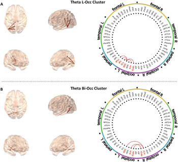
When comparing FC values in the theta band (4-8 Hz) we obtained two significant FC clusters: the theta left occipital cluster (Theta L-Occ) and the theta bilateral occipital cluster (Theta Bi-Occ). The theta L-Occ cluster was formed by 5 significant links (accumulated cluster T = 11.150; cbpt p-value = 0.002), all of them including the left inferior occipital cortex and connecting to the left lingual gyrus, the left calcarine cortex, the left cuneus, the left precuneus and the left posterior cingulate cortex (PCC) respectively (figure 1.1). These areas are either part of the DMN or the visual cortex (Hudspeth, Jessell, Kandel, Schwartz & Siegelbaum, Reference Hudspeth, Jessell, Kandel, Schwartz and Siegelbaum2013; Fransson & Marrelec, Reference Fransson and Marrelec2008). Bilinguals showed higher FC in all these links with respect to their highly educated healthy monolingual counterparts (figure 2). Additionally, the Theta Bi-Occ cluster included three significant links (accumulated cluster T = 11.490; cbpt p-value = 0.002) in which bilingual older adults exhibited higher FC values compared to monolinguals (figure 2). Two of these links connected the right lingual gyrus with the the left inferior occipital cortex and the left calcarine cortex, the third link involved the left lingual gyrus and the right calcarine cortex (figure 1.1). These are again areas involved in visual processing (Hudspeth et al., Reference Hudspeth, Jessell, Kandel, Schwartz and Siegelbaum2013).
Figure 1.1 Shows the regions included in the two clusters (A: Theta L-Occ; B: Theta Bi-Occ) in the Theta band in which bilinguals exhibit greater FC than monolinguals.

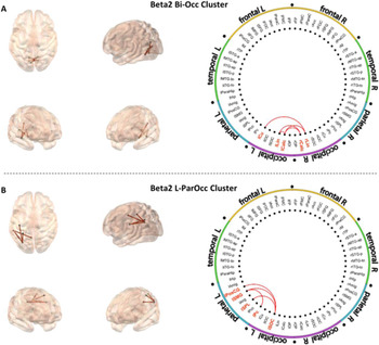
In the alpha band (8-12 hz) there were no significant FC differences. Contrarily, we obtained a significant left parieto-occipital cluster (Beta1 L-ParOcc henceforth) in the beta1 band (12-20 Hz), comprising four links (accumulated cluster T = 13.150; cbpt p-value = 0.001). Three of these links included left the superior occipital cortex connecting it to the left postcentral gyrus, the left supramarginal gyrus and the left superior parietal lobe respectively, while the fourth linked the left supramarginal gyrus with the left cuneus (figure 1.2). These areas are related to visual and somatosensory processing (Hudspeth et al., Reference Hudspeth, Jessell, Kandel, Schwartz and Siegelbaum2013). The FC over these regions was enhanced in the bilingual group (figure 2).
Figure 1.2 Shows the regions included in the cluster in the Beta1 band (Beta1 L-ParOcc) in which bilinguals exhibit greater FC than monolinguals.

Lastly, we obtained two significant FC clusters in the beta2 range (20-30 Hz) where bilinguals also exhibited increased synchronization values when compared to monolinguals (figure 2). The first was a bilateral occipital cluster (Beta2 Bi-Occ) that comprised 5 links (accumulated cluster T = 10.570; cbpt p-value = 0.003). The hyper-synchronized links connected the left lingual gyrus with the left and right calcarine, the right lingual gyrus to the left calcarine and the left cuneus and, lastly, the left calcarine to the right calcarine cortex (figure 1.3). As mentioned before, these areas play a role in visual processing (Hudspeth et al., Reference Hudspeth, Jessell, Kandel, Schwartz and Siegelbaum2013). Finally, we observed a left parieto-occipital cluster showing significant FC differences (Beta2 L Par-Occ) (accumulated cluster T = 11.640; cbpt p-value = 0.008) included 4 links connecting the left superior occipital cortex to the left postcentral gyrus, the left supramarginal gyrus and the left superior parietal lobe, and a fourth link associating the left precuneus with the left supramarginal gyrus (figure 1.3), areas that are involved in visual and somatosensory processing among other roles (Hudspeth et al., Reference Hudspeth, Jessell, Kandel, Schwartz and Siegelbaum2013).
Figure 1.3 Shows the regions included in the two clusters in the Beta2 band (A: Beta2 Bi-Occ; B: Beta2 L-ParOcc) in which bilinguals exhibit greater FC than monolinguals.

Figure 2 presents violin plots displaying the distribution of individual FC values in each cluster.

Correlation analyses
We first conducted Pearson correlations between mean FC of each significant cluster and age, to discard any possible contribution of this variable to the results, and confirmed that none of the clusters demonstrated a significant relationship with age in any of the groups (α = 0.050). Furthermore, we ensured that there were no significant differences in grey matter volume between bilinguals and monolinguals in any of the structures included in the analyses that could potentially bias the results (α = 0.050).
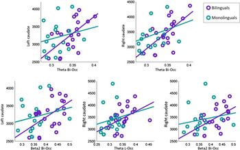
In order to study the possible functional meaning of these FC changes observed in bilingual older adults, we decided to conduct a set of correlations between FC values and the volume of certain structures linked to bilingualism (i.e., anterior cingulate cortex; ACC), caudate, putamen and pallidum. The complete set of results for bilinguals is shown in table 3. Two significant correlations remained significant after FDR correction only in the bilingual group. We observed a strong positive association between mean Theta Bi-Occ connectivity with the left caudate volume (rho = 0.600; p = 0.004) and the right caudate volume (rho = 0.640; p = 0.002). It is remarkable, however, that the left and the right caudate volumes were also significantly associated with the mean FC of other clusters such as Theta L-Occ and Beta2 Bi-Occ at a less conservative threshold (uncorrected α < 0.050). These correlations highlighted a remarkable positive association (0.470 < rho < 0.530) between the FC among these subsets of regions and caudate nuclei volumes bilaterally. Figure 3 presents scatter plots of the above-mentioned associations for both groups.
Figure 3 presents the scatter plots of the significant correlations (p <0.05) between caudate volume and FC values in each cluster.

Correlations between Grey Matter Structures Volumes and Functional Connectivity Values in Bilinguals
Table 3 presents Pearson correlation coefficients (rho) and p-values (p) for the correlations between selected grey matter structures (where Caud, Put and Pal stand for caudate, putamen and pallidus respectively and ACC stands for anterior cingulate cortex; laterality is indicated by an r -right- or -l- left) and average functional connectivity (FC) values for each cluster (*alpha < 0.05; **FDR corrected)

None of the correlations calculated in the monolingual group of older adults reached significance, not even employing the uncorrected threshold. Furthermore, the age of acquisition of the second language in the bilingual group was not significantly associated with grey matter volumes nor with FC (α < 0.050).
Discussion
Approximately half of the world's population is bilingual (European Commission Special Eurobarometer, 2012). Bilingualism has been suggested to modify brain structure and function, to enhance cognition and even to delay the onset of dementia (Adesope et al., Reference Adesope, Lavin, Thompson and Ungerleider2010; Craik, Bialystok & Freedman, Reference Craik, Bialystok and Freedman2010; Grundy et al., Reference Grundy, Anderson and Bialystok2017). Nevertheless, such claims have given rise to controversy among the scientific community, since other research groups failed to replicate those findings (Cox et al., Reference Cox, Bak, Allerhand, Redmond, Starr, Deary and MacPherson2016; Zahodne et al., Reference Zahodne, Schofield, Farrell, Stern and Manly2014; Zatorre et al., Reference Zatorre, Belin, Crane, Gruhl, Erosheva, Gibbons, McCurry, Rhoads, Nguyen, Arani, Masaki and White2001). The aim of the present research work was to unravel the influence of bilingualism on the functional network organization in healthy highly-educated older adults using MEG. Our presumption was that a better understanding of how bilingualism shapes the brain could shed light on this debate. This would enable the emergence of a plausible explanation with regards to how this skill results in the beneficial effects that certain studies have reported.
With this purpose, we compared the brain connectivity at rest of a group of monolingual and a group of bilingual healthy older adults. The performed analyses exposed five significant FC clusters in three different frequency ranges in which bilinguals exhibited greater FC than monolinguals (figures 1.1, 1.2 and 1.3). Interestingly, most of the areas within the clusters in which bilinguals exhibited greater FC than monolinguals are involved in language processing, which seems to support the relevance of our results. One of the regions showing increased FC in bilinguals, the supramarginal gyrus, has been reported to play a role in the network involved in word processing (Oberhuber, Hope, Seghier, Parker Jones, Prejawa, Green & Price, Reference Oberhuber, Hope, Seghier, Parker Jones, Prejawa, Green and Price2016; Zou, Abutalebi et al., Reference Zou, Abutalebi, Zinszer, Yan, Shu, Peng and Ding2012) and language switching (Luk, Green, Abutalebi & Grady, Reference Luk, Green, Abutalebi and Grady2012; Moritz-Gasser & Duffau, Reference Moritz-Gasser and Duffau2009; Olsen, Pangelinan, Bogulski, Chakravarty, Luk, Grady & Bialystok, Reference Olsen, Pangelinan, Bogulski, Chakravarty, Luk, Grady and Bialystok2015; Price, Green & Von Studnitz, Reference Price, Green and Von Studnitz1999). Similarly, the superior parietal lobe is well-known for its role in the circuit of phonological processing (Wong et al., Reference Wong, Yin and O'Brien2016; Zatorre et al., Reference Zatorre, Belin, Crane, Gruhl, Erosheva, Gibbons, McCurry, Rhoads, Nguyen, Arani, Masaki and White2001). Suh, Yoon, Lee, Chung, Cho and Park (Reference Suh, Yoon, Lee, Chung, Cho and Park2007) report that sentence processing in bilinguals (both in L1 and L2) involves a network composed of the inferior frontal gyrus, the inferior parietal lobe and several occipital areas, such as the cuneus and the lingual gyrus. Also, the superior parietal cortex, as well as the superior occipital cortex (together with other temporal and frontal structures), are part of the network described by García-Pentón, Pérez Fernández, Iturria-Medina, Gillon-Dowens and Carreiras (Reference García-Pentón, Pérez Fernández, Iturria-Medina, Gillon-Dowens and Carreiras2014) involved in visual word recognition, reading and semantic processing. Therefore, our results (i.e., increased FC between most of these areas) could represent the neurophysiological substrate of improved language abilities in the bilingual senior population. This hypothesis could be supported by previous findings reporting that FC at rest between several regions involved in the FC changes that we described (including the supramarginal gyrus, the postcentral gyrus, the superior parietal cortex, the lingual gyrus and several areas within the occipital lobe) correlated with reading abilities in bilingual younger adults (Zhang, Li, Chen, Xue, Lu, Mei, Xue, Xue, He, Chen, Wei & Dong, Reference Zhang, Li, Chen, Xue, Lu, Mei, Xue, Xue, He, Chen, Wei and Dong2014). Thus, our results strongly suggest that bilingualism directly affects FC within networks involved in language processing.
Strikingly, bilingual older adults did not show changes in frontal structures, in contrast to previous studies (García-Pentón et al., Reference García-Pentón, Pérez Fernández, Iturria-Medina, Gillon-Dowens and Carreiras2014; Olsen et al., Reference Olsen, Pangelinan, Bogulski, Chakravarty, Luk, Grady and Bialystok2015; Perani et al., Reference Perani, Farsad, Ballarini, Lubian, Malpetti, Fracchetti, Magnani, March and Abutalebi2017). However, our functional results are in line with Grundy et al.’s (Reference Grundy, Anderson and Bialystok2017) proposition, stating that during the first stages of L2 acquisition bilinguals rely more on frontal structures but over time they devote more resources to posterior and subcortical circuits. According to this hypothesis, FC changes over posterior areas could be interpreted as a reorganization of functional networks driven by speaking two languages. This goes in line with the suggestion by Grant, Dennis and Li (Reference Grant, Dennis and Li2014) to study FC patterns of the bilingual brain focusing not only on the frontal lobe but also considering temporal, parietal, occipital and subcortical regions. As an example, Berroir, Ghazi-Saidi, Dash, Adrover-Roig, Benali and Ansaldo (Reference Berroir, Ghazi-Saidi, Dash, Adrover-Roig, Benali and Ansaldo2017) report that, during a Simon task in an fMRI scanner, monolinguals recruited motor, visual and executive function areas, while bilinguals relied solely on visuospatial processing. Similar results had been previously found by Ansaldo, Ghazi-Saidi and Adrover-Roig (Reference Ansaldo, Ghazi-Saidi and Adrover-Roig2015), who concluded that bilinguals do not activate a circuit that is particularly vulnerable to the aging process. Gold et al. (Reference Gold, Kim, Johnson, Kryscio and Smith2013) also proposes that bilingualism protects against age-related changes in fMRI measurements, such as the over-recruitment of frontal areas (during a perceptual switching task). Furthermore, it is important to mention that some key structures in the field of bilingualism such as the caudate or putamen nuclei among others are not present in our FC results. Beyond the above-mentioned reasons explaining the absence of frontal regions involvement, it is crucial to bear in mind that such deep structures cannot be detected with MEG, which is mainly sensitive to tangential sources located in the cortex. Therefore, we cannot rule out that such deep structures could also be involved in network reorganization in healthy highly educated older adults, in addition to the regions described in our results.
Additionally, bilingualism has been related to later onset of Alzheimer's disease (Alladi, Bak, Duggirala, Surampudi, Shailaja, Shukla, Chaudhuri & Kaul, Reference Alladi, Bak, Duggirala, Surampudi, Shailaja, Shukla, Chaudhuri and Kaul2013). AD is commonly regarded as a disconnection syndrome (Delbeuck, Linden & Collette, Reference Delbeuck, Linden and Collette2003), and it is known to particularly impair FC over posterior brain regions such as occipital and parietal regions, reducing the strength of hub regions over these brain areas (Jones, Knopman, Gunter, Graff-Radford, Vemuri, Boeve, Petersen, Weiner & Jack, Reference Jones, Knopman, Gunter, Graff-Radford, Vemuri, Boeve, Petersen, Weiner and Jack2015; Nakamura, Cuesta, Kato, Arahata, Iwata, Yamagishi, Kuratsubo, Kato, Bundo, Diers, Fernández, Maestú & Ito, Reference Nakamura, Cuesta, Kato, Arahata, Iwata, Yamagishi, Kuratsubo, Kato, Bundo, Diers, Fernández, Maestú and Ito2017; Yu, Engels, Hillebrand, van Straaten, Gouw, Teunissen, van der Flier, Scheltens & Stam, Reference Yu, Engels, Hillebrand, van Straaten, Gouw, Teunissen, van der Flier, Scheltens and Stam2017). These brain regions suffer alterations very early in the course of the disease, even in the preclinical stages (López-Sanz et al., Reference López-Sanz, Bruña, Garcés, Martín-Buro, Walter, Delgado, Montenegro, Lopez-Higes, Marcos and Maestú2017a; López-Sanz, Garcés, Álvarez, Delgado-Losada, López-Higes & Maestú, Reference López-Sanz, Garcés, Álvarez, Delgado-Losada, López-Higes and Maestú2017b). Interestingly, our results could bridge the gap between the above-mentioned findings, providing a plausible neurophysiological substrate for this protective effect. According to this hypothesis, it could be argued that bilingualism enhances FC within posterior brain regions in healthy older adults. Later on, should neurodegeneration initiate, such stronger FC would be hypothesized to delay 2-4 functional changes associated with AD symptomatology, (i.e., posterior disconnection) which could in turn result in later disease onset. In this vein, the enhanced FC values over posterior brain regions observed in healthy bilingual older adults could be interpreted as an increased brain reserve capacity (Satz, Reference Satz1993) or a sign of network flexibility due to increased cognitive reserve (Stern, Reference Stern2002). However, this hypothesis should be confirmed by future work using longitudinal designs. Although changes in posterior regions were not easily foreseeable in the specific context of bilingualism, the fact that aging is also typically accompanied by FC decreases over these same regions (Damoiseaux, Beckmann, Arigita, Barkhof, Scheltens, Stam, Smith, & Rombouts, Reference Damoiseaux, Beckmann, Arigita, Barkhof, Scheltens, Stam, Smith and Rombouts2008) supports the interpretation of the observed network reorganization as a potential brain protective mechanism against healthy and pathological aging.
Beyond that, in the present study, FC values in Theta Bi-Occ cluster were strongly correlated with caudate volumes bilaterally. Remarkably, these correlations were only significant for the bilingual subsample. A significant uncorrected correlation was also evident for Beta2 Bi-Occ cluster with bilateral caudate and Theta L-Occ cluster with right caudate (and a tendency was observed with left caudate). The caudate nucleus is a key structure in the field of bilingualism, and has been widely studied, mainly for its involvement in executive function (Pliatsikas & Luk, Reference Pliatsikas and Luk2016). Allegedly, this region plays an important role when switching between two languages (Abutalebi, Annoni, Zimine, Pegna, Seghier, Lee-Jahnke, Lazeyras, Cappa & Khateb, Reference Abutalebi, Annoni, Zimine, Pegna, Seghier, Lee-Jahnke, Lazeyras, Cappa and Khateb2008; Luk et al., Reference Luk, Green, Abutalebi and Grady2012; Moritz-Gasser & Duffau, Reference Moritz-Gasser and Duffau2009), also in bimodal bilinguals (Zou, Ding, Abutalebi, Shu & Peng, Reference Zou, Ding, Abutalebi, Shu and Peng2012). More specifically, the head of the left caudate is part of a language control network (Abutalebi & Green, Reference Abutalebi and Green2016; Friederici, Reference Friederici2006), and it is particularly involved when a cognitive process cannot be automatically carried out. Furthermore, caudate activation has been found to be higher when switching to a less dominant language (Abutalebi, Brambati, Annoni, Moro, Cappa & Perani, Reference Abutalebi, Brambati, Annoni, Moro, Cappa and Perani2007). In our study, only late bilinguals were included, and Spanish was the dominant language for all of them (most of them reported having acquired proficiency in their L2 for professional reasons). Thus, we propose that this lack of codominance between L1 and L2 in our sample could be enhancing the role of the caudate nucleus in language processing. The fact that we observed strong correlations between FC values and caudate volumes only in the bilingual group supports the relationship between FC changes and network reorganization in bilinguals. Along with our results, previous studies have shown increased FC between the left caudate and language processing regions in bilinguals (Li, Abutalebi, Zou, Yan, Liu, Feng, Wang, Guo & Ding, Reference Li, Abutalebi, Zou, Yan, Liu, Feng, Wang, Guo and Ding2015). This is a very relevant finding, since the development of a gating system in the striatum to the prefrontal cortex has been hypothesized to underpin the improved executive function that bilinguals show (Stocco & Prat, Reference Stocco and Prat2014).
Focusing on the role of education, bilingual participants have been said to potentially have higher educational levels. Although this is not necessarily the case of older simultaneous bilinguals in Spain (mainly those who were born in bilingual regions), in general, sequential bilinguals within our age range are those who had access to a better education (bear in mind that L2 acquisition was not mandatory at the time) and those who held highly qualified job positions. For that reason, educational attainment has been suggested as a relevant confounding factor in bilingual research. Evidence so far has not been able to solve this question. For example, while Liu, Liu, Yip, Meguro and Meguro (Reference Liu, Liu, Yip, Meguro and Meguro2017) and Gollan, Salmon, Montoya and Galasko (Reference Gollan, Salmon, Montoya and Galasko2011) describe a protective effect of speaking several languages only among low educated older adults, Alladi et al. (Reference Alladi, Bak, Duggirala, Surampudi, Shailaja, Shukla, Chaudhuri and Kaul2013) claim that bilingualism delays the onset of dementia independently of other cofounding factors. In this regard, it is important to highlight that in this study we constrained the sample to healthy highly educated older adults with the intention to reveal the effect that bilingualism exerts on the brain FC beyond the already widely studied effect of educational attainment (Arenaza-Urquijo, Landeau, La Joie, Mevel, Mézenge, Perrotin, Desgranges, Bartrés-Faz, Eustache & Chételat, Reference Arenaza-Urquijo, Landeau, La Joie, Mevel, Mézenge, Perrotin, Desgranges, Bartrés-Faz and Chételat2013; Bastin, Yakushev, Bahri, Fellgiebel, Eustache, Landeau, Scheurich, Feyers, Collette, Chételat & Salmon, Reference Bastin, Yakushev, Bahri, Fellgiebel, Eustache, Landeau, Scheurich, Feyers, Collette, Chételat and Salmon2012; Lopez, Aurtenetxe, Pereda, Cuesta, Castellanos, Bruña, Niso & Maestu, Reference Lopez, Aurtenetxe, Pereda, Cuesta, Castellanos, Bruña, Niso, Maestu and Bajo2014). In other words, we were interested in unveiling the potential contribution of bilingualism given an already high educational level. At this respect, we have been able to describe a neurophysiological signature specific to bilingualism. However, we cannot dismiss the possibility that there was an actual causal relationship between being bilingual and acquiring a higher educational level.
Finally, L2 age of acquisition did not correlate either with FC values or grey matter volumes. However, it is relevant to emphasize that only sequential bilinguals were included in this work. Thus, individuals who acquired an L2 very early in life were not considered in this study. Additionally, most bilinguals in this sample were firstly exposed to their L2 in school, but they only developed L2 proficiency and began to actively and or/socially use their L2 later in life, when their professional or personal context required so. Beyond AoA, bilingual experience is a relevant factor (Luk & Bialystok, Reference Luk and Bialystok2013) and therefore we consider that “degree of L2 social usage” or “age of L2 immersion” could be more closely related to the functional and structural changes that we reveal in this study. More accurate information regarding these two variables should be collected in future studies in order to perform such analyses.
Also, as has already been said in this article, bilingualism is a complex skill that encompasses several aspects. In this regard, a potential limitation of our study was not to incorporate a specific test for L1 and L2 proficiency. This would enable the analysis of how proficiency in L2 relates to the functional differences that we have reported in highly educated bilingual older adults.
To the best of our knowledge, this is the first MEG study addressing the effect of bilingualism at the neurophysiological level. Also, the fact that we focused on a specific subpopulation of bilinguals (late bilinguals having been introduced to L2 in a formal setting) allows us to more accurately compare our results with those from other studies and unravel presumed discrepancies. Rigorous inclusion criteria are critical for more accurate interpretations in the field of bilingualism, although at the cost of sample representativeness. Yet, our results are promising and support the relevance of bilingualism as it enhances posterior functional circuits typically impaired in aging (Jones et al., Reference Jones, Machulda, Vemuri, McDade, Zeng, Senjem, Gunter, Przybelski, Avula, Knopman, Boeve, Petersen and Jack2011). Based on these results, policies promoting the use of a second language in typically monolingual populations could be beneficial to promote healthy aging.
Disclosure statement
The authors have no conflicts of interest to disclose.
Acknowledgements
This study was funded by the Spanish Ministry of Economy and Competitiveness under Grant PSI2012-38375- C03-03. Complementary, it was supported by a postdoctoral fellowship to Pablo Cuesta (FJCI-2015-26755) and María Eugenia López (IJCI-2016-30662), and a predoctoral fellowship to David López Sánchez from the Spanish Ministry of Economy and Competitiveness (PSI2012-38375-C03-01), and a predoctoral fellowship to Jaisalmer de Frutos by La Caixa Foundation. Jaisalmer de Frutos Lucas and David Lopez Sanz equally contributed to this manuscript and therefore share first authorship.