1. Introduction
The fact that photosynthetic organisms can evolve oxygen has been known since 1772 when Preistley (Reference Preistley1772) demonstrated that green plants could ‘purify the air-made foul by animals’. Soon after this the Dutchman, van Ingen-Housz (Reference Van Ingen-Housz1779) showed that light was required for O2 generation to occur, while de Saussure (Reference De Saussure1804) realized that water was involved in the process. However, there was no evidence to suggest that oxygen was derived from water and the more popular view during the early part of the 20th century was that the gas came from CO2, the other reactant of photosynthesis. For more details of the history see Rabinowitch (Reference Rabinowitch1945) and Hill (Reference Hill, Forti, Avron and Melandri1972). It was not until 1937 that Hill (Reference Hill1937) conclusively demonstrated that photosynthetic O2 generation could occur in the absence of CO2 reduction. Using isolated chloroplasts and artificial electron acceptors such as ferrioxalate and ferricyanide, he demonstrated that the photoreduction of these compounds was at the expense of the oxidation of water. Further support for the concept that oxygen was derived from the water came from van Neil (Reference Van Neil1941) and Ruben et al. (Reference Ruben, Randle, Kaman and Hyde1941). Since these early experiments, there has been considerable effort to understand how photosynthetic organisms can efficiently bring about the splitting of water and in so doing maintain our oxygenic atmosphere while at the same time supply reducing equivalents to power virtually all life on our planet.
2. Photosystem II (PSII)
It has long been known that the catalytic water-splitting site contains four Mn ions bound within an enzyme known as PSII (Debus, Reference Debus1992). PSII is a pigment-binding, multisubunit protein complex embedded in the lipid environment of the thylakoid membranes of plants, algae and cyanobacteria (Barber, Reference Barber2003; Reference Barber2009). Today we have a very good understanding of the structure and functioning of this key and unique enzyme (Barber, Reference Barber2016a). However, understanding the precise details of how the cluster of Mn ions interact and catalyse the water-splitting reaction remains a major challenge of bioinorganic chemistry. The importance of this cannot be overstated since splitting water into its constituents is thermodynamically and chemically demanding and takes place in the delicate protein environment of PSII. Because of this, PSII has to repair itself from time to time with a half-time of about 30 min in bright illumination (Barber & Andersson, Reference Barber and Andersson1992).
The thermodynamic potential for the water-splitting reaction (Eq. (1)) is E O = 1·23 eV at pH 0, which decreases to about 0·88 eV at a less acid pH estimated for PSII water-splitting reaction (e.g. 6·0)
 $$2\hbox{H}_{2} \hbox{O} \buildrel {^{{\rm 4}\;h\nu}} \over \longrightarrow \hbox{O}_{2} + 4\hbox{H}^{+} + 4\hbox{e}$$
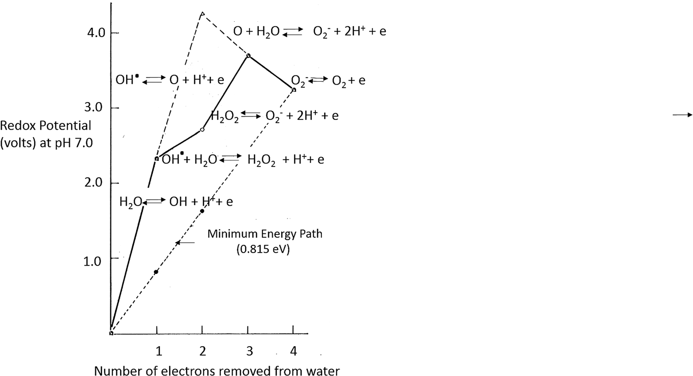
Note from Fig. 1 that at neutral pH, the one-electron potential for the oxidation of water to a hydroxyl radical (OH•) is 2·23 eV, which is over 1·5 eV more oxidizing than a four-electron concerted oxidation (0·815 eV) shown as the minimum energy path in Fig. 1. In PSII, it is this four-electron oxidation concerted oxidation pathway which is employed to split water and generate dioxygen. This is because the first step of the alternative hydroxyl (OH•) route requires energy well in excess of that contained in a quantum of long wavelength visible light (680 nm) and involves hydrogen peroxide and oxygen radicals, which would attack the protein environment.
An energy diagram showing the redox potential of the one-electron steps for the oxidation of water to O2 at pH 7·0. Solid line denotes a pathway via H2O2 as the two-equivalent reduction stage. Solid and dashed lines are pathways via ‘H2O + O’ as the two-equivalent reduction stage. Dotted line is the minimum energy pathway with the four electron removal from two H2O molecules driven by four equi-energetic steps of 0·815 eV therefore an overall minimum energy requirement of 3·26 V for the production of one molecule of dioxygen. It is this four equi-energetic steps taken by the S-state cycle of PSII but with about 1 V available for each step. This difference, therefore, allows the small overpotential to be satisfied by the available total photon energy in the region of 4 V.

2.1. S-state cycle and the Mn4Ca2+O X catalytic centre
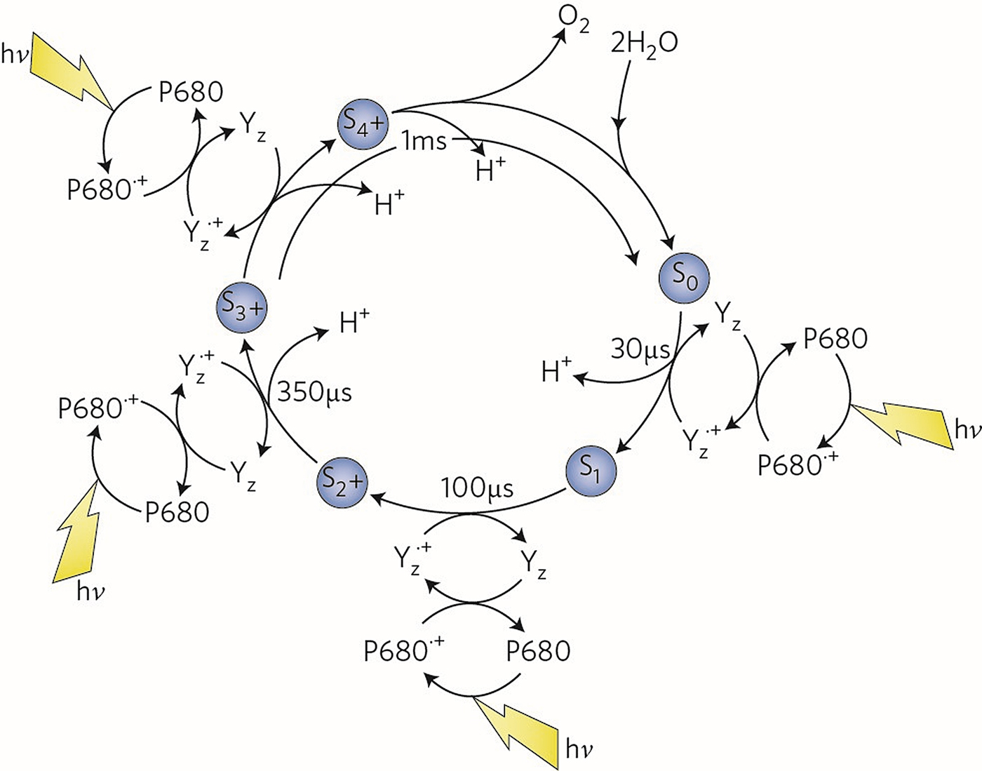
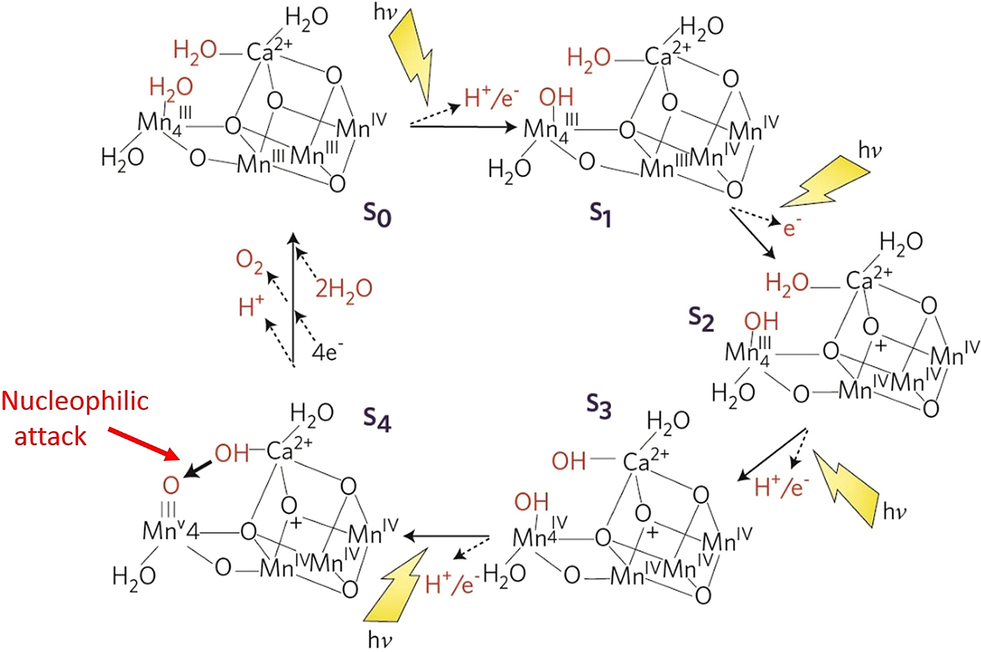
The function of the Mn-cluster is to accumulate the four oxidizing equivalents required to split two water molecules and generate a molecule of dioxygen (Barber, Reference Barber2003). The final step leading to O–O bond formation has to be very fast involving two closely located activated oxygen atoms. In this way, the generation of reactive oxygen species is avoided and the triplet state form of O2 established. Each oxidation step at one catalytic centre is driven by the absorption of one photon, which was elegantly shown by monitoring oxygen release in a series of single turnover flashes of light where the maximum yield of oxygen followed a period of four (Joliot et al. Reference Joliot, Barbieri and Chabaud1969). This finding which gave rise to a scheme known as the S-state cycle (S0 to S4) is depicted in Fig. 2 (Kok et al. Reference Kok, Forbush and McGLOIN1970). This cycle is powered by the initial oxidation of a chlorophyll known as P680 generated by light-driven primary charge separation in the reaction centre of PSII, which is coupled to a redox-active tyrosine (YZ) serving as an intermediate electron carrier between P680+ and the Mn-cluster.
The S-state cycle showing how the absorption of four photons of light (hv) by the reaction centre primary oxidant P680 drives the splitting of two water molecules and formation of O2 through a consecutive series of five intermediates (S0, S1,S2, S3 and S4). Protons (H + ) are released during each step of this cycle except for the S1 to S2 transition. Electron donation from the Mn4Ca2+ cluster to P680•+ is aided by the redox-active tyrosine YZ. Each step involves a single oxidation of a Mn ion in the cluster, starting at SO with 3 × MnIII plus MnIV advancing to S3 with 4 × MnIV. The exact oxidation state of S4 is unknown but could be 3 × MnIV plus MnV or 3 × MnIV plus MnIV-oxyl radical (see below). Also shown are half-times for the various steps of the cycle (taken from Barber, Reference Barber2017).
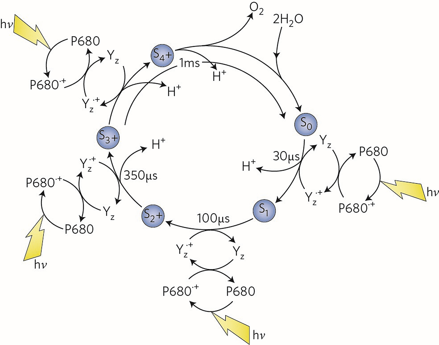
Each photon absorbed provides about 1 V of oxidizing potential per S-state transition since the oxidizing potential of P680+ is about 1·25 V. This means that the overpotential for each step of the catalytic cycle is relatively small. As shown in Fig. 2, as the catalytic cycle proceeds from S0 to S4, protons are released at each S-state step except for the S1 to S2 transition where the metal cluster accumulates one positive charge. The redox-active YZ is a neutral tyrosyl radical where its phenolic proton is donated to a nearby base, D1His190, to which it is H-bonded. Therefore, the neutral YZ radical is an ideal candidate for facilitating a proton-coupled electron transfer (PCET) from the catalytic centre, as originally suggested by Hoganson & Babcock (Reference Hoganson and Babcock1997). An important study (Haumann et al. Reference Haumann, Liebisch, Muller, Barra, Grabolle and Dau2005), indicated the coupling is sequential, with a strictly alternating removal of electrons and protons rather than hydrogen atom transfer.
A whole range of techniques, particularly magnetic resonance spectroscopy, have shown that each step in this cycle involves an increase in the oxidation of a Mn ion, starting at S0 with 3 × MnIII plus MnIV advancing to S3 with 4 × MnIV (Cox et al. Reference Cox, Retegan, Neese, Pantais, Boussac and Lubitz2014). The precise details of the final oxidation state, S4, are not clear because O–O bond formation has to be very fast to avoid side reactions.
Not surprisingly, it has been one of the greatest challenges of photosynthesis research to determine the structure of the Mn-containing cluster of the PSII oxygen-evolving complex (OEC) and thus reveal the molecular mechanism of the water-splitting reaction. Over the years there have been many postulates of its structure mainly derived from X-ray absorption fine-structure (XAFS) spectroscopy (Yachandra, Reference Yachandra2002) and electron paramagnetic resonance (EPR) studies (Britt et al. Reference Britt, Campbell, Peloquin, Gilchrist, Aznar, Cicus, Robble and Messinger2004; Haddy, Reference Haddy2007). However, in 2004 a complete fully refined structure of PSII was determined at 3·5 Å resolution using X-ray crystallography by my colleagues and me at Imperial College London and published in Science (Ferreira et al. Reference Ferreira, Iverson, Maglaoui, Barber and Iwata2004).
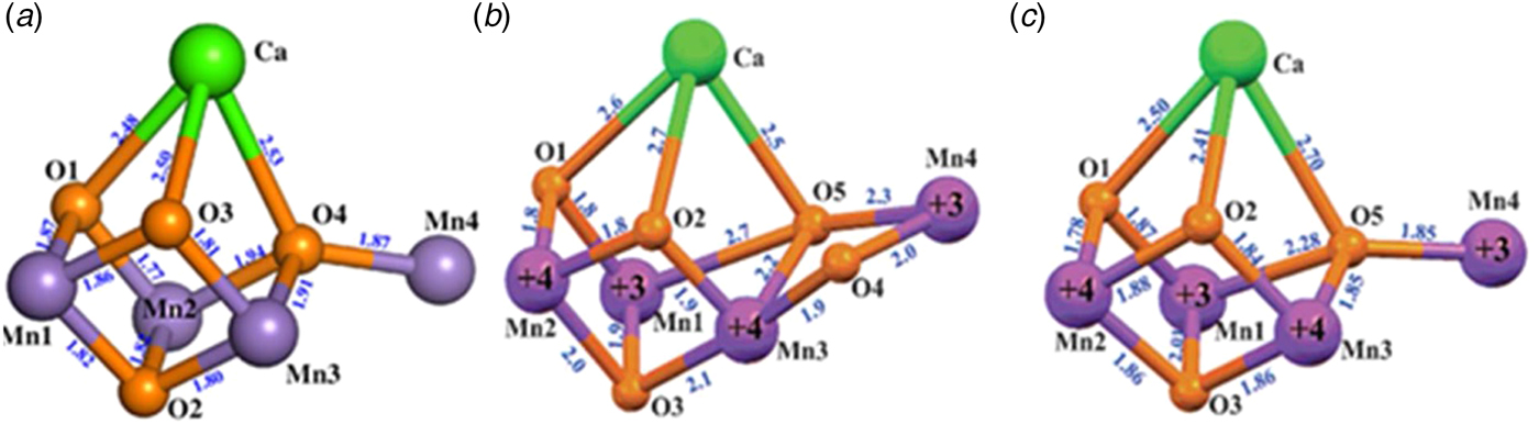
We were able to assign, for the first time, over 5000 amino acid side chains of this huge dimeric membrane complex (700 kD) with each monomer consisting of 19 different protein subunits. In so doing we answered many outstanding questions as well as revealing a wide range of important details hitherto unknown. The native electron density map, together with anomalous diffraction data collected at wavelengths of 1·89 and 2·25 Å, provided electron density profiles for Mn and Ca2+, respectively. This information was then used to build a model of the metal-cluster. The anomalous electron density attributed to the four Mn ions was ‘pear-shaped’ indicative of the 3 + 1 organization and thus one Mn was assigned to the small domain and three in the large globular domain, whereas the 2·25 Å wavelength map covered one metal ion in the large domain of the native density. From these data the three Mn ions (Mn1, Mn2 and Mn3) and the Ca2+, located in the large domain, were modelled with a cubane geometry having bridging oxygens, an organization which was compatible with the native electron density. The fourth Mn ion (Mn4), located in the small domain was modelled so that it was linked to the cubane by one of its bridging oxygens (dangler Mn4) (see Fig. 3a ). Based on values determined by XAFS measurements and comparison with other Mn-containing proteins, the three Mn-di-μ-oxy-Mn bonds of the cubane were spaced at 2·7 Å, while the three Mn-di-μ-oxy-Ca2+ bonds were separated by 3·4 Å. The dangler Mn4 was positioned 3·3 Å from the closest Mn ion of the cubane and about 4 Å from the Ca2+. The 3 + 1 organization of the Mn-cluster was predicted by Peloquin & Britt (Reference Peloquin and Britt2001) based on the degree of spin coupling detected by resonance spectroscopy and they introduced the term ‘dangler’ for the less coupled Mn ion (Mn4) (Peloquin & Britt, Reference Peloquin and Britt2001).
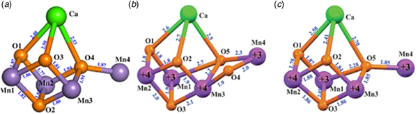
(a) Comparison of the Mn4Ca2+O4 cubane model from coordinates (PDB 1S5L) deposited by Ferreira et al. in Reference Ferreira, Iverson, Maglaoui, Barber and Iwata2004 with (b) the 1·9 Å structure of the Mn4Ca2+O5 cluster from Umena et al. (Reference Umena, Kawakami, Shen and Kamiya2011) (PDB 3WU2) and (c) the chemically synthesized Mn4Ca2+O4-cubane by Zhang et al. (Reference Zhang, Chen, Dong, Shen, Dau and Zhao2015). Note that unfortunately, the numbering of the Mn and O are different in (b) and (c) to that originally given in (a) (taken from Barber, Reference Barber2016b).
At that time there was no similar chemical structure known in biochemistry or chemistry and therefore the model was greeted with some uncertainty, especially since it had been conceived from relatively low-resolution data. But the main opposition to this model was the argument that the metal cluster, and surrounding amino acid ligands, would be seriously disturbed by radiation damage during data collection (Yano et al. Reference Yano, Kern, Irrgang, Latimar, Bergmann, Glatzel, Pushkar, Loll, Sauer, Messinger and Yachandra2005). Indeed the cubane structure did not seem to be consistent with polarized XAFS measurements made at much lower radiation levels (Yano et al. Reference Yano, Kern, Sauer, Latimer, Pushkar, Biesiadka, Loll, Saenger, Messinger, Zouni and Yachandra2006). Unfortunately, the claim that the Ferreira et al. cubane model was invalid because of significant radiation damage caused a substantial level of confusion for several years, both for specialists and non-specialists. However, it has now become clear that the 2004 cubane model of Ferreira et al. was valid within the limitation of its resolution and therefore the extensive radiation damage argument and associated alternative postulated XAFS structures for the Mn-cluster (Yano et al. Reference Yano, Kern, Sauer, Latimer, Pushkar, Biesiadka, Loll, Saenger, Messinger, Zouni and Yachandra2006) turned out to be wrong. However, doubts in the interpretation of the polarized XAFS data were presented by Sproviero et al. (Reference Sproviero, Gascon, McEVOY, Brudvig and Batista2008) who concluded that the polarized XAFS spectra were consistent with the cubane model of Ferreira et al., which they went on to refine using QM/MM calculations.
In 2011, 7 years after the coordinates of the cubane model by Ferreira et al. were deposited, the uncertainty of the cubane model was finally lifted with the report of 1·9 Å PSII crystal structure from Umena et al. (Reference Umena, Kawakami, Shen and Kamiya2011). At this resolution, the electron densities of individual metal ions could be resolved and bridging oxygens inferred. As shown in Fig. 3b , the resulting model was overall similar to that of Ferreira et al. (Reference Ferreira, Iverson, Maglaoui, Barber and Iwata2004) except an additional oxo bridge was proposed to link the dangler Mn outside the Mn3Ca2+ cubane (Mn4) to a Mn ion of the cubane (Mn3). Importantly this additional oxo-bridge had already been predicted by Siegbahn (Reference Siegbahn2008) and by Dau et al. (Reference Dau, Grundmeier, Loja and Haumann2008) based on a modification of the original 2004 cubane model using Density Function Theory (DFT) and other quantum mechanical methods. Nevertheless, the 1·9 Å structure of Umena et al. (Reference Umena, Kawakami, Shen and Kamiya2011), together with high-resolution diffraction data obtained by femtosecond X-ray free-electron laser (XFEL) pulses (Suga et al. Reference Suga, Akita, Hirata, Ueno, Murakami, Nakajima, Shimizu, Yamashita, Yamamoto, Ago and Shen2015, Reference Suga, Akita, Sugahara, Kubo, Nakajima, Nakane, Yamashita, Umena, Nakabayashi, Yamane, Nakano, Suzuki, Masuda, Inoue, Kimura, Nomura, Yonekura, Yu, Sakamoto, Motomura, Chen, Kato, Noguchi, Tono, Joti, Kameshima, Hatsui, Nango, Tanaka, Naitow, Matsuura, Yamashita, Yamamoto, Nureki, Yabashi, Ishikawa, Iwata and Shen2017; Young et al. Reference Young, Ibrahim, Chatterjee, Gul, Fuller, Koroidov, Brewster, Tran, Alonso-Mori, Kroll, Michels-Clark, Laksmono, Sierra, Stan, Hussein, Zhang, Douthit, Kubin, de Lichtenberg, Long Vo, Nilsson, Cheah, Shevela, Saracini, Bean, Seuffert, Sokaras, Weng, Pastor, Weninger, Fransson, Lassalle, Bräuer, Aller, Docker, Andi, Orville, Glownia, Nelson, Sikorski, Zhu, Hunter, Lane, Aquila, Koglin, Robinson, Liang, Boutet, Lyubimov, Uervirojnangkoorn, Moriarty, Liebschner, Afonine, Waterman, Evans, Wernet, Dobbek, Weis, Brunger, Zwart, Adams, Zouni, Messinger, Bergmann, Sauter, Kern, Yachandra and Yano2016) have provided reliable atomic structures of the OEC at cryo-and room-temperatures, particularly in its dark-stable S1 state. Therefore this has given validity to the detailed quantum mechanical calculations for the OEC and its functioning (Cox et al. Reference Cox, Retegan, Neese, Pantais, Boussac and Lubitz2014; Lohmiller et al. Reference Lohmiller, Krewald, Navarro, Retegan, Rapatskiy, Nowaczyk, Boussac, Neese, Lubitz, Pantazis and Cox2014; Luber et al. Reference Luber, Rivalta, Umena, Kawakami, Shen, Kamiya, Brudvig and Batista2011; Yamanaka et al. Reference Yamanaka, Isobe, Kanda, Saito, Umena, Kawakami, Shen, Kamiya, Okumur, Nakamura and Yamaguchi2011) and more rigorous interpretation of experimental results (Ames et al. Reference Ames, Pantazis, Krewald, Cox, Messinger, Lubitz and Neese2011; Bovi et al. Reference Bovi, Narzi and Guidoni2013; McConnell et al. Reference McConnell, Grigoryants, Scholes, Myers, Chen, Whittaker and Brudvig2012; Rapatskiy et al. Reference Rapatskiy, Cox, Savitsky, Ames, Sander, Nowaczyk, Rogner, Boussac, Neese, Messinger and Lubitz2012).
2.2. Synthesized cubanes mimicking the Mn4Ca2+O X cluster
Of considerable importance is the report that the PSII cubane cluster can be synthesized as a Mn4Ca2+O4 molecule (Zhang et al. Reference Zhang, Chen, Dong, Shen, Dau and Zhao2015) whose structure is very similar to that of the OEC (compare Figs 3c with 3d ) and remarkably similar to the original model of Ferriera et al. (compare Figs 3c with 3a ). As with the Ferreira et al. structure, the synthetic Mn4Ca2+O4 molecule was also missing the additional oxo-bridge to Mn4. The chemical synthesis of the Mn4Ca2+O4 structure in organic solvent shows that this cluster does not require protein to assemble and as long as there is an appropriate source of ligands, the structure is stable in an oxidized state. Moreover, cyclic voltammetry conducted in 1,2-dichloroethane showed that it can advance through the S-state cycle to S3 (Zhang et al. Reference Zhang, Chen, Dong, Shen, Dau and Zhao2015). However, no oxygen evolution was detected when a small amount of water was present. Nevertheless, this extraordinary finding may have implications for the evolutionary origin of the OEC and provides some explanation for the robustness of the in vivo cluster to site-directed mutations (see below) and X-ray radiation damage.
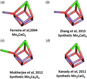
Before the synthesis of the Mn4Ca2+O4 cluster by Zhang et al. (Reference Zhang, Chen, Dong, Shen, Dau and Zhao2015), Christou and colleagues (Mukherjee et al. Reference Mukherjee, Stull, Yano, Stamatato, Pringouri, Stich, Abbroud, Britt, Yachandra and Christou2012) reported the synthesis of a Mn3Ca2O4 molecule which is structurally similar to that of Mn4Ca2+O4 (see Fig. 4c ). Another important study was the synthesis of a Mn3CaO4 cubane by Agapie and colleagues (Kanady et al. Reference Kanady, Tsui, Day and Agapie2011) (see Fig. 4d ). However, neither showed redox properties like that of the Mn4CaO4 synthesized by Zhang et al. (Reference Zhang, Chen, Dong, Shen, Dau and Zhao2015).
Diagrammatic comparison of synthesized cubane structures (b), (c) and (d) with the Ferreira et al. X-ray model (a). Mn purple, Ca2+ green and red are oxo bonds (taken from Barber, Reference Barber2016b).

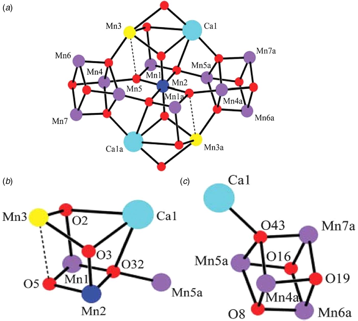
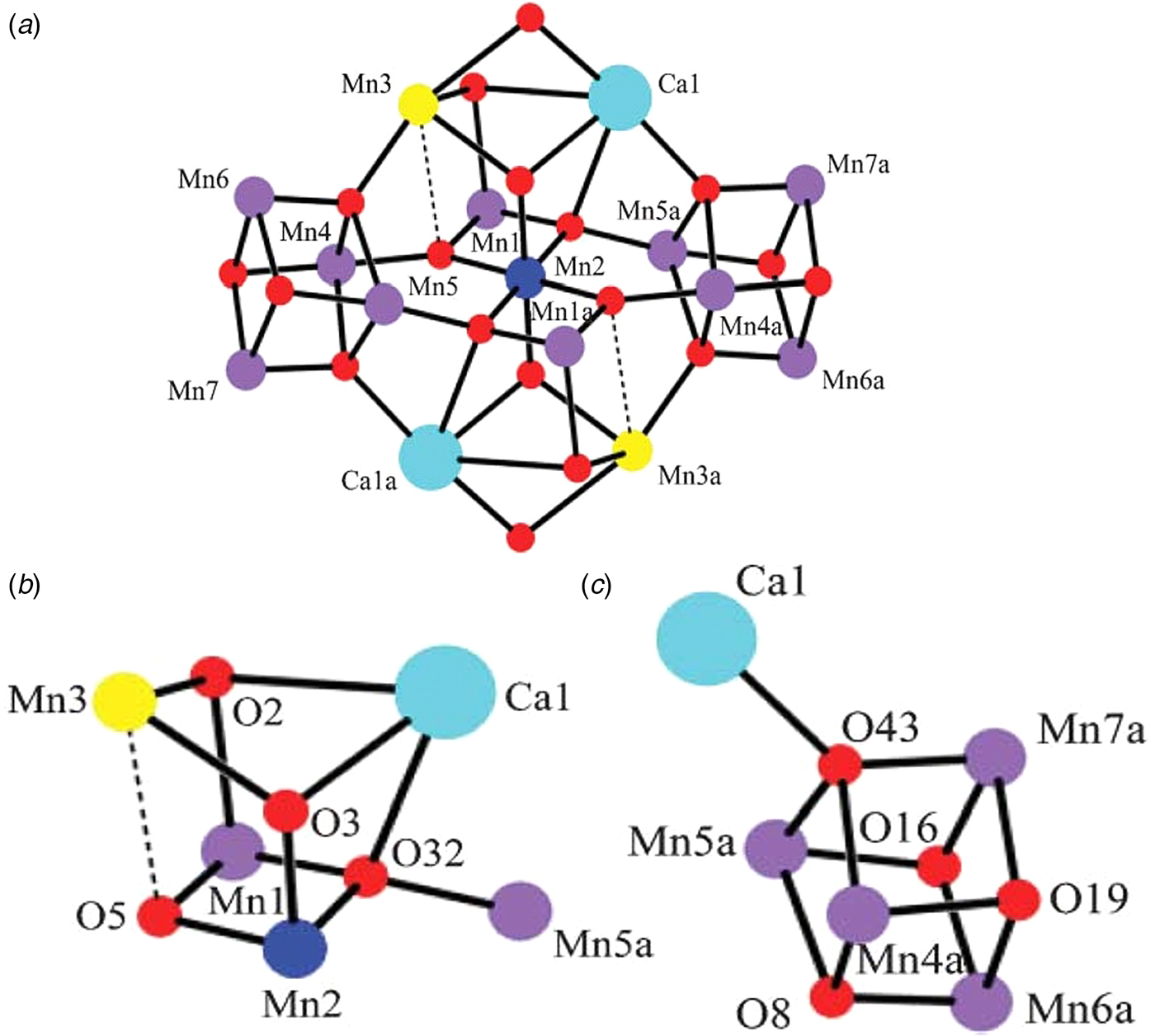
The fact that a Mn4Ca2+O4 structure like the one proposed by Ferreira et al. can also exist in the lattice of a macromolecular Mn–Ca molecule was shown by Misra et al. (Reference Misra, Wernsdorfer, Abboud and Christou2005). They synthesized a [Mn13Ca2O10(OH)2(OMe)2(O2CPh)18(H2O)4] molecule in a highly oxidized state (Fig. 5a ) which consisted of two Mn4Ca building blocks. As can be seen in Fig. 5, one form is the well-known Mn4O4 cube (Ruettinger et al. Reference Ruettinger, Ho and Dismukes1999) with a Ca attached to the cube by one of its bridging oxos (Fig. 5c ), while the other building block (Fig. 5b ) has a similar cubane structure to that of Mn4CaO4 of PSII. This study was the first to show that Mn and Ca can form discrete molecular complexes and also gave credibility to the Mn4CaO4 cubane model of Ferreira et al. (Reference Ferreira, Iverson, Maglaoui, Barber and Iwata2004) well before the publication of the 1·9 Å structure of Umena et al. (Reference Umena, Kawakami, Shen and Kamiya2011).
(a) [Mn13Ca2O10(OH)2(OMe)5(O2CPh)18(H2O)4] macromolecule synthesized by Misra et al. (Reference Misra, Wernsdorfer, Abboud and Christou2005) composed of two building blocks shown in (b) and (c). Ca is turquoise, Mn purple and yellow and oxygen in red.
3. Mechanism of water splitting
3.1. Base-catalysed nucleophilic attack mechanism
Prior to the 2004 crystal structure, there had been many postulates of how PSII splits water to form dioxygen. One idea suggested by Messinger et al. (Reference Messinger, Badger and Wydrzynski1995) and Pecararo et al. (Reference Pecararo, Baldwin, Caudl, Hsieh and Law1998) was that O–O bond formation in PSII resulting from a based catalysed nucleophilic attack of a hydroxyl group onto an electrophilic terminal oxo derived from the deprotonation of the second substrate water molecule. In particular, Pecararo et al. (Reference Pecararo, Baldwin, Caudl, Hsieh and Law1998) conducted studies on synthesized Mn complexes and despite there being no crystal structure of the Mn4Ca2+ cluster at that time they proposed that a ‘nucleophilic attack by a calcium-ligated hydroxide on an electrophilic oxo group ligated to a high-valent manganese to achieve the critical O–O bond formation’. The high oxidation state was taken to be MnV in line with the suggestion of Limburg et al. (Reference Limburg, Brudvig and Crabtree1997), but could also be a MnIV-oxyl radical (Brudvig, Reference Brudvig2008). The Ca2+ would act as a weak Lewis acid and align a specific H2O molecule for an efficient nucleophilic attack of an OH on the highly electrophylic terminal oxo of Mn4. This mechanism was also favoured by Britt et al. (Reference Britt, Campbell, Peloquin, Gilchrist, Aznar, Cicus, Robble and Messinger2004) based on EPR studies.
The cubane structure of the Mn4Ca2+O4 cluster of PSII derived from X-ray crystallography in 2014 by my colleagues and I was consistent with this proposed mechanism and discussed in our papers at that time (Barber et al. Reference Barber, Ferreira, Maghlaoui and Iwata2004; Ferreira et al. Reference Ferreira, Iverson, Maglaoui, Barber and Iwata2004). Importantly, the dangler Mn4 was found to be immediately adjacent to the Ca2+ and close to side chains of several key amino acids, including the redox-active tyrosine, YZ , and therefore we suggested that Mn4 and Ca2+ form a ‘‘catalytic’ surface outside the cubane for binding the two substrate water molecules and their subsequent oxidation. The progression through the S-state cycle would lead to deprotonation of these water molecules and the formation of a highly electrophilic MnV-oxo or MnIV-oxyl radical, and to a nucleophilic hydroxyl on the Ca2+ in the former case and an oxo–oxyl coupling mechanism in the latter so as to generate a dioxygen molecule (not shown in Fig. 6).
Diagrammatic representation of a mechanistic scheme for water splitting and dioxygen formation in PSII reproduced from Barber (Reference Barber2017). The substrate water molecules and products of the oxidation reactions for each S-state are shown in red. Intermediates that may exist between the S-state transitions (Dau & Haumann, Reference Dau and Haumann2007) are not depicted nor is the possible peroxide intermediate just prior to O–O bond formation. The essence is that a proton (H+) and an electron (e−) are removed by PECT for the flash-induced S0 to S1, S2 to S3 and S3 to S4 light-driven transitions but only an electron is removed during the S1 to S2 transition resulting in the accumulation of a positive charge in the metal cluster as shown. The final S3 to S4 flash-induced transition progresses to a highly electrophilic terminal oxo (electron deficient) ideally poised for a nucleophilic attack by a hydroxyl coordinated to the nearby Ca ion (electron rich) (taken from Barber, Reference Barber2017).
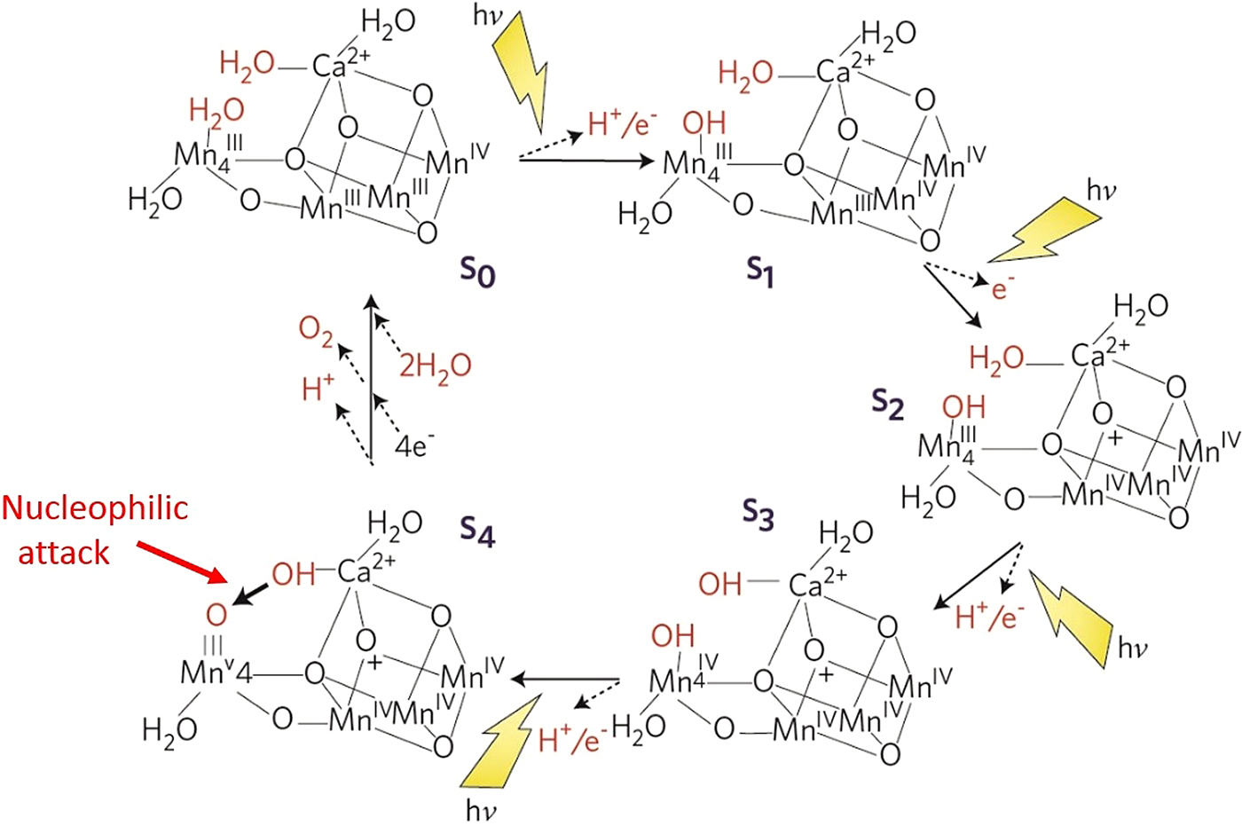
Thus as shown in Fig. 6, the formation of the O–O bond of dioxygen would result from the based catalysed nucleophilic attack of hydroxyl onto the electrophilic oxo of Mn4 with a likely peroxide intermediate. The electrophilicity of MnV-oxo would be enhanced by the high oxidation level of the cubane (3 × MnIV) plus a net positive charge (formed during the S1 to S2 transition) acting together as an ‘oxidizing battery’. The deprotonation of the substrate waters would be aided by nearby bases such as CP43Arg357 and by the weak Lewis acidity of Ca2+. Indeed, there is an extensive H-bonding network leading from Yz to the lumenal side of OEC (Ferreira et al. Reference Ferreira, Iverson, Maglaoui, Barber and Iwata2004; Murray & Barber, Reference Murray and Barber2007; Umena et al. Reference Umena, Kawakami, Shen and Kamiya2011). However, as stated above, the dangler Mn4 and Ca2+ each bind two water molecules according to the high-resolution structure of PSII (Umena et al. Reference Umena, Kawakami, Shen and Kamiya2011) (see Fig. 6). This may indicate a ‘carousel mechanism’ providing efficient delivery of substrate water molecules to the active sites and allow maximum turnover rates for the S-state cycle rather than being restricted by the rate of water diffusion to the catalytic site just prior to the formation of the S0-state. This can explain the action of ammonia, often considered to be an analogue of water. It has been shown that NH3 binds strongly to Mn4 (Oyala et al. Reference Oyala, Stich, Debus and Britt2015) and does not inhibit production of O2 but rather slows the turnover of the S-state cycle (Boussac e t al. Reference Boussac, Rutherford and Styring1990; Navarro et al. Reference Navarro, Ames, Nilsson, Lohmiller, Pantazis, Rapatskiy, Nowaczyk, Neese, Boussac, Messinger and Lubitz2013). This would be consistent with it inhibiting the carousel process but not the chemistry of O–O bond formation. The idea of a carousel mechanism was first proposed by Batista and colleagues but for a different mechanism for the S2 to S3 transition (Askerka et al. Reference Askerka, Wang, Vinyard, Brudvig and Batista2016; Vinyard & Brudvig, Reference Vinyard and Brudvig2017) and supporting in part the proposals described below in Section 4.4.
Although the electrophilic oxo in the S4 state is shown attached to MnV it could equally be an energy equivalent terminal MnIV-oxyl, which would be available for an oxo–oxyl coupling mechanism.
Fig. 6 is a mechanistic scheme for water splitting and dioxygen formation in PSII based on the acid–base, hydroxyl/water nucleophilic attack mechanism for O–O bond formation discussed above and taken from Barber (Reference Barber2017).
4 Indirect support for the nucleophilic mechanism for O–O bond formation in PSII
Although the nucleophilic attack mechanism for O–O bond formation in PSII makes good chemical sense (Vinyard et al. Reference Vinyard, Khan and Brudvig2015) and fits well with the geometry and positioning of the metal cluster in the complex, there is no direct experimental evidence for it and there are a number of alternative mechanisms being advocated recently with focus on the O5 oxygen linking Mn4 to cubane (see Fig. 3b ) (Askerka et al. Reference Askerka, Wang, Vinyard, Brudvig and Batista2016; Cox et al. Reference Cox, Retegan, Neese, Pantais, Boussac and Lubitz2014; Perez-Navarro et al. Reference Perez-Navarro, Neese, Lubitz, Pantazis and Cox2016; Siegbahn, Reference Siegbahn2013; Suga et al. Reference Suga, Akita, Hirata, Ueno, Murakami, Nakajima, Shimizu, Yamashita, Yamamoto, Ago and Shen2015, Reference Suga, Akita, Sugahara, Kubo, Nakajima, Nakane, Yamashita, Umena, Nakabayashi, Yamane, Nakano, Suzuki, Masuda, Inoue, Kimura, Nomura, Yonekura, Yu, Sakamoto, Motomura, Chen, Kato, Noguchi, Tono, Joti, Kameshima, Hatsui, Nango, Tanaka, Naitow, Matsuura, Yamashita, Yamamoto, Nureki, Yabashi, Ishikawa, Iwata and Shen2017; Umena et al. Reference Umena, Kawakami, Shen and Kamiya2011; Young et al. Reference Young, Ibrahim, Chatterjee, Gul, Fuller, Koroidov, Brewster, Tran, Alonso-Mori, Kroll, Michels-Clark, Laksmono, Sierra, Stan, Hussein, Zhang, Douthit, Kubin, de Lichtenberg, Long Vo, Nilsson, Cheah, Shevela, Saracini, Bean, Seuffert, Sokaras, Weng, Pastor, Weninger, Fransson, Lassalle, Bräuer, Aller, Docker, Andi, Orville, Glownia, Nelson, Sikorski, Zhu, Hunter, Lane, Aquila, Koglin, Robinson, Liang, Boutet, Lyubimov, Uervirojnangkoorn, Moriarty, Liebschner, Afonine, Waterman, Evans, Wernet, Dobbek, Weis, Brunger, Zwart, Adams, Zouni, Messinger, Bergmann, Sauter, Kern, Yachandra and Yano2016). However, below I present some convincing indirect evidence, which supports the nucleophilic mechanism for water splitting by PSII as detailed in Fig. 6.
4.1. Comparison with Fe–Ni carbon monoxide dehydrogenase (CODH)
There are anaerobic prokaryotic organisms which use the oxidation of carbon monoxide as an energy source to split water. The enzyme that catalyses this reaction is CODH. There are some interesting similarities between PSII and the Fe–Ni CODH, which supports the nucleophilic attack mechanism for photosynthetic O–O bond formation (Barber, Reference Barber2017). In CODH, the energy of the oxidation of a CO molecule to CO2 is used to split one water molecule to produce carbon dioxide and two reducing equivalents.
 $$\hbox{CO} + 2\hbox{H}_{2} \hbox{O} \to \hbox{CO}_{2} + 2\hbox{e}^{\rm -} + 2\hbox{H}^{+} $$
This chemistry is the well-known ‘water-gas shift reaction’ discovered in 1780 by the Italian physicist, Fontana (Reference Fontana1780), and today adopted as a large-scale industrial process to make pure hydrogen gas from carbon monoxide. This commercial process requires shifts in reaction temperature from high (approximately 400 °C with an iron-chromium oxide catalyst) to low (approximately 225 °C with copper-based catalysts) (Smith et al. Reference Smith, Loganathan and Shantha2010).
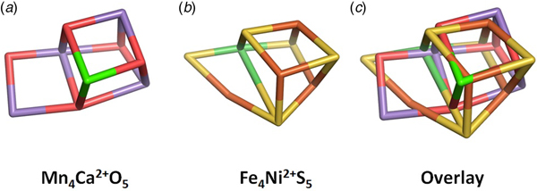
In contrast, in the case of Fe–Ni CODH, the generated reducing equivalents are produced at ambient temperatures and are used to drive the reductive chemistry of the organism rather than producing hydrogen gas, usually by electron transport involving nearby Fe4–S4 centres (Gong et al. Reference Gong, Hao, Wei, Ferguson, Tallant, Krzycki and Chan2008; Svetlitchnyi et al. Reference Svetlitchnyi, Dobbek, Meyer-Klaucke, Meins, Thiele, Romer, Huber and Meyer2004). Remarkably, the catalytic centre of Fe–Ni CODH has a similar geometry to that of Mn4CaO5 of PSII (Fig. 7). X-ray crystallography at high resolutions (Dobbek et al. Reference Dobbek, Svetlitchnyi, Gremer, Huber and Meyer2001; Jeoung & Dobbek, Reference Jeoung and Dobbek2007) has shown that Fe–Ni CODH consists of a Fe3NiS4 cubane with a fourth ‘dangler’ Fe attached to the cubane via a bridging S of the cubane and an additional S bridge to make a Fe4NiS5 cluster as shown in Fig. 7b .
Comparison of (a) the Mn4Ca2+O5 cluster of PSII at 1·9 Å using PDB 3WU2 (Umena et al. Reference Umena, Kawakami, Shen and Kamiya2011) with (b) Fe4Ni2+S5 cluster of CO dehydrogenase at 1·03 Å using PDB 4UDX (Fesseler et al. Reference Fesseler, Jeoung and Dobbek2015). (c) Overlay of the two structures with a RMSD of 0·65 between the metal atoms. Oxo bonds in red, sulphur bonds in yellow, manganese ions in purple, calcium and nickel ions in green as labelled and iron ions in orange (taken from Barber, Reference Barber2017).
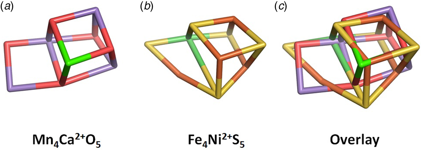
Despite the two catalytic centres being composed of different elements, the overlay of their structures, shown in Fig. 7c , is remarkably good with a root mean square deviation (RMSD) of 0·65 between the metal atoms. In fact, they are the only known examples of metallo-catalytic centres having heterocubanes with an additional metal in exo.
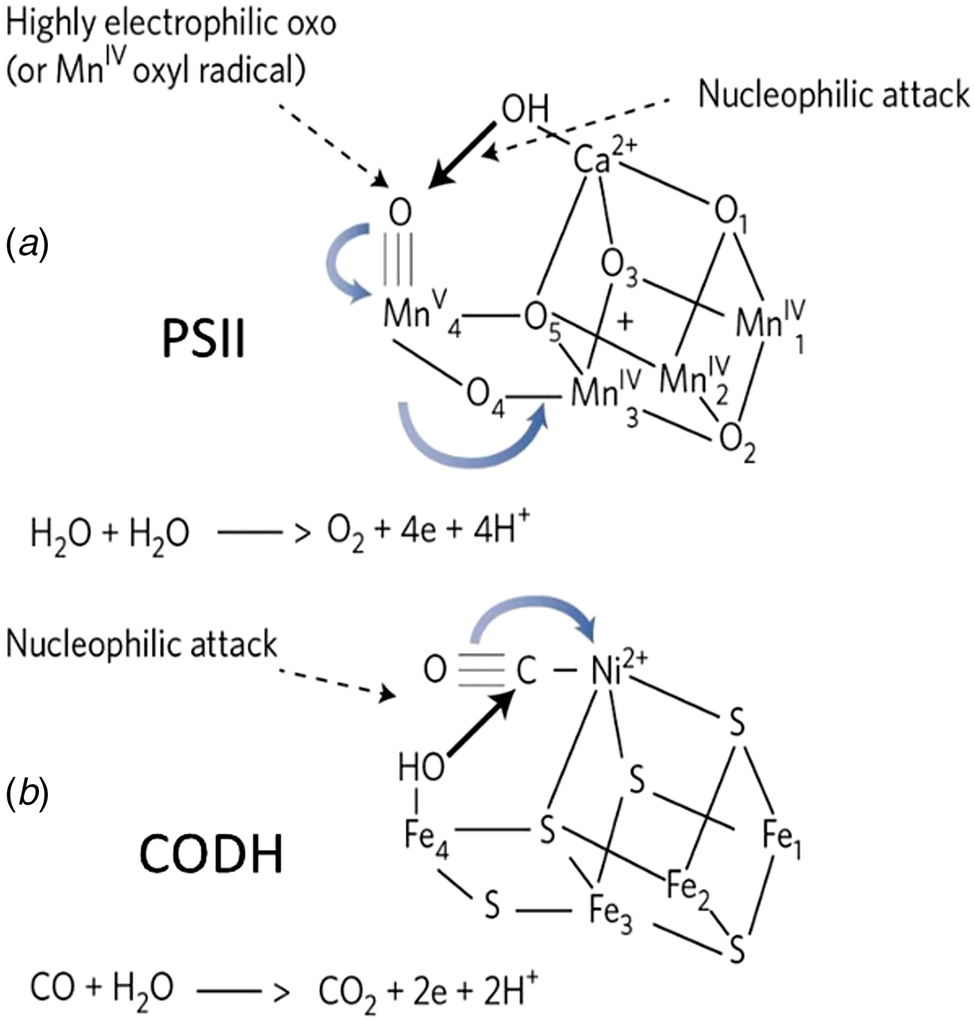
It is known that in CODH, the CO is activated by binding to the Ni2+ of the cubane, which causes a shift in the coordination of the Ni atom from square-planar to a square-pyramidal geometry (Jeoung & Dobbek, Reference Jeoung and Dobbek2007) and increases the electrophilicity of CO. Similarly, water is activated by forming a complex with the dangler Fe42+. This induces a movement of the cysteine Ni ligand leading to the possible formation of a carboxyl bridge between the Ni2+and Fe4. Thus, the mechanism is a base-catalysed nucleophilic attack by the Fe4 bound water/hydroxyl group onto CO (see Fig. 8). A decarboxylation of the intermediate leads to CO2 release and the reduction of the metal cluster (Jeoung & Dobbek, Reference Jeoung and Dobbek2007). Oxidation back to the active state involves electron transfer to nearby Fe4–S4 centres including ferredoxin or to a closely bound acetyl-CoA synthase depending on species (Gong et al. Reference Gong, Hao, Wei, Ferguson, Tallant, Krzycki and Chan2008).
A structurally based diagrammatic comparison of a base nucleophilic attack of a hydroxyl onto an electrophile leading to oxygen atom transfer for (a) O–O formation in the S4 state of PSII and (b) CO conversion to CO2 in CODH. Note that the numbering of Fe ions in (b) are different to those in PDB 4UDX (Fesseler et al. Reference Fesseler, Jeoung and Dobbek2015) so as to compare with the numbering of the Mn ions in (a) taken from PDB 3WU2 (Umena et al. Reference Umena, Kawakami, Shen and Kamiya2011). Unfortunately, the numbering of the Mn4Ca2+O5 in PDB 3WU2 (Umena et al. Reference Umena, Kawakami, Shen and Kamiya2011) differs from that of the earlier Mn4Ca2+O4 in PDB 1S5L (Ferreira et al. Reference Ferreira, Iverson, Maglaoui, Barber and Iwata2004). Note that in the S4 state there is the storage of a net positive charge generated during the S1 to S2 transition (see Fig. 6) (taken from Barber, Reference Barber2017).
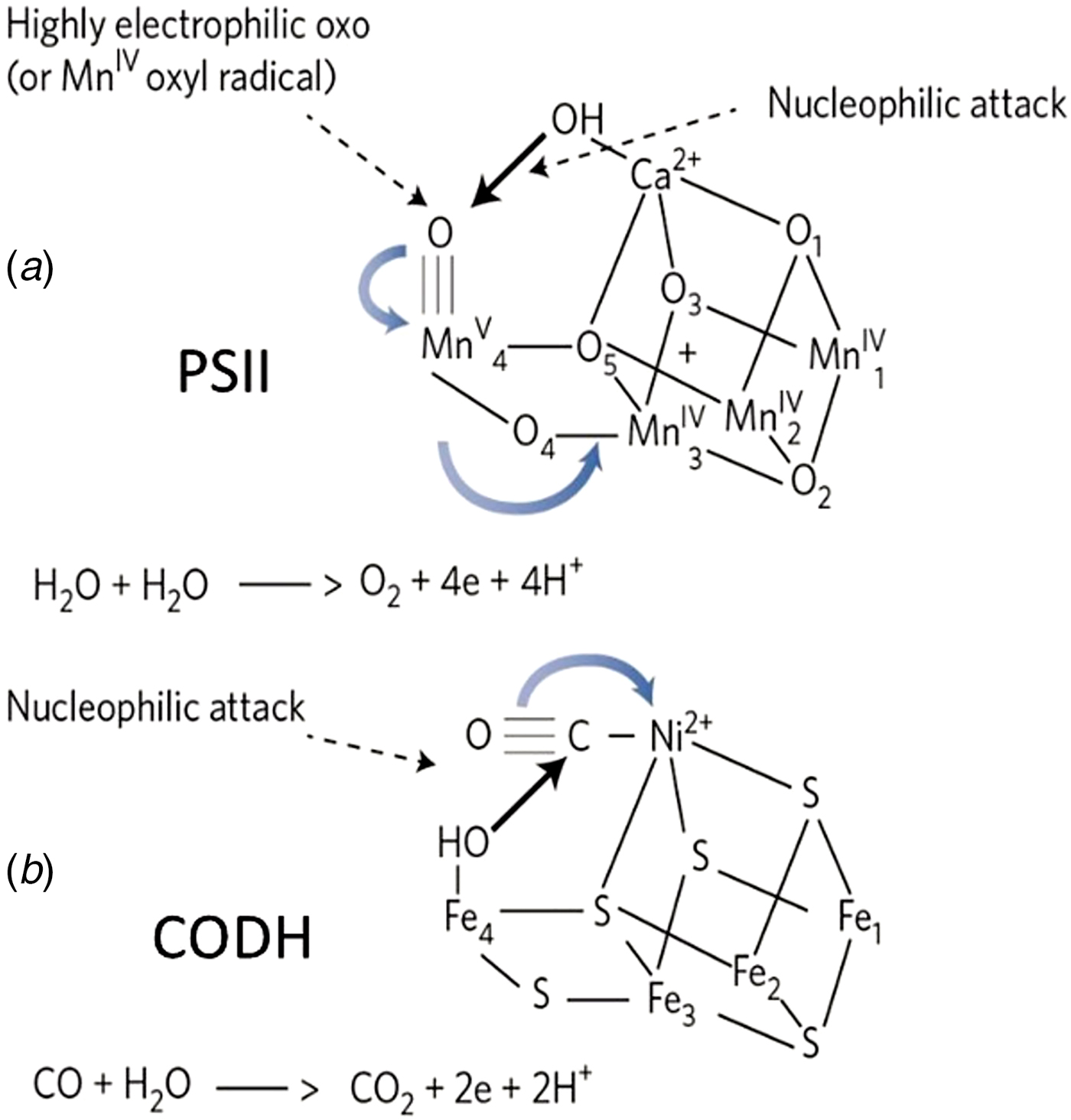
Given the striking geometrical similarities between the catalytic centres of CODH and PSII, together with the fact they both catalyse the removal of reducing equivalents from water, it would be reasonable to conclude that there may be common features in their catalytic mechanisms. Of course PSII water splitting involves high-valent oxidative chemistry, while CODH is low-valent organometallic chemistry. Moreover, the water-splitting reaction of PSII is non-reversible while that of CODH is. For these reasons, I wish to emphasize that the comparison of PSII with Fe–Ni CODH does not imply any direct evolutionary link but a similarity in their function to extract reducing equivalents from the water.
4.2. Ligands to the Mn4Ca2+O5 cluster
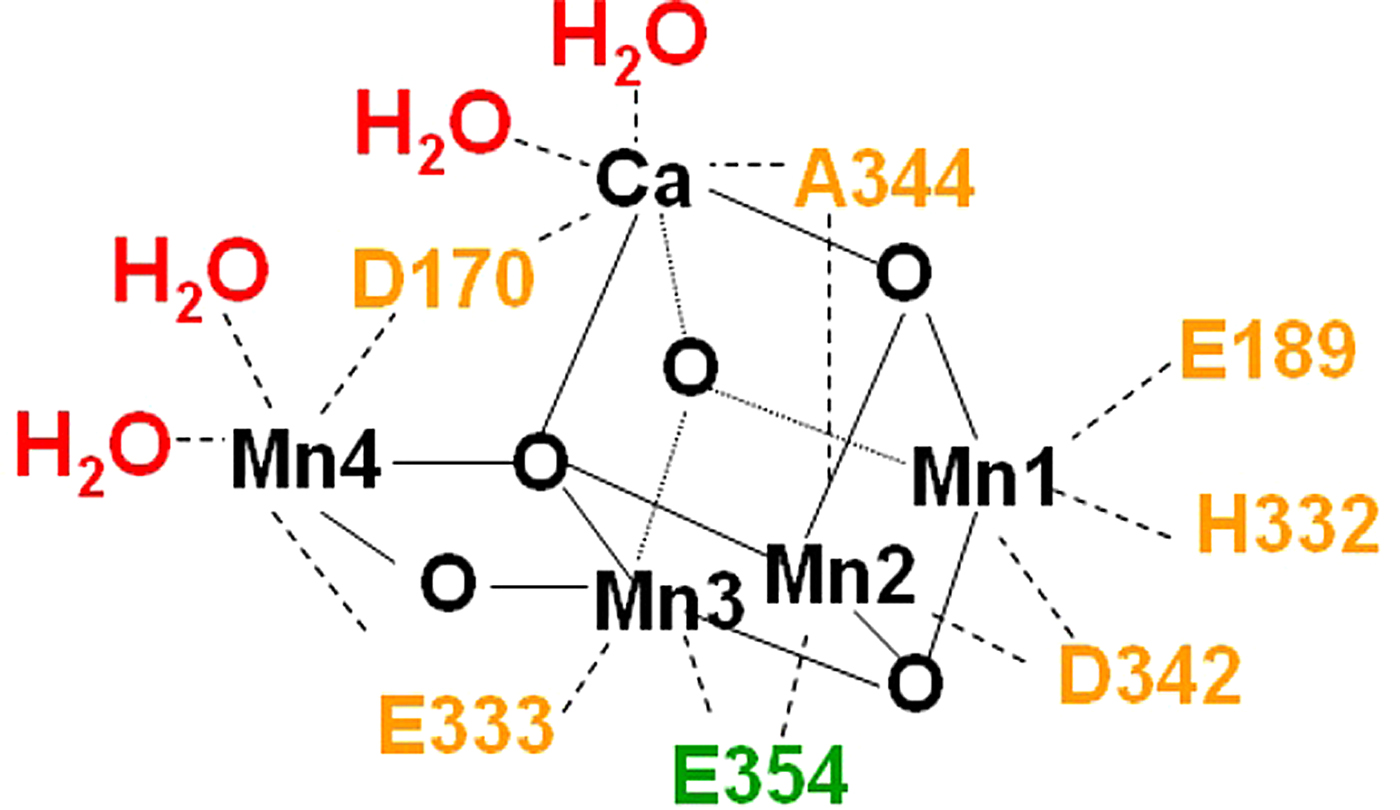
The Ferreira et al. structure (Reference Ferreira, Iverson, Maglaoui, Barber and Iwata2004) identified seven amino acids as ligands to the Mn4Ca-cluster, six from the D1 reaction centre protein: Asp170, Glu189, His332, Ala344, Glu333 and Asp342 and Glu354 of the inner antenna chlorophyll-binding protein, CP43. That these seven amino acids are ligated to the Mn4Ca-cluster has been confirmed in the 1·9 Å structure of Umena et al. (Reference Umena, Kawakami, Shen and Kamiya2011) and the precise details of the ligation pattern revealed (see Fig. 9). The coordination properties of the three Mn ions of the cubane are totally satisfied by amino acid ligands. However, as stated above, Umena et al. (Reference Umena, Kawakami, Shen and Kamiya2011) assigned two water ligands for the non-cubane dangler Mn4 and also for the Ca ion as shown in Fig. 9. Ferreira et al. (Reference Ferreira, Iverson, Maglaoui, Barber and Iwata2004) had previously suggested that water molecules could act as ligands to Mn4 and Ca2+ and that this may be the site for dioxygen formation.
Diagrammatic representation of the amino acid side chains acting as ligands to the Mn4CaO5 cluster of the OEC together with four water molecules providing ligands to Ca and Mn4 (constructed from Umena et al. crystal structure (2011)). D1 residues shown in orange and green depict the CP43 residue Glu354 (taken from Barber, Reference Barber2016a).
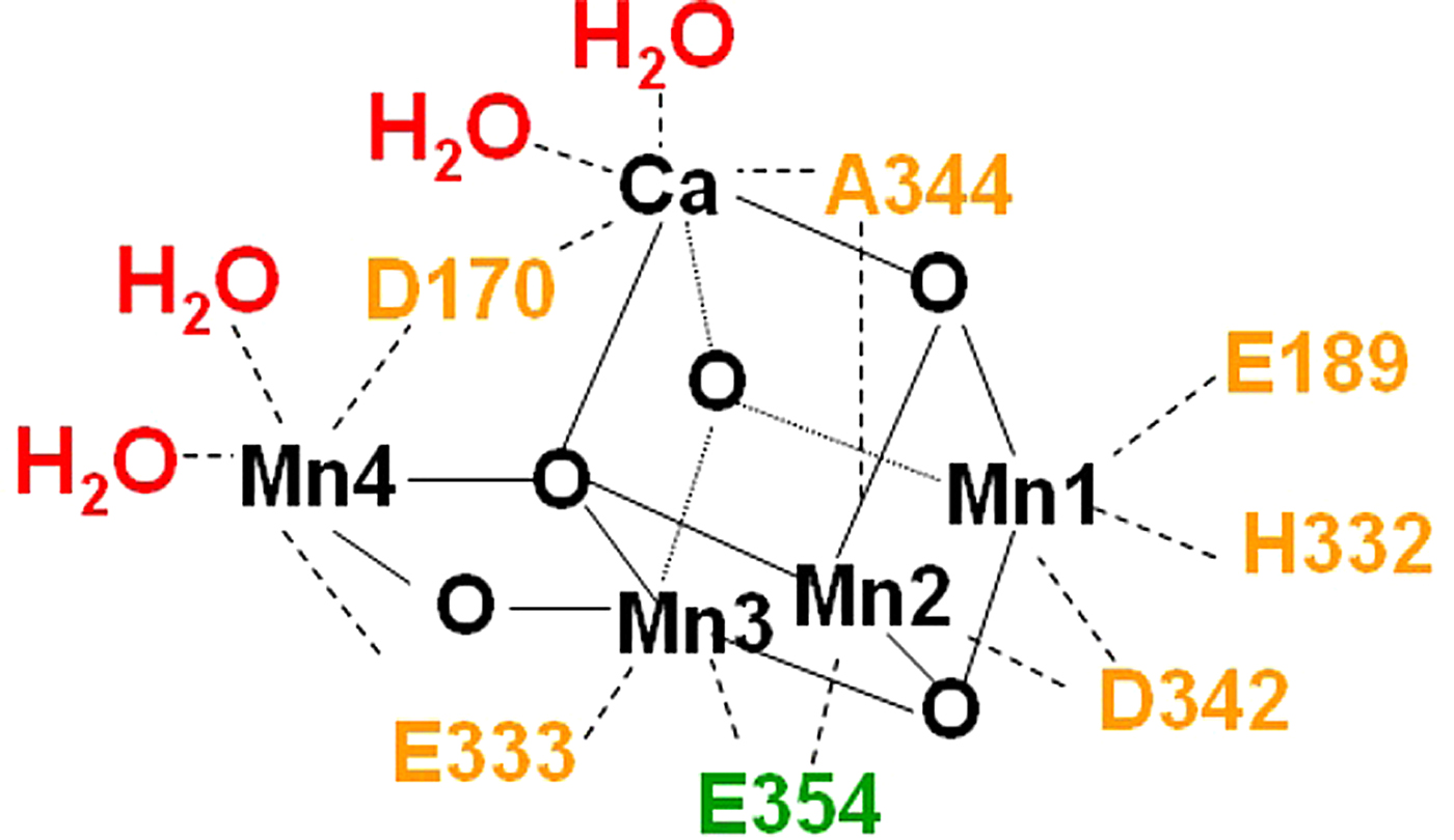
The amino acids which serve as ligands to the metal cluster are highly conserved in PSII over the whole cyanobacterial, plant and algal kingdoms (Murray, Reference Murray2012). Yet, there are a large number of reports that their replacement by other residues using site-directed mutagenesis does not inhibit oxygen evolution completely and sometimes not at all (Debus, Reference Debus2008). One example is the replacement of the negatively charged D1-Glu189 by positively charged Lys, which made no difference to the rate of oxygen evolution (Clausen et al. Reference Clausen, Winkler, Hays, Hundelt, Debus and Junge2001). This was also true for other D1-Glu189 mutants leading to the logical conclusion that it cannot be a ligand to the Mn4Ca-cluster. Another example is the replacement of acidic CP43-Glu354 by neutral glutamine where O2 evolution persisted at a reduced rate of 10–18% compared with the wild-type (Service et al. Reference Service, Yano, McCONNELL, Hwang, Niks, Hille, Wydrzynski, Burnap, Hillier and Debus2010). It is also possible to totally remove CP43 and yet assemble the OEC (Buchel et al. Reference Buchel, Barber, Ananyev, Eshaghi, Watts and Dismukes1999). Thus compared with many other metallo-enzymes, the assembled Mn4CaO5 cluster is rather robust to changes in its first coordination sphere. There are exceptions since, for example, D1-Asp170 also acts as a high-affinity site for Mn2+ binding (Diner et al. Reference Diner, Nixon and Farchaus1991) so that the photo-oxidation of P680 to P680+ can facilitate its conversion to Mn3+. In this way, as long as CaCl2 is present, the Mn4CaO5 is assembled step-by-step to form the OEC, a process known as photoactivation (Ananyev & Dismukes, Reference Ananyev and Dismukes1996).
4.3. Synthesized chemical model systems
Ruthenium model chemistry was the first to achieve homogeneous water oxidation and has subsequently become the best characterized. However, functional manganese model complexes have also been synthesized and have the added advantage of being more biologically relevant to the OEC.
4.3.1. Mechanism of O2 production from organo-Ru complexes
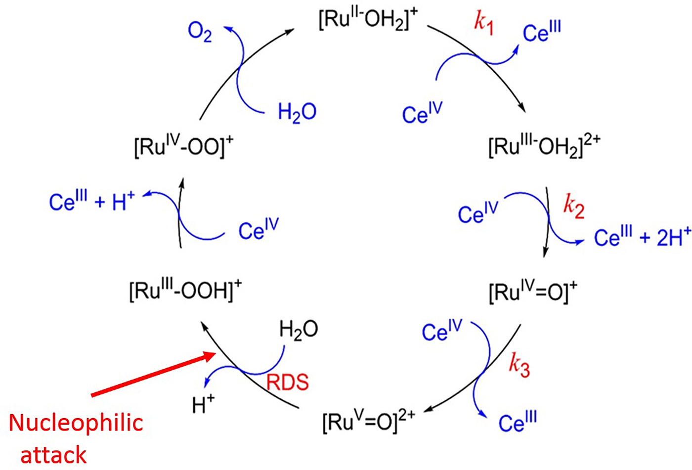
The blue dimer, cis,cis-[(bpy)2(H2O)RuIIIORuIII(H2O)(bpy)2]4+ (where bpy = 2,2′-bipyridine) was the first designed, well-defined molecule known to function as a catalyst for water oxidation (Gersten et al. Reference Gersten, Samuels and Meyer1982). As in the case of PSII, it met the stoichiometric requirements for water oxidation by utilizing PCET reactions in which both electrons and protons are transferred. This avoided charge build-up, allowing for the accumulation of multiple oxidative equivalents at the Ru–O–Ru core. Since then a wide range of both dinuclear Ru and mononuclear Ru organo-complexes have been synthesized, which show high rates of O2 evolution using Ce4+ as an oxidant. A significant number of them, particularly the mononuclear Ru molecules, generate the O–O bond by nucleophilic attack of an electrophilic oxo of RuV = O (Duan et al. Reference Duan, Wang, Li, Li and Sun2015; Murakami et al. Reference Murakami, Hong, Suenobu, Yamaguchi, Ogura and Fukuzumi2011; Sala et al. Reference Sala, Maji, Bofill, Garcia-Anton, Escriche and Llobet2013; Tong et al. Reference Tong, Duan, Xu, Privalov and Sun2011). This mechanism and the rate of turnover are dependent on the design of the ligands (Concepcian et al. Reference Concepcian, Jurss, Brennaman, Hoertz, Patrocinio, Murakami Iha, Templeton and Meyer2009). One example is [RuII(bpc)(bpy)(OH2)]+ (where bpc = 2,2′-bipyridine-6-carboxylate and bpy = 2,2′-bipyridine). A proposed mechanism for these types of mononuclear Ru catalysts at low pH is shown in Fig. 10. The [RuII-OH2]+ is first oxidized to [RuIII-H2]2+, followed by a 2H+/1e PCET oxidation step generating [RuIV = O]+. [RuIV = O]+ is not reactive towards the water and therefore has to be further oxidized to generate the active species [RuV = O]2. Thereafter, water nucleophilic attack on the RuV oxo occurs, forming the hydroperoxo intermediate [RuIII-OOH]+, and oxygen evolves from [RuIV-OO]2+. The carbonate ligand (bpc) enhances electron transfer steps without obviously influencing the PCET (Duan et al. Reference Duan, Wang, Li, Li and Sun2015).
(a) Mechanistic scheme for the generation of dioxygen by treating [RuII(bpc)(bpy)(OH2)]+ (where bpy = 2,2-bipyridine and bpc = 2,2-bipyridine-6-carboxylate) with CeIV. A water nucleophilic attack onto RuV = O is proposed for the rate determining step (RDS) (taken from Duan et al. Reference Duan, Wang, Li, Li and Sun2015 with permission).

This type of rational design of Ru-complexes with a general bipyridine–dicarboxylate ligand motif has produced many molecular catalysts having exceptionally high turnover frequencies (TOF). For example, the mononuclear ruthenium complex [Ru(bda)(isoq)2] (where bda = 2,2′-bipyridine-6,6-dicarboxylic acid and isoq = isoquinoline) has a high catalytic activity for water oxidation when using CeIV as the oxidant under acidic conditions (Duan et al. Reference Duan, Wang, Li, Li and Sun2015). However, higher rates of O2 formation are achieved which are much faster than PSII (1000 s−1), with turnover rates of 8000 s−1 at pH 7·0 and 50 000 s−1 at pH 10 and is combined with a high chemical stability (Matheu et al. Reference Matheu, Ertem, Benet-Buchholz, Coronado, Batista, Sala and Llobet2015). These very impressive turnover rates are obtained with Ru complexes having the general formula [Ru n (tda)(py)] m+ with tda being [[2,2 : 6′,2″-terpyridine]-6,6″-dicarboxylate]. Again the mechanism is thought to involve a nucleophilic attack by water onto the oxo of RuV as in Fig. 10. The high rates are attributed to: (i) easy access to the high oxidation states, provided by the tda−2 ligand through the anionic nature of the carboxylates and the capability of stabilizing a coordination of seven to Ru in high oxidation states and (ii) the ability of the dangling carboxylate to be a proton acceptor in an intramolecular fashion, while the incoming substrate water molecule undergoes the nucleophilic attack.
4.3.2. Mechanism of O2 production from organo-Mn complexes
In the context of the water/hydroxide nucleophyllic attack mechanism for PSII O–O bond formation, a number of efforts have been made to prepare MnV oxo complexes, which relate to OEC of PSII. The groups of both Crabtree and Brudvig (Cady et al. Reference Cady, Crabtree and Brudvig2008; Limburg et al. Reference Limburg, Brudvig and Crabtree1997; Reference Limburg, Vrettos, Liable-Sands, Rheingold, Crabtree and Brudvig1999; Reference Limburg, Vrettos, Chen, De Paula, Crabtree and Brudvig2001; Vrettos et al. Reference Vrettos, Limberg and Brudvig2001) and Pecararo et al. (Reference Pecararo, Baldwin and Gelasco1994, Reference Pecararo, Baldwin, Caudl, Hsieh and Law1998) have provided evidence for MnV oxo intermediates related to the OEC that were very short lived. One catalyst, [Mn2 III/IV(-O)2(terpy)2(OH2)2](NO3)3 (where terpy = 2,2′:6′2″-terpyridine) has provided a model system for the OEC of PSII (Limburg, et al. Reference Limburg, Vrettos, Liable-Sands, Rheingold, Crabtree and Brudvig1999; Reference Limburg, Vrettos, Chen, De Paula, Crabtree and Brudvig2001). These studies used a range of added oxidants and often involved the radical coupling of two adjacent oxos.
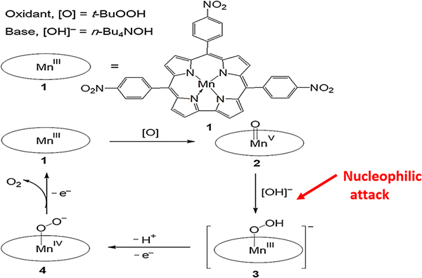
However, both porphyrins and corroles and some related complexes have been shown to produce dioxygen by a nucleophilic mechanism by generating a MnV oxo as the electrophilic species (Gao et al. Reference Gao, Liu, Wang, Na, Åkermark and Sun2007, Reference Gao, Åkermark, Liu, Sun and Åkermark2009) (see Fig. 11). A hydroxyl nucleophilic attack of water on the MnV oxo yielded dioxygen (steps 2 to 3 in Fig. 11) as deduced from DFT calculation, spectroscopy and isotopic O18 exchange experiments.
Mechanism for a base-induced hydroxyl nucleophilic attack on a MnV = O of nitrophenylcorrole (structure is shown and depicted as MnIII in the scheme) using t-BuOOH as the oxidant and n-Bu4NOH as the nucleophilic base (taken from Gao et al. Reference Gao, Åkermark, Liu, Sun and Åkermark2009 with permission).

In another study, it was shown that both mononuclear and dinuclear ‘Pacman’ MnIV corrole complexes could act as a catalyst for water splitting (Gao et al. Reference Gao, Liu, Wang, Na, Åkermark and Sun2007). In principle, the dinuclear Mn complexes could react via oxygen–oxygen radical coupling, but in these systems this route was found to be energetically less favourable than the nucleophilic attack mechanism.
4.4. Alternative mechanisms are less compelling
An alternative to the base-catalysed nucleophilic mechanism for O–O bond formation in PSII, is radical pair coupling, which was proposed by Babcock and colleagues (Hoganson & Babcock, Reference Hoganson and Babcock1997; Tommas & Babcock, Reference Tommas and Babcock1998) prior to the elucidation of the OEC structure. DFT calculations and X-ray diffraction data have proved to be remarkably good for studying the OEC as emphasized by Siegbahn who predicted, using the Ferreira et al. cubane model as the starting point, the existence and structure of the Mn4Ca2+O5 cluster three years before the 1·9 Å crystal structure of Umena et al. was published (Siegbahn, Reference Siegbahn2008). This powerful DFT approach has also been used extensively by Siegbahn (Reference Siegbahn2006, Reference Siegbahn2009, Reference Siegbahn2013) and Li and Siegbahn (Reference Li and Siegbahn2015) to explore the mechanism of water oxidation and particularly the nature of S4 in the S-state cycle.
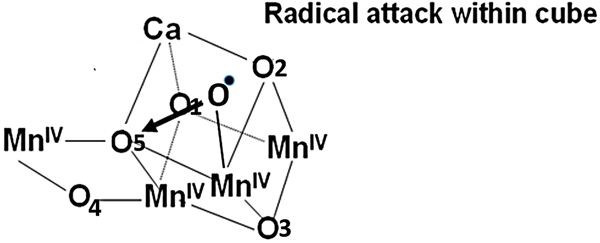
Initially, Siegbahn favoured a surface radical coupling mechanism involving a terminal MnIV-oxyl radical (Siegbahn & Crabtree, Reference Siegbahn and Crabtree1999) but according to his calculation, this mechanism has a higher energy barrier than an oxyl–oxo coupling mechanism. He therefore concluded that the most likely mechanism is the formation of a terminal oxyl radical within the cubane at S4, which attacks a nearby bridging oxo, O5 to form the O–O bond (see Fig. 12). This proposed mechanism requires significant conformational changes due to the insertion of a substrate water molecule into the cubane during the S2 to S3 transition and reconstruction of the cubane during the S4 to S0 transition. However, a recent XFEL study (Young et al. Reference Young, Ibrahim, Chatterjee, Gul, Fuller, Koroidov, Brewster, Tran, Alonso-Mori, Kroll, Michels-Clark, Laksmono, Sierra, Stan, Hussein, Zhang, Douthit, Kubin, de Lichtenberg, Long Vo, Nilsson, Cheah, Shevela, Saracini, Bean, Seuffert, Sokaras, Weng, Pastor, Weninger, Fransson, Lassalle, Bräuer, Aller, Docker, Andi, Orville, Glownia, Nelson, Sikorski, Zhu, Hunter, Lane, Aquila, Koglin, Robinson, Liang, Boutet, Lyubimov, Uervirojnangkoorn, Moriarty, Liebschner, Afonine, Waterman, Evans, Wernet, Dobbek, Weis, Brunger, Zwart, Adams, Zouni, Messinger, Bergmann, Sauter, Kern, Yachandra and Yano2016) obtained room temperature diffraction data, which did not provide any evidence of structural changes in the S3 state as required by the Siegbahn oxyl–oxo coupling mechanism. Thus, in my opinion, this alternative mechanism is unlikely although very recently a XFEL study did detect some structural changes close to O5, which was suggested to be involved in O–O bond formation (Suga et al. Reference Suga, Akita, Sugahara, Kubo, Nakajima, Nakane, Yamashita, Umena, Nakabayashi, Yamane, Nakano, Suzuki, Masuda, Inoue, Kimura, Nomura, Yonekura, Yu, Sakamoto, Motomura, Chen, Kato, Noguchi, Tono, Joti, Kameshima, Hatsui, Nango, Tanaka, Naitow, Matsuura, Yamashita, Yamamoto, Nureki, Yabashi, Ishikawa, Iwata and Shen2017) and also see Vinyard & Brudvig (Reference Vinyard and Brudvig2017) for a balanced review of this mechanism.
A mechanism for O–O bond formation during the S4 to S0 transition postulated by Siegbahn (Reference Siegbahn2006; Reference Siegbahn2008; Reference Siegbahn2009; Reference Siegbahn2013) whereby formation of a terminal oxyl radical originating from a second substrate water incorporated into the cubane during the S2 to S3 transition leads to a radical attack on an adjacent oxo ligand (O5) incorporated within the Mn3CaO4-cubane from the first substrate water during the S4 to S0 transition.

5. Artificial photosynthesis
While some progress has been made in mimicking photosynthesis in artificial systems there is as yet no system identified which is both efficient and robust for incorporation into a working technology for capturing and storing solar energy in chemical bonds on a large scale as does natural photosynthesis.
To date the main focus of research has been to design and synthesize molecular catalysts, which can be linked to a light-driven charge separation system (Tran et al. Reference Tran, Wong, Barber and Loo2012). Dyes have been used for the latter but inorganic semiconductors offer a more realistic and robust approach for providing the redox potentials necessary to split water and power reductive chemistry, particularly the production of hydrogen gas.
Insights gleaned from the recent structural determination of PSII have initiated considerable efforts to identify artificial catalysts for water splitting using solar energy (Eisenberg & Gray, Reference Eisenberg and Gray2008). It has been demonstrated that catalysts based on Mn or on Mn doped with Ca2+ are capable of water splitting leading to the generation of dioxygen (Limburg et al. Reference Limburg, Vrettos, Liable-Sands, Rheingold, Crabtree and Brudvig1999; Najafpour, Reference Najafpour2011; Najafpour et al. Reference Najafpour, Ehrenberg, Wiechen and Kurz2010; Reference Najafpour, Rahimi, Aro, Lee and Allakhverdiev2012; Tagore et al. Reference Tagore, Crabtree and Brudvig2008; Zaharieva et al. Reference Zaharieva, Najafpour, Wiechert, Haumann, Kurz and Dau2011). Frei's group reported that nanostructured manganese oxide clusters supported on mesoporous silica efficiently evolved oxygen in aqueous solution under mild conditions (Jiao & Frei, Reference Jiao and Frei2010). The recently synthesized Mn4Ca2+ O4-cluster (Zhang et al. Reference Zhang, Chen, Dong, Shen, Dau and Zhao2015) shows charge storage under strong oxidation conditions similar to those observed in the natural PSII-cluster progressing to S3 but has yet to be shown that it can catalyse the generation of oxygen from water.
Complementing the work on Mn has been the earlier discovery mentioned above, that ruthenium-based catalysts, such as the ‘blue dimer’ can photo-oxidize water to dioxygen (Gersten et al. Reference Gersten, Samuels and Meyer1982; Liu et al. Reference Liu, Concepcion, Jurss, Cardolaccia, Templeton and Meyer2008) and the recent spectacular work of Duan et al. (Reference Duan, Bozoglian, Mandal, Stewart, Privalov, Llobet and Sun2012, Reference Duan, Wang, Li, Li and Sun2015) and Matheu et al. (Reference Matheu, Ertem, Benet-Buchholz, Coronado, Batista, Sala and Llobet2015) have led to rationally designed Ru-based catalysts with unprecedented turnover rates far higher than PSII oxygen production. But perhaps one of the most practical catalysts for water splitting is based on cobalt, which is a relatively abundant element. Kanan & Nocera (Reference Kanan and Nocera2008) have described a self-assembling catalyst composed of Co and phosphate ions which can produce molecular oxygen from water at neutral pH with a low overpotential akin to that which operates in the OEC of PSII. Dau and co-workers (Risch et al. Reference Risch, Khare, Zaharieva, Gerencser, Cherney and Dau2009) have revealed important structural details and properties of this Co-based catalyst and found it to have a molecular organization remarkably similar to the Mn3Ca-cubane of PSII. Du & Eisenberg (Reference Du and Eisenberg2012) have reviewed recent progress and future challenges of using Co as a water-splitting catalyst compared with other transition metals.
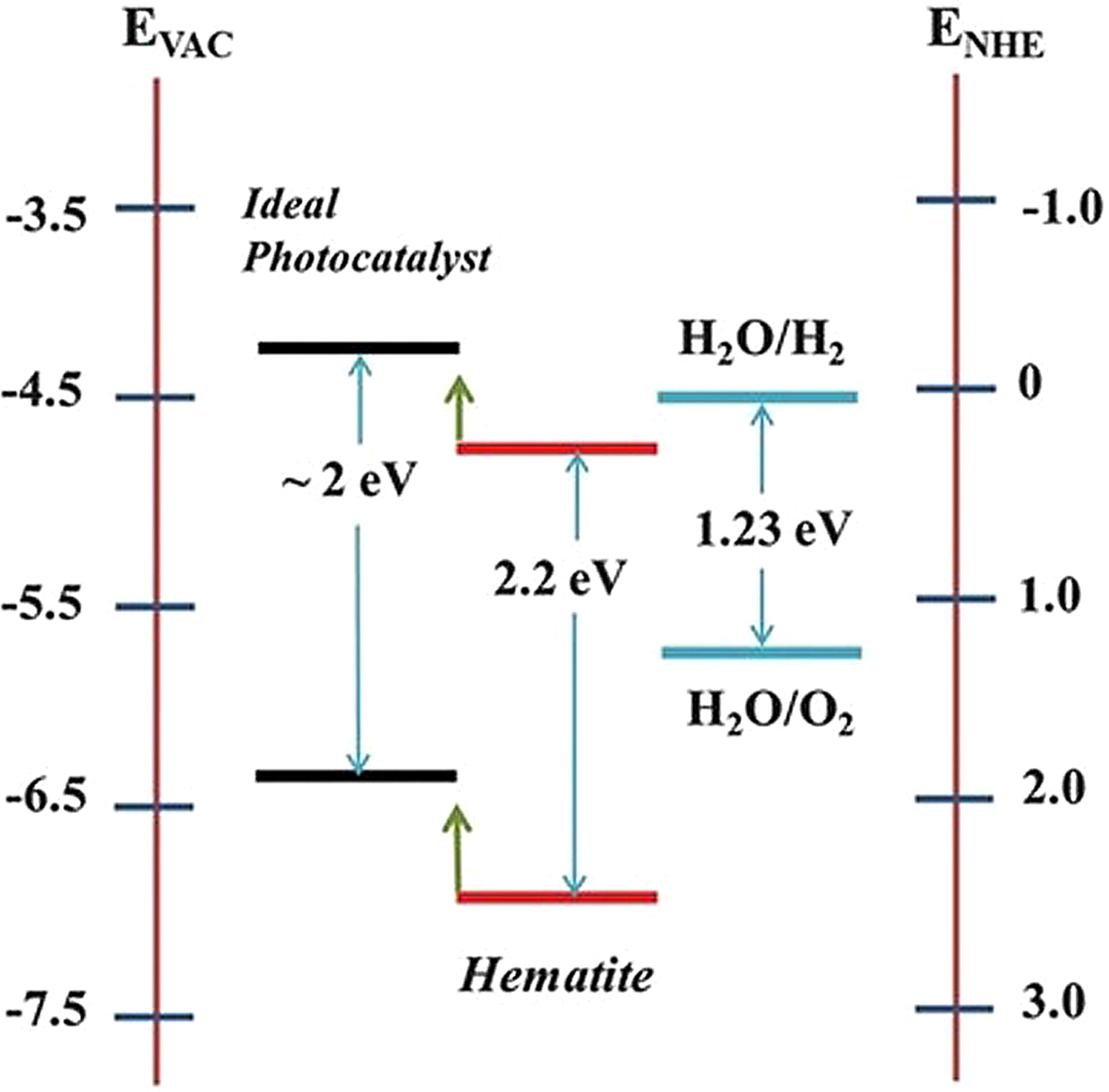
Hematite (α-Fe2O3) is a semiconductor, which is also capable of photoelectrochemically splitting water into molecular oxygen. It has a favourable optical band gap (2·2 eV, see Fig. 13), excellent chemical stability in aqueous environments, natural abundance and low cost (Bassi et al. Reference Bassi, Wong and Barber2014; Sivula et al. Reference Sivula, Le Formal and Graetzel2011). Indeed, hematite has been theoretically predicted to achieve a water oxidation efficiency of 16·8% (Murphy et al. Reference Murphy, Barnes, Randeniy, Plumb, Grey, Horne and Glasscock2006). However, the reported efficiencies of hematite are lower than this predicted value, mainly due to the very short lifetime of photogenerated charge carriers, short hole diffusion length (2–4 nm), slow oxygen evolution reaction kinetics, low flat band potential and significant reduction in the absorption cross-section at wavelengths approaching the band gap value. Another fundamental limitation of the hematite system is the need for externally applied bias because the conduction band of hematite is lower than the potential required to reduce protons to hydrogen (see Fig. 13). Nevertheless, it is a system which is receiving considerable attention at the present time and the recent construction of a tandem cell incorporating perovskite and Co-doped hematite nanorods and requiring no external electrical bias for photo-driven water splitting (Gurudayal et al. Reference Gurudayal, Sabba, Kumar, Wong, Barber, Graetzel and Mathews2015), is an example of the potential for this type of photoanode for constructing an ‘artificial leaf’ for solar fuel production.
Schematic representation of band levels for hematite and for an ideal photocatalyst for water oxidation to hydrogen and oxygen.

The next step will be to couple these oxygen-producing systems to non-platinum catalysts, which will use the protons and high-energy electrons derived from the water-splitting reaction to produce hydrogen gas or reduce CO2. In the case of the former, considerable progress is being made (Tran & Barber, Reference Tran and Barber2012, Wang et al. Reference Wang, Chen and Sun2012), in part by mimicking the natural hydrogenase enzymes found in a wide variety of microorganisms (Tran et al. Reference Tran, Artero and Fontecave2010). Also a number of inorganic catalysts have been identified with activities, which are almost as good as platinum (Tran & Barber, Reference Tran and Barber2012). One such class of catalysts is based on sulphides of molybdenum and tungsten (Merki & Hu, Reference Merki and Hu2011; Tran et al. Reference Tran, Chaim, Boix, Ren, Pramana, Fize, Artero and Barber2013; Reference Tran, Tran, Orio, Torelli, Truon, Nayuki, Sasaki, Chiam, Yi, Honma, Barber and Artero2016; Zong et al. Reference Zong, Han, Ma, Yan, Wu and Li2011) and another is an alloy composed of nickel, molybdenum and zinc (Reece et al. Reference Reece, Hamel, Sung, Jarvi, Esswein, Pijpers and Nocera2011).
A successful coupling of catalysts using a semiconductor for light capture and charge separation was reported by Nocera and co-workers (Reece et al. Reference Reece, Hamel, Sung, Jarvi, Esswein, Pijpers and Nocera2011). They used a triple junction amorphous Si wafer as the semiconductor, the CoPi catalyst for water splitting and the NiMoZn alloy for the cathodic hydrogen-producing catalyst.
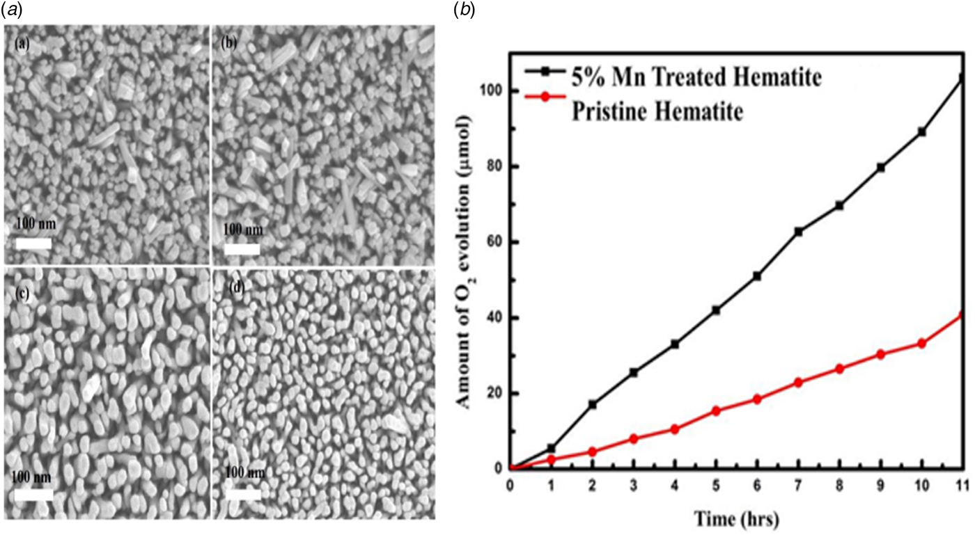
This demonstration is a significant step towards the development of an efficient, robust, low-cost and scalable photocatalytic device for water splitting at neutral pH to generate molecular hydrogen using only sunlight as the energy source. However, in many ways hematite is even more attractive. It can both act as a light-harvesting system and a generator of oxygen when illuminated as shown in Fig. 14b . Here an array of hematite nanorods were used (Fig. 14a ). Nano-structured hematite photoanode has been shown to have a slow first-order reaction at low light intensities, which changed to a third-order mechanism with high light when the surface hole density is sufficient to allow the oxidation of nearest-neighbour metal atoms (Le Formal et al. Reference Le Formal, Pastor, Tilley, Mesa, Pendlebury, Graetzel and Durrant2015). It was speculated that this first-order reaction involves a high-energy single-hole oxidation to OH and subsequent hydrogen peroxide formation as indicated in Fig. 1 while the third-order reaction could be an expression of a nucleophilic mechanism involving OH. The nucleophilic attack would be onto electrophilic terminal oxos of high valence Fe, possibly FeV via the formation of a peroxide intermediate. If this is correct, then it is another indirect example giving credence to the proposed mechanism for water splitting in PSII as shown in Fig. 6.
(a) Top view FESEM images of (a) pristine hematite after annealing at 550 °C, (b) 5% Mn-treated hematite after annealing at 550 °C (square cross-section), (c) pristine hematite after annealing at 750 °C and (d) 5% Mn-treated hematite after annealing at 750 °C (circular cross-section). (a) Time progression of light-induced oxygen evolution of 5% Mn-treated hematite and pristine hematite photoanodes at an applied potential of 1·23 V versus RHE. Both sets of measurements were conducted under AM 1·5 G 100 mW cm−2 illumination in 1 M NaOH electrolyte solution. (b) Time progression of light-induced oxygen evolution of 5% Mn-treated hematite and pristine hematite photoanodes at an applied potential of 1·23 V versus RHE. Both sets of measurements were conducted under AM 1·5 G 100 mW cm−2 illumination in 1 M NaOH electrolyte solution (taken from Gurudayal et al. Reference Gurudayal, Chaim, Kumar, Bassi, Seng, Barber and Wong2014).

6. Conclusions
Several lines of evidence suggest that in its oxidized state, the Mn4Ca2+O4 unit of the water-splitting catalytic centre of PSII is a stable chemical and robust entity able to assemble in the absence of protein. The additional oxo (O4) bridging between dangler Mn4 and the cubane is not present in the synthesized cluster suggesting it is more labile and for this reason may be involved in the chemistry of O–O bond formation. Nevertheless, the robustness of the Mn4Ca2+O4 unit suggests that it maintains its structure throughout the S-state cycle. If this is the case then it would rule out the Siegbahn mechanism, which involves the insertion of substrate water molecules into the cubane structure during the S2 to S3 transition and during the re-formation of cubane geometry associated with the S4 to S0 step of the S-state cycle. These large conformational changes in the catalytic centre will be energy demanding and thus likely to be incompatible with the low overpotential of water-splitting reaction of PSII. On the other hand, the nucleophilic mechanism presented in Fig. 6 occurs on the surface of the Mn4Ca2+O5 cluster and therefore involves no large energy demanding conformational changes of the cubane. Moreover, this pathway for O–O bond formation seems well established in synthesized complexes of Ru and Mn activated by the addition of chemical oxidants. The similarity of PSII with Fe–Ni CODH to split water to obtain reducing equivalents is also persuasive of a nucleophilic surface mechanism.
However, identifying the precise mechanism of O–O bond formation in PSII will be a significant challenge since the reactants, H2O, OH and O differ only by one or two protons. Although recent studies directed at this challenge using femtosecond XFEL diffraction are technically impressive the resolutions of the data obtained (approximately 2–2·5 Å), is far too low to distinguish between these reactants which will need to be at least 1 Å or less.
Acknowledgements
I wish to acknowledge all those who have worked on various aspects of PSII research in my laboratory at Imperial College London over the years and a special thank you goes to Dr James Murray who has helped me with some of the figures used in this article.