Introduction
To date, most epidemiological observations that have shown a link between prenatal life and later chronic disease have used birthweight as a marker of early life. A baby’s birthweight reflects its success in obtaining nutrients from its mother.Reference Harding 1 The source of these nutrients is not only the mother’s daily diet, but her nutritional stores and metabolism, which are the product of her lifetime nutrition.Reference Jackson 2 We have previously reviewed how the nutrition and growth of girls during infancy, childhood and at puberty influence chronic disease in the next generation.Reference Barker, Osmond, Kajantie and Eriksson 3 A baby’s nutrition also depends on the placenta’s ability to transport nutrients to it from the mother.Reference Harding 1 We here use new data from the Helsinki Birth Cohort to review three questions: (1) What measures of placental size can be used as markers of its function? (2) Is a large placenta always beneficial? (3) Are there sex differences in the maternal–placental phenotypes that predict chronic disease?
Measurements of placental size that reflect function
Low placental weight at birth has been shown to predict hypertension and coronary heart disease in later life.Reference Eriksson, Forsen, Toumilheto, Osmond and Barker 4 , Reference Forsén, Eriksson and Tuomilehto 5 Placental weight, however, does not distinguish the surface area of the placenta from its thickness. In order to increase the surface for nutrient and oxygen exchange, the placenta can increase the surface area of its villi, or expand its invasion across the surface of the uterine lining or invade the maternal spiral arteries more deeply. The long-term consequences may be different. Textbooks of midwifery written early in the last century describe the placental surface as being either ‘oval’ or ‘round’.Reference Anderson 6 , Reference Hinselmann 7 In order to measure the extent to which the surface was more oval than round, two so-called ‘diameters’ of the surface were routinely recorded in some hospitals in the past: a maximal diameter, the length, and a lesser one bisecting it at right angles, the width. These measurements are available for the Helsinki Birth Cohort that comprises 20,436 men and women born in the city during 1924–1944.Reference Barker, Thornburg, Osmond, Kajantie and Eriksson 8 The mean sizes of the length and breadth were 19.5 and 16.9 cm, and the sizes were highly correlated (correlation coefficient = 0.63). We used the length and breadth to estimate the surface area. How closely this area reflects the total surface area for maternal–fetal exchange through the gestational period is not known. A thin placenta with a large surface area could have a smaller villous exchange surface than a thick placenta with a small surface area. Nevertheless, it seems reasonable to suppose that the surface area measured in the Helsinki data correlates with the exchange area. We combined the two diameters and placental weight to estimate thickness.
Preeclampsia is associated with abnormal placentation as a result of impaired invasion of the maternal spiral arteries by the trophoblast at implantation.Reference Roberts and Cooper 9 We examined the effect of preeclampsia on placental area and thickness.Reference Kajantie, Barker, Osmond, Kajantie and Eriksson 10 In all, 6410 of the mothers of the younger part of the Helsinki birth cohort, born 1934–1944, had their blood pressures and the results of urinary protein tests recorded after 20 weeks of pregnancy; 284 of these pregnancies were complicated by preeclampsia. Compared to normotensive pregnancies, the placentas from these pregnancies had a reduced surface area, but the thickness was increased. We speculated that the increase in thickness in preeclampsia was compensatory for restricted expansion of the surface.
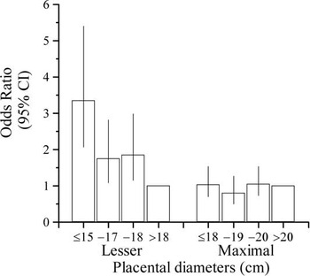
Placentas from pregnancies complicated by preeclampsia had a more oval surface than those from normotensive pregnancies because of a disproportionate reduction in the breadth. When the length and breadth were analyzed together, preeclampsia was not associated with the size of the length, but was strongly associated with a short breadth. This is shown in Figure 1. The relationship with the breadth was graded: the shorter the breadth, the greater the risk for, and severity of, preeclampsia. This association with short breadth depended on its absolute size rather on its size in relation to the size of the length. Processes that underlie the disease may therefore be closely linked to the absolute amount of placental tissue on the breadth of the placental surface. This link must be through a structure or function that it does not share with tissue along the length. We postulated that placental growth is polarized from the time of implantation, so that growth along the major axis, defined by the length, is qualitatively different from growth along the minor axis.Reference Kajantie, Barker, Osmond, Kajantie and Eriksson 10 One possibility is that the major axis is aligned to the rostro-caudal axis of the embryo, while the minor axis is more important for nutrient transfer to the fetus.
Odds ratios for preeclampsia according to the diameters of the placental surface.

Large placentas
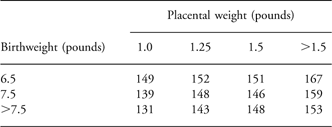
The ability of the placenta to transfer nutrients from the mother to the baby is reflected in its overall size.Reference Sibley 11 Babies whose birthweights are toward the lower end of the normal range generally have small placentas, and low placental weight is associated with hypertension in later life.Reference Eriksson, Forsen, Toumilheto, Osmond and Barker 4 However, a study of men and women born in a maternity hospital in Preston, UK, showed that high placental weight in relation to birthweight is also associated with later hypertension.Reference Barker, Bull, Osmond and Simmonds 12 Table 1 shows that whereas people who had both low placental weight and low birthweight had higher mean systolic blood pressures than those who had low placental weight but high birthweight, the highest mean systolic pressures occurred in people who had high placental weight but low birthweight. This observation has been replicated, and high placental weight in relation to birthweight has also been shown to predict coronary heart disease.Reference Eriksson, Forsen, Toumilheto, Osmond and Barker 4 , Reference Martyn, Barker and Osmond 13 An additional point made by Table 1 is that the ability of birthweight to predict blood pressure is enhanced if the size of the placenta is taken into account.Reference Barker, Bull, Osmond and Simmonds 12
Mean systolic blood pressure of men and women, aged 50 years, born at term

The associations with large placental size shown in Table 1 suggest that cardiovascular disease can be initiated through placental overgrowth as well as through restricted placental growth. Observations in sheep show that in response to undernutrition a fetus is able to extend the area of the placenta by expanding the individual cotyledons.Reference McCrabb, Egan and Hosking 14 , Reference McCrabb, Egan and Hosking 15 This increases the area available for nutrient and oxygen exchange, and results in a larger lamb than there would otherwise have been. This is profitable for the farmer, and manipulation of placental size by changing the pasture of pregnant ewes is standard practice in sheep farming.
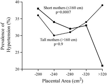
Placental overgrowth in response to undernutrition in mid-gestation can only occur, however, if the ewe was well nourished before conception.Reference McCrabb, Egan and Hosking 15 There is evidence of a similar phenomenon in humans. 2003 members of the Helsinki Birth Cohort were randomly selected to attend a clinic; 644 were being treated for hypertension.Reference Barker, Thornburg, Osmond, Kajantie and Eriksson 8 We found that the effects of placental size on hypertension depended on the mother’s socio-economic status, a marker of her current nutrition, and on her height, which reflected her nutrition and metabolism in childhood and adolescence.Reference Barker, Thornburg, Osmond, Kajantie and Eriksson 8 , Reference Tanner 16 In people whose mothers were of low socio-economic status or below average height, hypertension was associated with low placental weight and area. In people whose mothers were likely to have been the best nourished, being tall and middle class, hypertension was predicted by large placental size in relation to birthweight. Figure 2 shows the trends in the prevalence of hypertension in people divided according to whether their mother’s height was above or below the median of 160 cm. In people whose mothers were short, the prevalence of hypertension fell progressively with increasing placental area. In people with tall mothers, the association was U-shaped, the prevalence initially falling as area increased but thereafter rising. Therefore, while the effect of small placental area on hypertension was similar in the two maternal height groups, a large area was associated with almost double the prevalence of hypertension in the offspring of tall mothers when compared with the offspring of short mothers.
Prevalence of hypertension according to placental area in the offspring of short and tall mothers.

These different trends in hypertension with placental area in people born to short or tall mothers suggest a link between hypertension and the lifetime nutrition of the mother. Short maternal stature is a product of poor fetal or childhood nutrition, or recurrent exposure to infections, though there are also genetic influences.Reference Tanner 16 Protein metabolism is established in early life and is related to visceral mass. Short women have less visceral mass than tall women and have reduced rates of protein synthesis in pregnancy.Reference Jackson 2 , Reference Duggleby and Jackson 17 In short mothers, an increased placental surface for protein transport might offset reduced rates of synthesis.
Among people with tall mothers, there was no overall effect of placental area on hypertension (Fig. 2). When, however, these offspring were subdivided by the family’s socio-economic status, two opposing trends were revealed. In lower social class families, hypertension was associated with small placental area and low weight. In middle class families, hypertension was associated with large placental area and high placental weight in relation to birthweight. Tall mothers in middle class families are likely to have been the best nourished before conception. During pregnancy, however, they would have been subject to the food scarcities that occurred in Finland around the time of the Second World War.Reference Pesonen, Raikkonen and Heinonen 18 We speculate that, similar to sheep, their fetuses responded to this by expansion of the placental surface. Compensatory placental growth may be beneficial in some circumstances, but if the compensation is inadequate and the fetus continues to be undernourished, the need to share its nutrients with an enlarged placenta may become an added metabolic burden. A long-term cost of this added burden is hypertension, possibly as a result of impaired development of low priority organs like the kidney.Reference Barker, Bagby and Hanson 19
Among the offspring of tall, middle class mothers, hypertension was associated with above average birthweight. This suggests that, as in sheep, placental enlargement in response to fetoplacental malnutrition leads to increased body size at birth. Epidemiological studies that include large numbers of tall, middle class mothers may therefore fail to show the well-known association between low birthweight and later hypertension. A general point made by Figure 2 is that, just as the ability of birthweight to predict long-term outcomes is enhanced if the size of the placenta is taken into account, the ability of placental size to predict outcomes is enhanced if maternal body size is known.
Of the two diameters of the surface, breadth was more closely associated with hypertension.Reference Barker, Thornburg, Osmond, Kajantie and Eriksson 8 The interaction between the effects of mother’s height and placental area (Fig. 2) was reflected in a similar interaction between the effects of mother’s height and the breadth of the surface, but there was no interaction between mother’s height and the length. These findings are consistent with the idea that placental tissue along the breadth of the surface is more important for nutrient transfer to the fetus than tissue along the length, and expansion of the placental surface along its minor axis is one way in which a fetus may compensate for malnutrition.
The findings on hypertension show that the effects of placental size on the fetus are conditioned by the mother’s height. Studies of lung cancer show that they are also conditioned by the mother’s body mass index in pregnancy height/weight2, a marker for her glucose and lipid metabolism.Reference Eriksson, Thornburg, Osmond, Kajantie and Barker 20 , Reference Barker, Thornburg, Osmond, Kajantie and Eriksson 21
Sex differences in the placenta
Boys grow faster than girls from an early stage of gestation, even from before implantation, and this makes them more vulnerable if their nutrition is compromised.Reference Tanner 16 , Reference Pedersen 22 We have examined the differences in fetal and placental size at birth among boys and girls in the Helsinki Birth Cohort.Reference Forsén, Eriksson, Tuomilehto, Osmond and Barker 23 We found that the small differences in average body measurements concealed large differences in body proportions. At any placental weight, boys tended to be taller than girls; and boys’ placentas were smaller than girls’ placentas when related to the weight of the baby.Reference Forsén, Eriksson, Tuomilehto, Osmond and Barker 23 This suggests that boys’ placentas are more efficient but may have less reserve capacity, which therefore increases their vulnerability to undernutrition. Boys tend to have larger head circumferences at birth but are thinner than girls.Reference Eriksson, Kajantie, Osmond, Thornburg and Barker 24 This suggests that they may more readily trade off visceral development to protect brain growth. The growth of every human fetus is constrained by the limited capacity of the mother and placenta to deliver nutrients to it.Reference Ounsted, Scott and Ounsted 25 The male fetus, by growing more rapidly and investing in brain growth rather than placental growth, is adopting a more dangerous strategy that puts it at greater risk of becoming undernourished.
We found that the relationship between placental diameters and later hypertension was different in the two sexes.Reference Eriksson, Kajantie, Osmond, Thornburg and Barker 24 In women, hypertension was associated with short breadth and small placental area. In men, it was associated with large breadth in relation to birthweight. This suggests that compensatory expansion of the placental breadth may occur more readily in boys. Direct evidence for this comes from findings in the Dutch famine (van Abeleen et al., personal communication). The dangerous growth strategy of boys may therefore be compounded by the costs of compensatory placental enlargement in mid-late gestation.
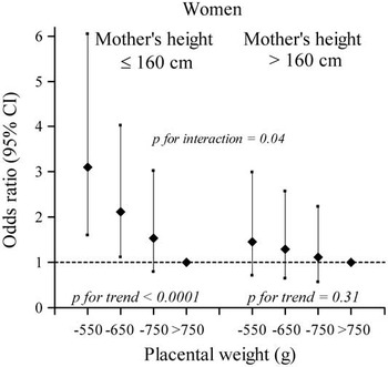
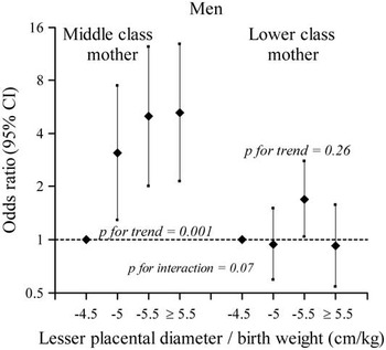
Among women the trends in hypertension with small placental area and low placental weight were stronger in those whose mothers were short (Fig. 3).Reference Eriksson, Kajantie, Osmond, Thornburg and Barker 24 The trends did not differ with the mother’s social class. In contrast, among men the trend in hypertension with a greater breadth in relation to birthweight was not influenced by the mother’s height but differed according to the mother’s social class. Figure 4 shows that the trend was confined to men whose mothers were middle class (Fig. 4). An interpretation of these findings is that during development in the womb, girls respond more to their mother’s lifetime nutrition and metabolism, indicated by her height, whereas boys respond more to their mother’s current diet, indicated by her social class. The greater effect of the Dutch famine on the numbers of boys supports this,Reference Ravelli, van der Meulen, Osmond, Barker and Bleker 26 as do the greater effects of experimental alterations in maternal diet on male animals.Reference Ozaki, Nishina, Hanson and Poston 27 – Reference Grigore, Ojeda and Alexander 29 While boys’ responsiveness to their mothers’ current diet enables them to capitalize on improving food supply, and promotes their agenda of rapid growth, it makes them vulnerable to food shortages.
Odds ratios for hypertension in women according to mother’s height and placental weight.

Odds ratios for hypertension in men according to the mother’s socio-economic status and the ratio of the lesser placental diameter to birthweight.

Conclusions
Birthweight is a crude marker of fetal nutrition. The same weight may result from a variety of different nutritional circumstances. A priori, it was likely that epidemiological associations between birthweight and later disease would not always be consistent. To some extent, these inconsistencies can be reduced by using additional measures of body size at birth, including length and head circumference. This study suggests that the size and shape of the placental surface is a new epidemiological marker. There is already a body of knowledge about the maternal determinants of normal placental structure.Reference Coall, Charles and Salafia 30 The biological functions that underlie the associations between different maternal–placental phenotypes and later disease now need to be explored.
This study leads to four conclusions about the maternal–placental programming of chronic disease: (1) Growth of the placental surface is polarized from the time of implantation, so that growth along the major axis is qualitatively different from growth along the minor axis. (2) The human fetus may attempt to compensate for undernutriton by expansion of the placental surface along its minor axis. (3) The effects of placental size on long-term health are conditioned by the mother’s nutritional state, as indicated by her socio-economic status and height. (4) The maternal–placental programming of chronic disease differs in boys and girls.
Acknowledgements
This study was supported by the British Heart Foundation, the Academy of Finland, the Paivikki and Sakari Sohlberg Foundation, the Finnish Diabetes Research Foundation, the Finnish Foundation for Cardiovascular Research, the Finnish Medical Society Duodecim, Yrjo Jahnsson Foundation, Finska Lakaresällskapet.