

Introduction
Graftonite, ideally Fe3(PO4)2, and beusite, ideally Mn3(PO4)2, form a continuous solid solution: (Fe2+,Mn2+,Ca)3(PO4)2. In addition to triphylite and sarcopside, minerals of the graftonite–beusite series are major accessory, primary phosphate minerals occurring as intergrowths in characteristic nodules in P-bearing pegmatites, especially of the beryl–columbite–phosphate subtype of the rare-element class (Penfield, Reference Penfield1900; Brooks and Shipway, Reference Brooks and Shipway1960; Fransolet, Reference Fransolet1977; Keller et al., Reference Keller, Fontan, Velasco Roldan, Melgarejo and Draper1977; Černý et al., Reference Černy, Selway, Ercit, Breaks, Anderson and Anderson1998; Smeds et al., Reference Smeds, Uher, Černý, Wise, Gustafsson and Penner1998; Guastoni et al., Reference Guastoni, Nestola, Mazzoleni and Vignola2007; Škoda et al., Reference Škoda, Staněk and Čopjaková2007; Łodziński and Sitarz, Reference Łodziński and Sitarz2009; Vignola et al., Reference Vignola, Diella, Oppizzi, Tiepolo and Weiss2008, Reference Vignola, Diella, Ferrari and Fransolet2011a,Reference Vignola, Fransolet, Guastoni and Appianib; Tait et al., Reference Tait, Hawthorne and Wise2013; Włodek et al., Reference Włodek, Grochowina, Gołębiowska and Pieczka2015). Graftonite has also been found in amphibolite-facies metamorphosed iron formations (Stalder and Rozendaal, Reference Stalder and Rozendaal2002) and as a constituent of iron meteorites (Olsen and Fredriksson, Reference Olsen and Fredriksson1966; Bild, Reference Bild1974; Chen and Xie, Reference Chen and Xie1996; Floss, Reference Floss1999; Steele et al. Reference Steele, Olsen, Pluth and Davis1991, Olsen et al. Reference Olsen, Kracher, Davis, Steele, Hutcheon and Bunch1999; Sharygin et al., Reference Sharygin, Karmanov and Podgornykh2016).
Calcium has long been considered as a minor constituent substituting for Fe2+ and Mn2+ in the graftonite–beusite series. However, structural studies (Wise et al., Reference Wise, Hawthorne and Černý1990; Tait et al., Reference Tait, Hawthorne and Wise2013) have shown that in the graftonite–beusite structure there is complete order of Ca at the M(1) site, followed firstly by Mn2+ and then by Fe2+. Remaining Mn and Fe are disordered over the M(2) and M(3) sites in accord with the scheme M(3) > M(2). These findings have enabled a new classification of the graftonite-group minerals (Hawthorne and Pieczka, Reference Hawthorne and Pieczka2018), earlier considered as the ‘graftonite–beusite series’. The classification was approved by the International Mineralogical Association (IMA) Commission on New Minerals, Nomenclature and Classification (CNMNC), as the IMA-CNMNC nomenclature voting proposal 17-H.
This paper presents new mineral descriptions for graftonite-(Ca) and graftonite-(Mn), two of three new mineral species, distinct in the new classification and approved in July 2017 by the IMA-CNMNC (IMA 2017-048 and 2017-050, respectively). Type samples of the both minerals are from two phosphate-bearing pegmatites in Poland; from Lutomia and Michałkowa villages in the gneissic Góry Sowie block. The names graftonite-(Mn) and graftonite-(Ca) are because the minerals are the M(1)Mn- and M(1)Ca analogues of graftonite, M(1)FeM(2),M(3)Fe2(PO4)2. The holotype and co-type samples are deposited in the collection of the Mineralogical Museum of University of Wrocław (University of Wrocław, Faculty of Earth Science and Environmental Management, Institute of Geological Sciences, Mineralogical Museum, 50-205 Wrocław, Cybulskiego 30, Poland), with catalogue numbers MMWr IV7927 [graftonite-(Mn) holotype], MMWr IV7928 [graftonite-(Mn) co-type], and MMWr IV7674 [graftonite-(Ca) and maneckiite holotype] (Pieczka et al., Reference Pieczka, Hawthorne, Gołębiowska, Włodek and Grochowina2017a). The third new mineral species in the graftonite group, beusite-(Ca), with the holotype coming from Canada, is described in a companion paper (Hawthorne et al., Reference Hawthorne, Wise, Černý, Abdu, Ball, Pieczka and Włodek2018). Neither of these minerals was a valid unnamed mineral (Smith and Nickel, Reference Smith and Nickel2007; http://pubsites.uws.edu.au/ima-cnmnc/). Graftonite-(Mn) and graftonite-(Ca) are isostructural with graftonite and beusite, with Mn or Ca completely ordered at the M(1) site. In the classification of Strunz (Strunz and Nickel, Reference Strunz and Nickel2001), it is a class 8.A Phosphate without additional anions, without H2O. In the classification of Dana (Gaines et al., Reference Gaines, Skinner, Foord, Mason and Rosenwieg1997), it is a class 38 Anhydrous normal phosphate isostructural with graftonite (38.3.3.1) and beusite (38.3.3.2).
Geological setting
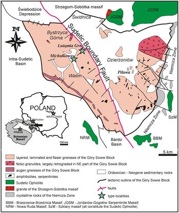
Graftonite-(Mn) and graftonite-(Ca) were collected in two phosphate-bearing pegmatites exposed in the Góry Sowie gneissic block, one of the main tectono-stratigraphic units (~650 km2) of the Sudetes, southwest Poland (northeast part of the Bohemian massif) (Fig. 1). The Góry Sowie gneissic block consists mainly of gneisses and migmatites with minor amphibolites, and is considered to be a fragment of the lower crust (for details see Mazur et al., Reference Mazur, Aleksandrowski, Kryza and Oberc-Dziedzic2006). The unit is a product of multistage evolution that culminated ~385–370 Ma in amphibolite-facies metamorphism and migmatization at temperatures of 775–910°C and pressures of 6.5–8.5 kbar, and was followed by rapid uplift and exhumation (Brueckner et al., Reference Brueckner, Blusztajn and Bakun-Czubarow1996; O'Brien et al., Reference O'Brien, Kröner, Jaeckel, Hegner, Żelaźniewicz and Kryza1997; Kryza and Fanning, Reference Kryza and Fanning2007). Anatectic melts, generated by partial melting of the metasedimentary–metavolcanic sequence, were injected as pegmatites and granite-like bodies during decompression, forming small concordant segregations and N-trending discordant dykes in migmatized gneisses and amphibolites (Kryza, Reference Kryza1981; van Breemen et al., Reference van Breemen, Bowes, Aftalion and Żelaźniewicz1988; Żelaźniewicz, Reference Żelaźniewicz1990; Bröcker et al., Reference Bröcker, Żelaźniewicz and Enders1998; Timmermann et al., Reference Timmermann, Parrish, Noble and Kryza2000; Aftalion and Bowes, Reference Aftalion and Bowes2002; Gordon et al., Reference Gordon, Schneider, Manecki and Holm2005). The latter sometimes form large systems of barren to rare-element pegmatites of different affiliations [NYF (niobium-yttrium-fluorine), LCT (lithium-caesium-tantalum) to mixed NYF + LCT], e.g. the Julianna system at Piława Górna (Pieczka et al., Reference Pieczka, Szuszkiewicz, Szełęg, Nejbert, Łodziński, Ilnicki, Turniak, Banach, Hołub, Michałowski and Różniak2013, Reference Pieczka, Szuszkiewicz, Szełęg, Ilnicki, Nejbert and Turniak2014, Reference Pieczka, Hawthorne, Cooper, Szełęg, Szuszkiewicz, Turniak, Nejbert and Ilnicki2015a,Reference Pieczka, Szuszkiewicz, Szełęg, Janeczek and Nejbertb, Reference Pieczka, Szełęg, Szuszkiewicz, Gołębiowska, Zelek, Ilnicki, Nejbert and Turniak2016, Reference Pieczka, Hawthorne, Ma, Rossman, Szełęg, Szuszkiewicz, Turniak, Nejbert, Ilnicki, Buffat and Rutkowski2017b; Szuszkiewicz et al., Reference Szuszkiewicz, Szełęg, Pieczka, Ilnicki, Nejbert, Turniak, Banach, Łodziński, Różniak and Michałowski2013, Reference Szuszkiewicz, Pieczka, Szełęg, Turniak, Ilnicki and Nejbert2016; Szełęg et al., Reference Szełęg, Zuzens, Hawthorne, Pieczka, Szuszkiewicz, Turniak, Nejbert, Ilnicki, Friis, Makovicky, Weller and Lemée-Cailleau2017).
Geological map of the Góry Sowie Block (after Szuszkiewicz et al., Reference Szuszkiewicz, Szełęg, Pieczka, Ilnicki, Nejbert, Turniak, Banach, Łodziński, Różniak and Michałowski2013) with localities of the Lutomia and Michałkowa pegmatites marked with green stars.
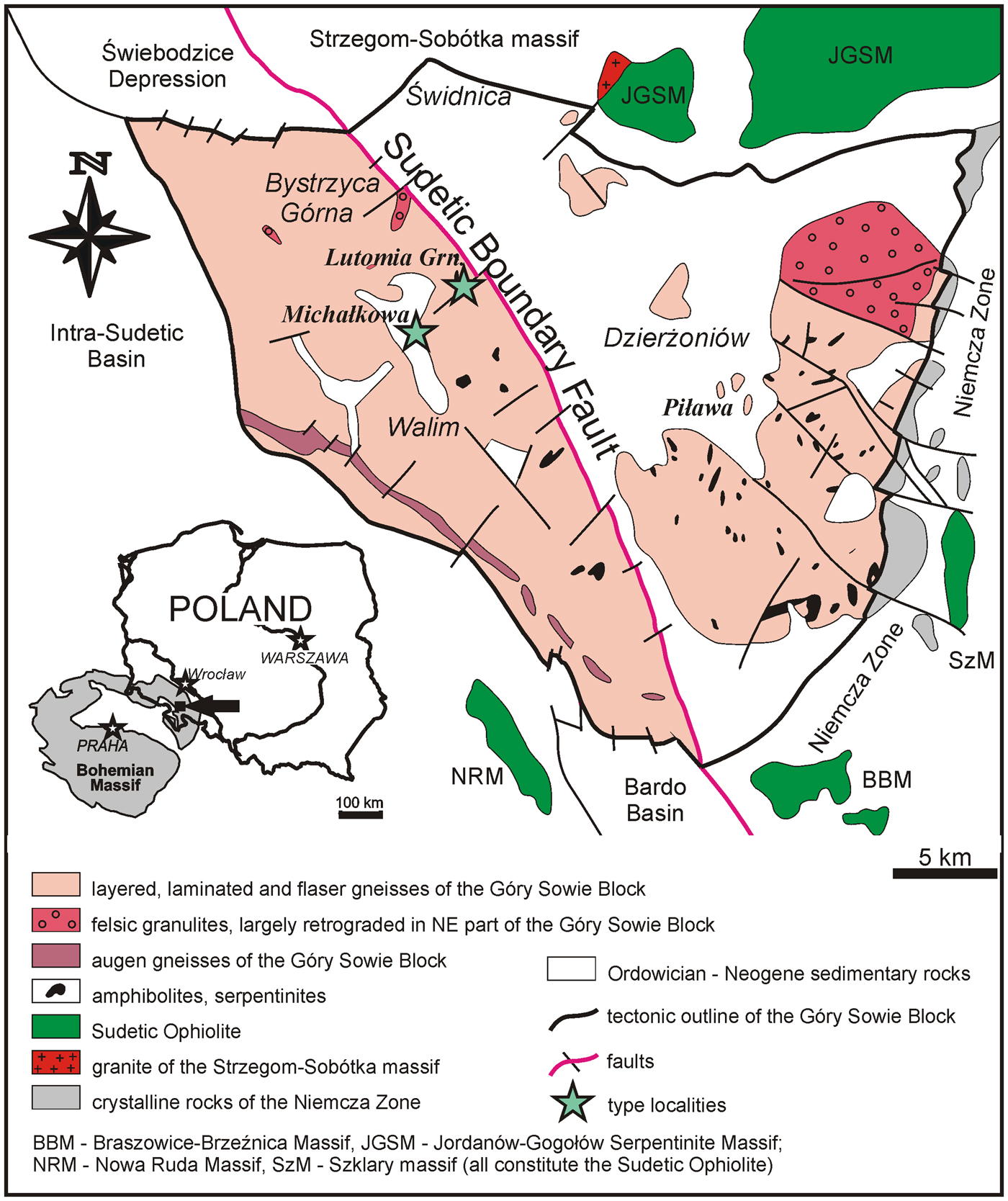
Graftonite-(Mn) was found in a pegmatite cropping out in a small abandoned quarry at Lutomia (50°45′N, 16°29′E), a village ~5 km southeast of Świdnica and ~60 km southwest of Wrocław. The pegmatite forms a relatively large, discordant vertical vein, ~20 m long and up to 1 m thick, hosted by migmatite and amphibolite, with local swellings up to 2 m across, and a few smaller offshoots. The pegmatite is of the beryl–columbite–phosphate subtype of rare-element pegmatites sensu Černý and Ercit (Reference Černý and Ercit2005) or phosphate subtype of rare-element pegmatites sensu Novák (Reference Novák2005). It shows, especially in the swellings, well-developed although not continuous zonation (wall zone – graphic zone – blocky feldspars zone – massive albite). The border and wall zones are composed of fine- to medium-grained plagioclase + quartz + ‘biotite’ (altered) ± K-feldspars ± apatite group ± almandine graphic zone of intergrowths of plagioclase + K-feldspar + quartz ± ‘biotite’/muscovite ± tourmaline ± phosphates (rare); blocky feldspar zone of coarse-grained K-feldspar + plagioclase + quartz ± ‘biotite’/muscovite + tourmaline + phosphates (rare nodules); and massive albite with rare phosphate nodules disseminated locally. No quartz core is observed. Beside the aforementioned rock-forming minerals, phosphate nodules, reaching to 5 cm × 3 cm × 3 cm in size, contain a large number of phosphate minerals, e.g. monazite-(Ce), xenotime-(Y), graftonite-(Mn), graftonite-(Ca), beusite-(Ca), sarcopside, triphylite partly oxidized topotactically to ferrisicklerite and heterosite, wolfeite, triploidite, stanĕkite, hagendorfite, ferrohagendorfite, alluaudite, fluorapatite and hydroxylapatite, whitlockite, kryzhanovskite, phosphoferrite, ludlamite, vivianite, fairfieldite, hureaulite, earlshannonite, whitmoreite, strunzite, ferrostrunzite, beraunite, dufrénite, landesite, jahnsite-(CaMnFe), -(CaMnMn) and -(MnMnMn), and occasionally also malhmoodite and zigrasite (Pieczka et al., Reference Pieczka, Gołębiowska and Skowroński2003; Włodek et al., Reference Włodek, Grochowina, Gołębiowska and Pieczka2015; Włodek and Pieczka, Reference Włodek and Pieczka2017). The following minerals were found as tiny inclusions in the phosphates or in the rock-forming minerals: fergusonite-(Y), gadolinite-(Y), allanite-(Ce), ilmenite, titanite, sillimanite (‘fibrolite’), uraninite, pyrite, arsenopyrite, löllingite, chalcopyrite, Cd-bearing wurtzite or sphalerite, chalcocite or covellite, cuprite, native copper, goethite and unidentified Mn oxides (Pieczka et al., Reference Pieczka, Włodek, Gołębiowska, Szełęg, Szuszkiewicz, Ilnicki, Nejbert and Turniak2015c; Włodek et al., Reference Włodek, Grochowina, Gołębiowska and Pieczka2015).
Graftonite-(Ca) was collected at Michałkowa (50°45′N, 16°27′E), in the middle part of the Góry Sowie Block, ~70 km southwest of Wrocław, on a small pegmatite dump, a relic of excavation in the ninteenth century as a source of quartz and feldspar. During this work, Websky (Reference Websky1868) found and described sarcopside (type locality), associated with hureaulite, vivianite and an apatite-group mineral. The dump is located on the left site of the Dział Michałowski Range, on the right side of the Młynówka stream, ~100 m from the Zagórze Śląskie – Lubachów – Michałkowa – Pieszyce road. The original pegmatite vein, ~10–12 m long and 2–4 m thick, with visible zoning (wall zone – graphic zone – blocky feldspar zone), was composed of albite, microcline, quartz, muscovite, ‘biotite’, schorl evolving to foitite, almandine, sillimanite, andalusite and phosphate minerals in nodules reaching a few centimetres in diameter (Łodziński and Sitarz, Reference Łodziński and Sitarz2009). Graftonite-(Ca) was found in an outer zone of a small phosphate nodule (~2 cm in diameter) collected on the dump. The nodule was composed mainly of lamellar intergrowths of the primary magmatic phosphates: sarcopside and graftonite-(Mn), with minor triphylite oxidized topotactically to ferrisicklerite and heterosite. In this zone, the magmatic phosphates underwent intensive Na and Ca metasomatism and were replaced by wolfeite, alluaudite, maneckiite, fluorapatite and hydroxylapatite, in places forming fine-grained mosaics (Pieczka et al., Reference Pieczka, Hawthorne, Gołębiowska, Włodek and Grochowina2017a). To date, ~50 phosphate minerals have been identified in the pegmatite (Grochowina, Reference Grochowina2016), including rare species such as maneckiite (type locality), wicksite, wyllieite, ferrowyllieite, qingheiite-(Fe), maghagendorfite, hagendorfite, ferrohagendorfite, johnsomervilleite, lazulite, gormanite, childrenite, souzalite and arrojadite-(KNa). Łodziński and Sitarz (Reference Łodziński and Sitarz2009) also mentioned the presence of qingheiite, rosemaryite and simferite in nodules of the pegmatite, but these phosphates have not yet been confirmed by chemical analysis or X-ray diffraction.
Physical and optical properties
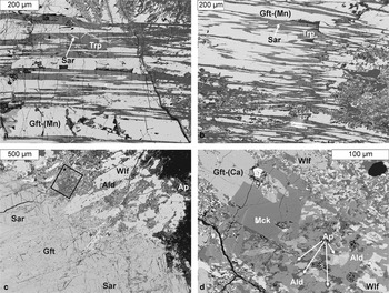
Graftonite-(Mn) occurs in the Lutomia pegmatite as lamellar intergrowths with triphylite or products of its topotactic oxidation: ferrisicklerite and heterosite, in the form of pinkish-brown lamellae up to 0.5 mm wide. Along fissures, the mineral and associated triphylite were Na- and Ca-metasomatized, commonly forming symplectic intergrowths of alluaudite-group minerals with wolfeite and stanĕkite, and sometimes with whitlockite. Sarcopside, up to 50 µm thick and several hundred μm long, as one or two independent sets of diagonally arranged and criss-crossing needles, occurs only locally as inclusions in triphylite and products of its topotactic oxidation. No direct intergrowths of graftonite-(Mn) with sarcopside were observed (Włodek et al., Reference Włodek, Grochowina, Gołębiowska and Pieczka2015). Towards the border of the nodule, the amount of graftonite-(Mn)–triphylite intergrowths generally decreases, and graftonite-(Mn) may occur in massive accumulations (Fig. 2a,b).
Back-scatter electron images. (a, b) Lamellar intergrowths of graftonite-(Mn) with triphylite (ferrisicklerite/heterosite) in the Lutomia pegmatite; (c) a fragment of a phosphate nodule from Michałkowa with graftonite-(Ca) (framed, d) intergrown with maneckiite in a mosaic of metasomatic alluaudite, wolfeite and hydroxylapaite. Abbreviations: Ald = alluaudite-group minerals, Ap = hydroxylapatite, Gft-(Ca) = graftonite-(Ca), Gft-(Mn) = graftonite-(Mn), Mck = maneckiite, Sar = sarcopside, Trp = triphylite and Wlf = wolfeite.
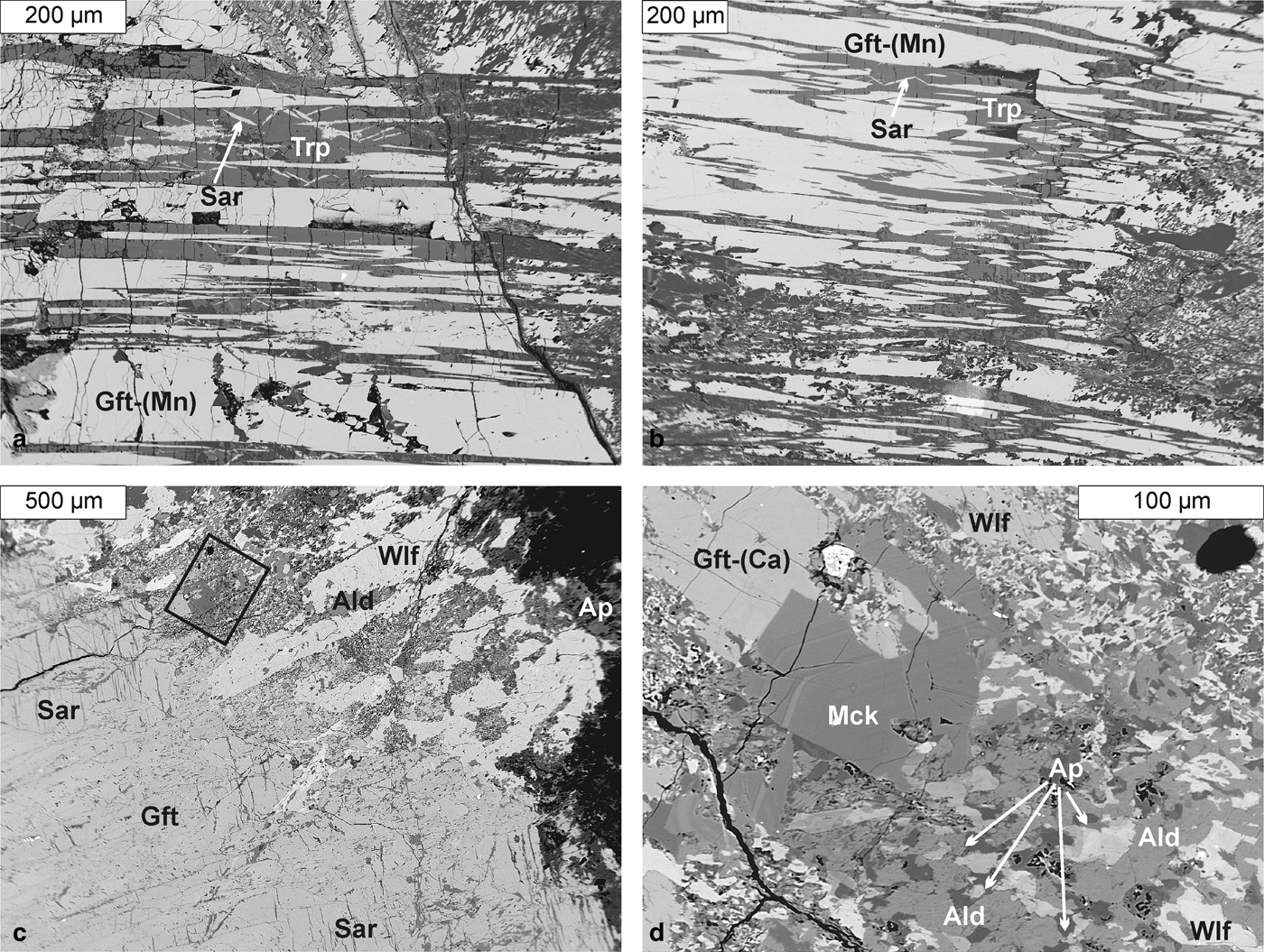
In the Michałkowa pegmatite, graftonite-(Mn) commonly evolves towards graftonite-(Ca). Graftonite with very high Ca content, close in composition to end-member graftonite-(Ca) is less common. One such crystal, ~70 µm × 100 µm, was found as an intergrowth with maneckiite, close to the border of the metasomatic phosphates with the primary magmatic phosphates of the unaltered interior of the nodule (Pieczka et al., Reference Pieczka, Hawthorne, Cooper, Szełęg, Szuszkiewicz, Turniak, Nejbert and Ilnicki2015a), overgrown by a mosaic of wolfeite, alluaudite and hydroxylapatite (Fig. 2c,d).
Graftonite-(Mn) is pinkish brown, whereas graftonite-(Ca) shows rather more brownish colouration. Both minerals have a colourless streak and vitreous lustre and are transparent. Neither of them is fluorescent. Both minerals have good cleavage along (010), irregular fracture and no observed parting. They are brittle and have a Mohs hardness of ~5 by analogy with graftonite. The densities, calculated on the basis of molar weights and unit-cell volumes, are 3.793 g/cm3 for the refined crystal of graftonite-(Mn) and 3.592 g/cm3 for graftonite-(Ca). Both minerals are non-pleochroic, colourless in all orientations, biaxial (+), with mean refractive indices α = 1.710(2) and 1.690(2), β = 1.713(2) and 1.692(2), and γ = 1.725(2) and 1.710(5), respectively, and 2Vmeas. = 54(2)° and 40.1(6)° and 2Vcalc. = 53.4° and 37 ° (wavelength 589 nm). The optical orientations are: X || b, Y ˄ a = 44.2° (in β obtuse), Z ˄ c = 35.0° (in β acute) for graftonite-(Mn), and X || b, Y ˄ a = 41.4° in β acute and Z ˄ c = 32.1° in β acute for graftonite-(Ca).
Chemical composition
Electron-microprobe analysis was undertaken at the Inter-Institute Analytical Complex for Minerals and Synthetic Substances at the University of Warsaw, using a Cameca SX 100 electron microprobe operating in wavelength-dispersive mode under the following conditions: accelerating voltage of 15 kV, beam current of 20 nA, beam diameter of 2 µm, peak count-time of 20 s, background time of 10 s. Standards (element, analytical lines, diffracting crystals and mean detection limits in wt.%) were as follows: YP5O14 (PKα, PET, 0.02); diopside (MgKα, TAP, 0.01) and (CaKα, PET, 0.02); rhodonite (MnKα, LIF, 0.06); hematite (FeKα, LIF, 0.06); and sphalerite (ZnKα, LIF, 0.06). The raw data were reduced with the ‘PAP’ routine of Pouchou and Pichoir (Reference Pouchou, Pichoir and Armstrong1985). The formulae were normalized to 8 O atoms per formula unit (apfu).
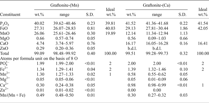
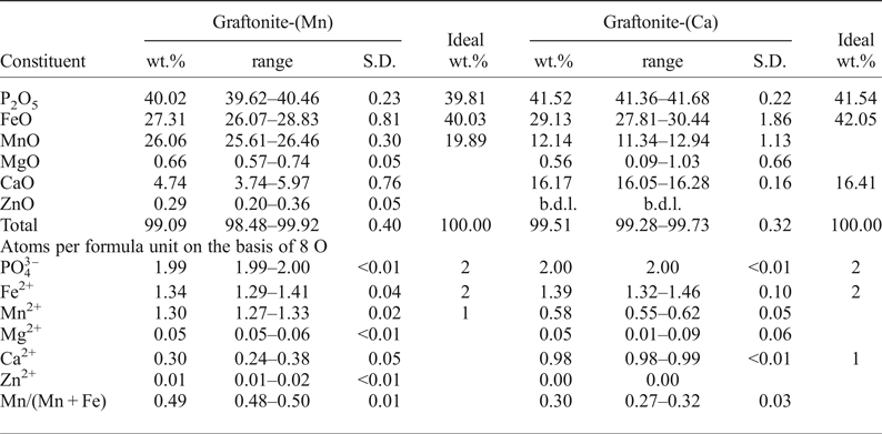
Chemical compositions are presented in Table 1. Average compositions of the holotype crystals correspond to following formulae: (Fe1.34Mn1.30Ca0.30Mg0.06Zn0.01)Σ3.00(PO4)1.99 and (Fe1.38Ca0.98Mn0.58Mg0.05)Σ2.99(PO4)2.00. Assuming complete order of Ca at the M(1) site and omitting minor inaccuracies in the contents of cations and PO4 anion, the optimized formulae are M(1)(Mn0.70Ca0.30)M(2),M(3)(Fe1.34Mn0.60Mg0.06Zn0.01)Σ3(PO4)2 and M(1)(Ca0.98Mn0.02)M(2),M(3)(Fe1.38Mn0.56Mg0.05)Σ3(PO4)2 for the holotype samples. The compositions indicate that the M(1) site should be dominated by Mn2+ in graftonite-(Mn) and by Ca2+ in graftonite-(Ca). In both minerals, the M(2) + M(3) sites are dominated by Fe2+, with a probable tendency to partial order of Mn2+ at the M(3) site and dominance of Fe2+ at the M(2) site.
Chemical compositions of graftonite-(Mn) and graftonite-(Ca)*.
*Number of analytical spots = 20 for graftonite-(Mn) and 2 for graftonite-(Ca).
b.d.l. = below detection limit; S.D. = standard deviation.
Raman spectroscopy
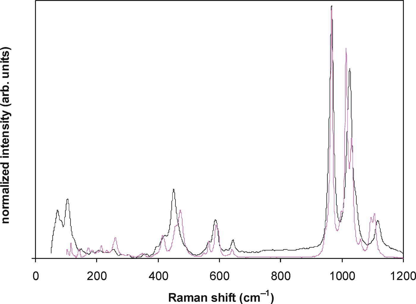
The Raman spectrum of graftonite-(Mn) was collected in back-scattered mode at the Faculty of Materials Science and Ceramics, AGH UST, Kraków, Poland, with a Horiba Labram HR spectrometer integrated with an Olympus BX 40 confocal microscope, equipped with a 532 nm Nd:YAG laser, (10 mW), grating 1800 gr/mm. The spectrum was recorded in the range 4000–50 cm–1 at a magnification of 100×, with resolution of 0.5 cm–1, an acquisition time of 600 s, and accumulation of 2 scans with an estimated spot size ~1 µm. The spectrum of graftonite-(Ca) was collected in back-scattered mode at the Department of Geological Sciences, University of Manitoba, Winnipeg, Canada, with a Horiba Jobin Yvon-LabRAM ARAMIS integrated confocal micro-Raman system equipped with a 460 mm focal length spectrograph and a multichannel air-cooled (–70°C) CCD detector. A magnification of 100× was used with an estimated spot size of 1 µm, a 1800 gr/mm grating, an excitation radiation of 532 nm, and a laser power between 5 and 12.5 mW. Calibration in both laboratories was done using the 520.7 cm–1 line of Si. The spectra are presented in Fig. 3.
Raman spectra of graftonite-(Mn) (black) and graftonite-(Ca) (violet).
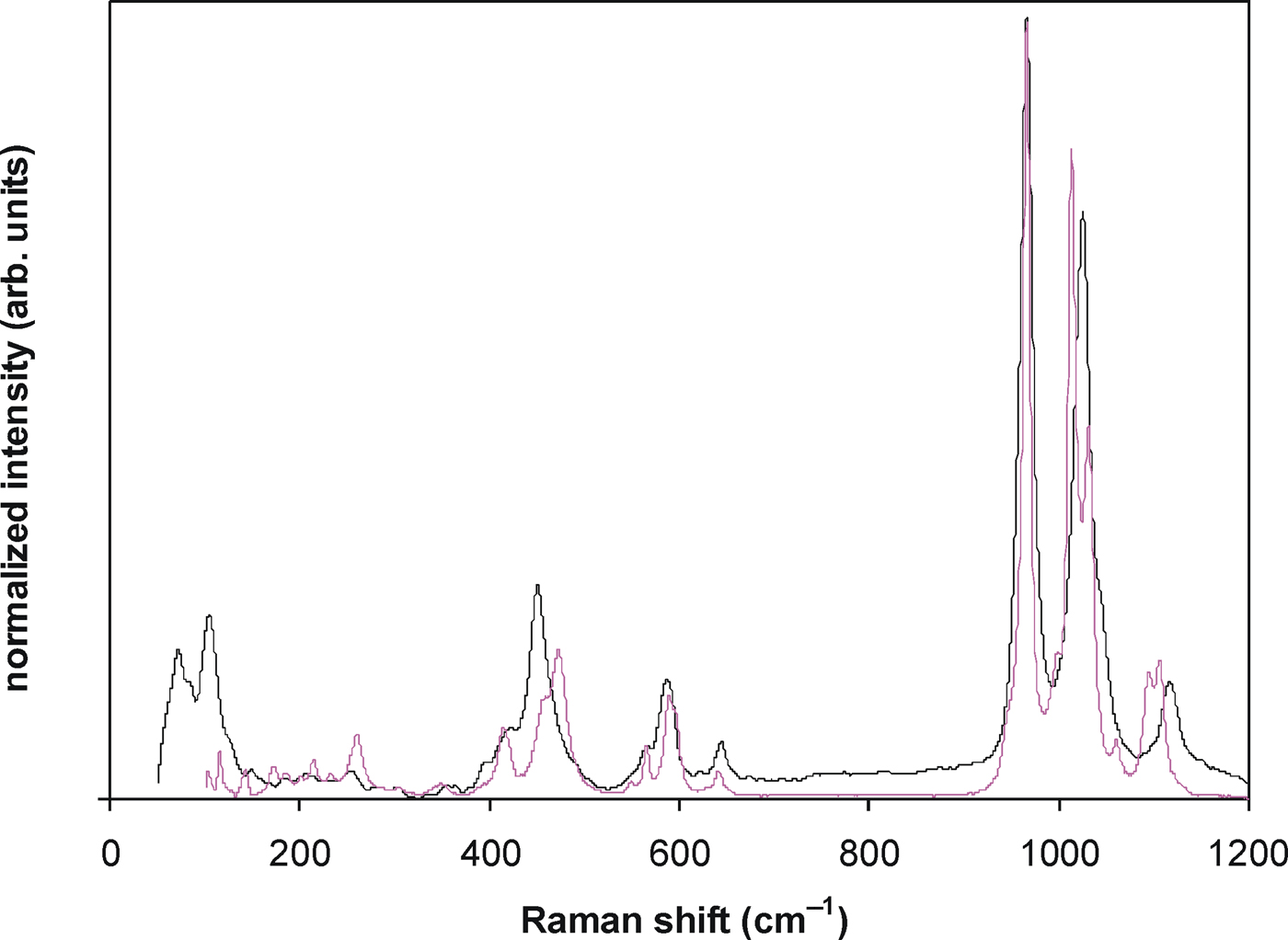
The peaks at ~966 (strong), 1025 (medium) and 1117 (weak) cm–1 in the Raman spectrum of graftonite-(Mn), and ~968, 1013 and 1032 cm−1 (strong) and 1098 and 1106 cm–1 (medium) in the spectrum of graftonite-(Ca) may be assigned to stretching vibrations of the PO4 groups. The peaks at 587, 566, 451 and 423 cm–1 [graftonite-(Mn)] and at 590, 472 and 458 cm–1 [graftonite-(Ca)] are due to the bending vibrations of PO4. The weaker peaks below ~400 cm–1 are due to deformations of the Ca and Mn polyhedra.
Mössbauer spectroscopy
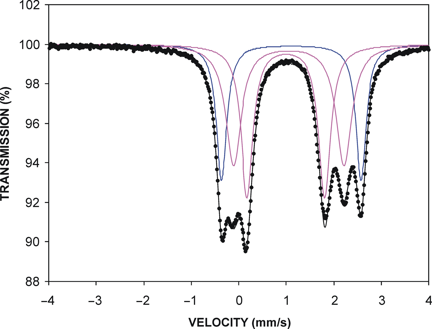
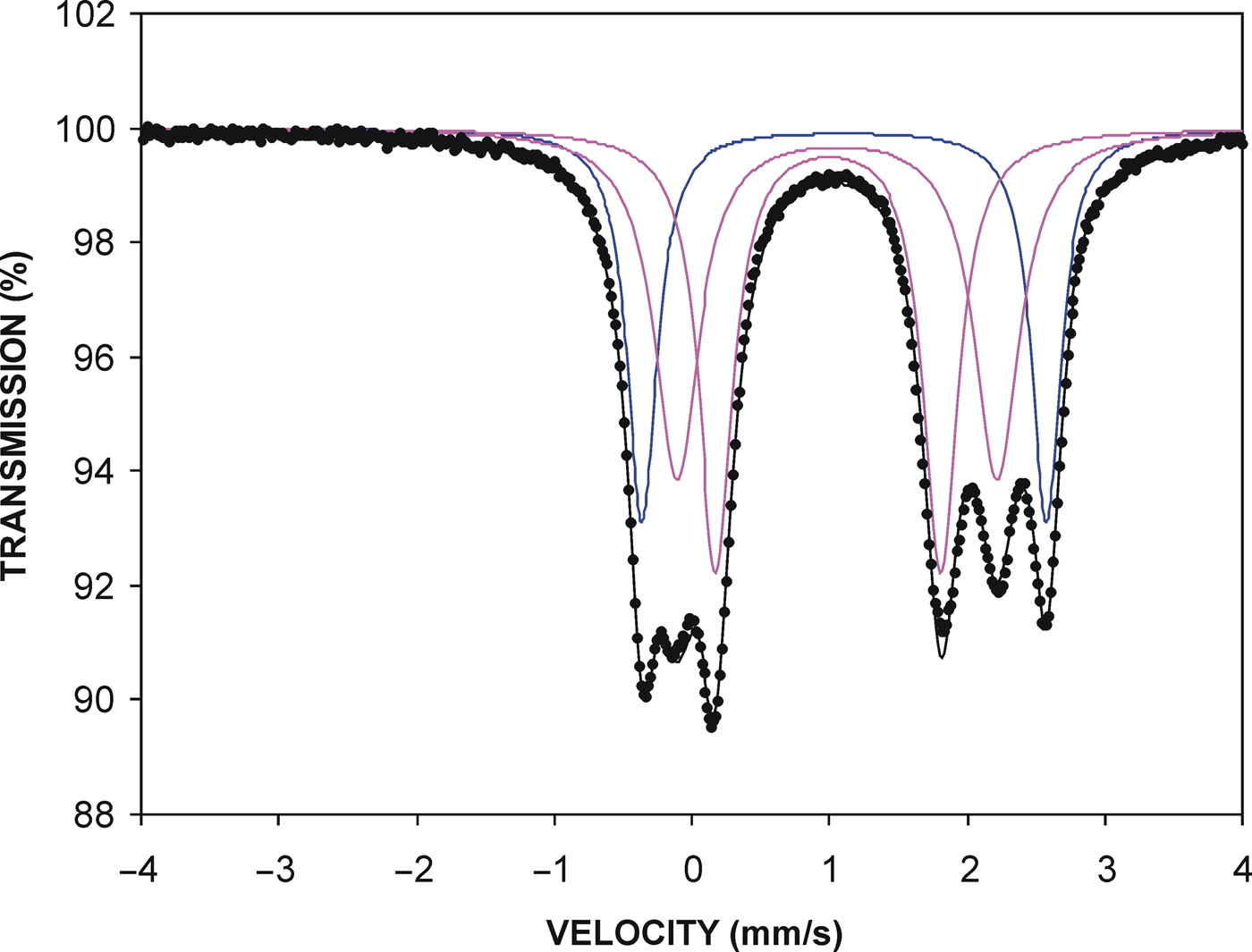
The 57Fe Mössbauer absorption spectrum of graftonite-(Mn) (Fig. 4) was collected at room temperature, at the Academic Center for Materials and Nanotechnology, AGH UST, Kraków, Poland, using a 57Co source in the Rh matrix. A Mössbauer spectrometer of an electromechanical type was used in the constant-acceleration mode. The 14.4 keV γ-rays were detected with a proportional counter. The velocity scale was calibrated at room temperature with a metallic iron foil. The spectrum was analysed by least-squares fitting, assuming three quadrupole-split spectrum components, corresponding to different Fe positions (Table 2).
Mössbauer spectrum of graftonite-(Mn); quadrupole-doublets are marked in violet. The quadrupole-doublet in blue originates from intergrown triphylite.
Resolution of Mössbauer spectrum of graftonite-(Mn).

IS – isomeric shift; QS – quadropole splitting; Γ = half of width at half of maximum.
*Data recalculated to 100%.
The outer doublet in the spectrum, with relative intensity 27.08% is due to triphylite. Two inner doublets, with comparable relative intensities of 50.8 and 49.2% (total recalculated to 100 relative%), are due to graftonite-(Mn). They indicate that in the Lutomia graftonite-(Mn), Fe is completely disordered over the M(2) and M(3) sites, which leads to the structural formula M(1)(Mn0.70Ca0.30)M(2)(Fe0.67Mn0.27Mg0.06Zn0.01)M(3)(Fe0.67Mn0.33)Σ3.00(PO4)2, corresponding to the general formula MnFe2(PO4)2 for the graftonite-(Mn) end-member.
Crystal structure
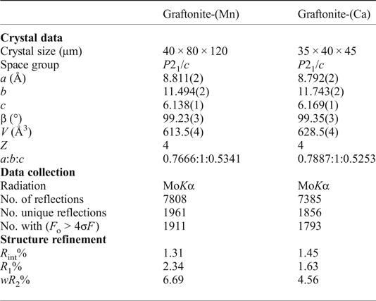
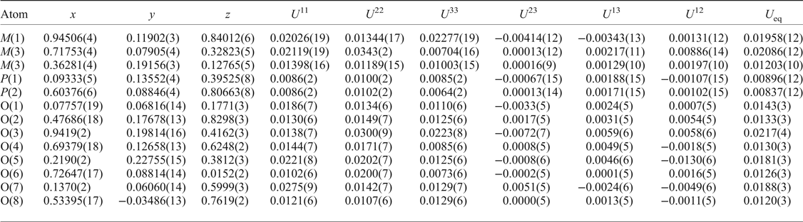
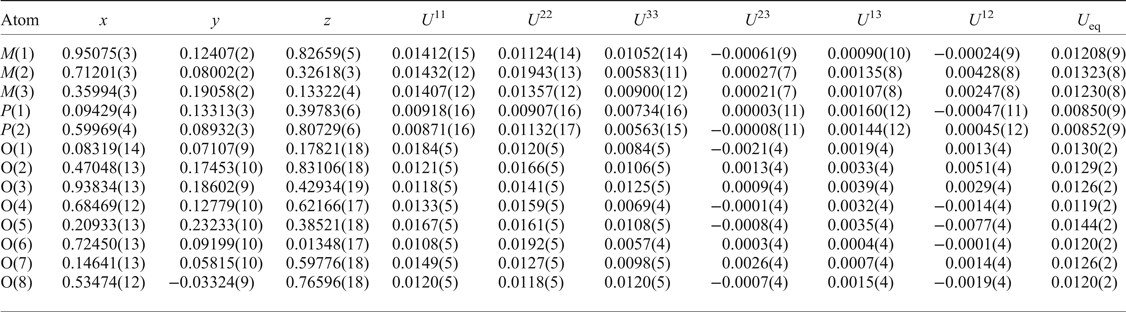
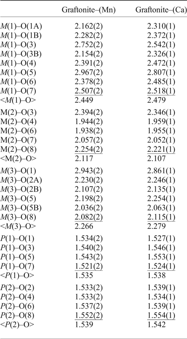
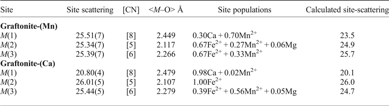
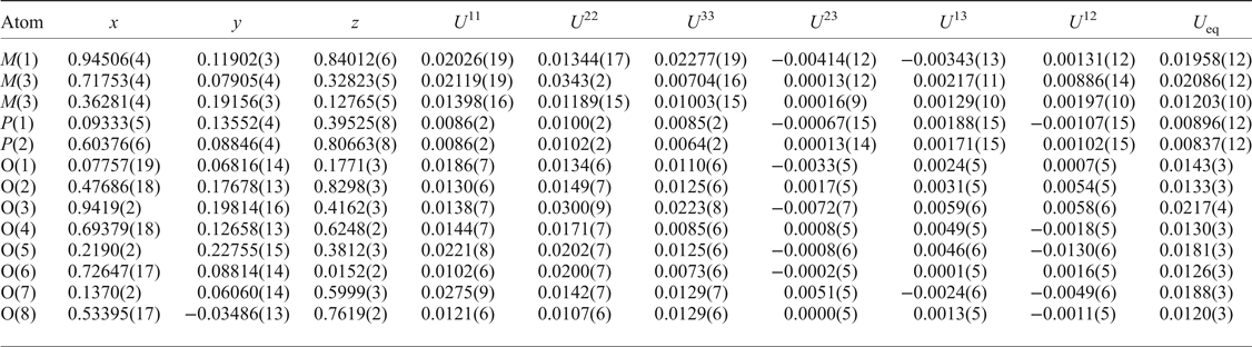
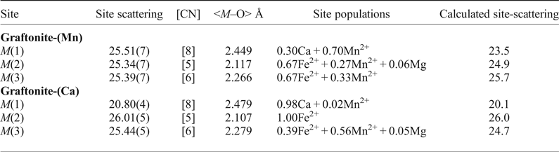
Single crystals of graftonite-(Mn) (40 µm × 80 µm × 120 µm) and graftonite-(Ca) (35 µm × 40 µm × 45 µm) were attached to tapered glass fibres and mounted on a Bruker APEX II ULTRA three-circle diffractometer equipped with a rotating-anode generator (MoKα X-radiation), multilayer optics and an APEX-II detector. For graftonite-(Mn), a total of 7808 intensities was collected to 62° (2θ) using 2 s per 0.3° frame; for graftonite-(Ca), a total of 7385 intensities was collected to 60° (2θ) using 16 s per 0.3° frame, with a crystal-to-detector distance of 5 cm for both crystals. Empirical absorption corrections (SADABS; Sheldrick, Reference Sheldrick2008) were applied and equivalent reflections were merged, resulting in 1961 unique reflections for graftonite-(Mn) and 1856 unique reflections for graftonite-(Ca). Unit-cell dimensions were obtained by least-squares refinement of the positions of 4073 reflections with I > 10σI for graftonite-(Mn), and of 9896 reflections for graftonite-(Ca) (Table 3). The structures were refined to R 1 indexes of 2.34% for graftonite-(Mn) and 1.63% for graftonite-(Ca). Atom positions and equivalent isotropic-displacement parameters are given in Tables 4 and 5, respectively, selected interatomic distances in Table 6, and refined site-scattering values (Hawthorne et al., Reference Hawthorne, Ungaretti and Oberti1995) and assigned site-populations in Table 7. Observed and calculated structure-factors and a crystallographic information file have been deposited with the Principal Editor of Mineralogical Magazine and are available as supplementary material (see below).
Miscellaneous information for graftonite-(Mn) and graftonite-(Ca).

R 1 = Σ(|F o| – |F c|)/Σ|F o|; wR 2 = [Σw( ${F}_{\rm o}^{2} $ –
 ${F}_{\rm c}^{2} $)2/Σw(
 ${F}_{\rm o}^{2} $)2]½, w = 1/[σ2(
 ${F}_{\rm o}^{2} $) + (0.0288P)2 + 0.79P] where P = (Max(
 ${F}_{\rm o}^{2} $,0) + 2
 ${F}_{\rm c}^{2} $).
Atom coordinates and displacement parameters for graftonite-(Mn).
Atom coordinates and displacement parameters for graftonite-(Ca).
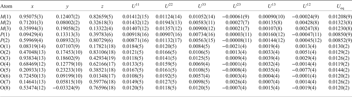
Interatomic distances (Å) in graftonite-(Mn) and graftonite-(Ca).
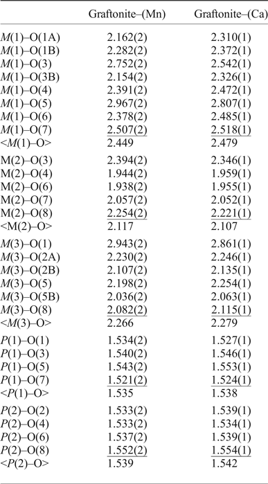
Site-scattering values (epfu) and site populations (apfu) in graftonite-(Mn) and graftonite-(Ca).
epfu – electrons per formula unit; apfu – atoms per formula unit; [CN] – coordination number.
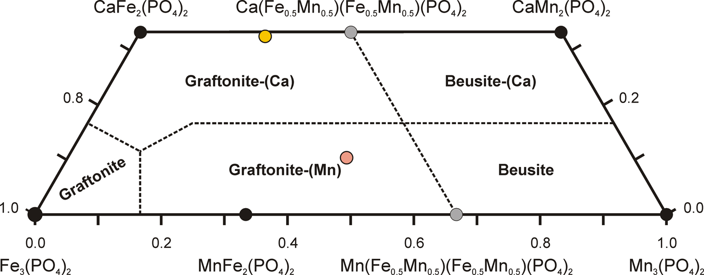
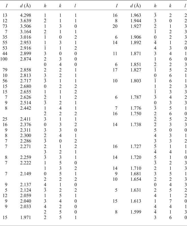
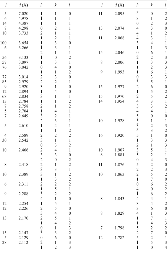
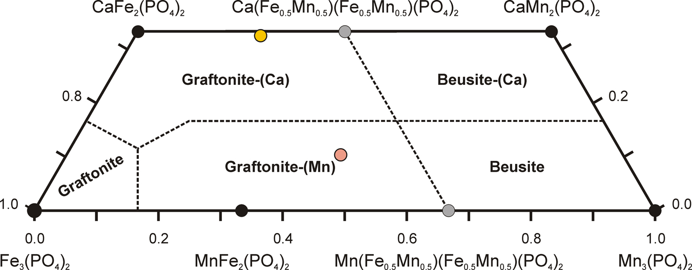
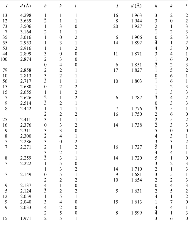
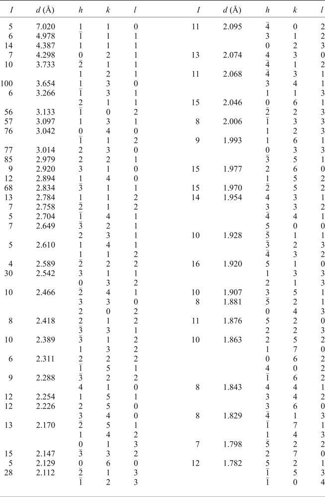
Graftonite-(Mn) and graftonite-(Ca) are isostructural with graftonite (Calvo, Reference Calvo1968) and beusite (Tait et al., Reference Tait, Hawthorne and Wise2013) (monoclinic system; space-group symmetry P21/c), with refined site-scattering values given in Table 6. These results are compatible with the compositions determined by electron-microprobe analysis, and show that the M(1) site is occupied dominantly by Mn2+ in graftonite-(Mn) and by Ca2+ in graftonite-(Ca), and the M(2) and M(3) sites are occupied by Fe2+ and Mn2+, with Fe2+ dominant over Mn2+ at the aggregate M(2) + M(3) sites (Fig. 5). The sizes and heterogeneity of the samples did not provide sufficient materials of the composition of graftonite-(Mn) and graftonite-(Ca) to allow collection of powder-diffraction data. Thus, we collapsed the single-crystal data to two-dimensional experimental diffraction patterns that simulate those of powder patterns (in much the same way as a Gandolfi camera). The patterns are shown in Tables 8 and 9.
Compositional plot for the Fe3(PO4)–Mn3(PO4)–CaFe2(PO4)–CaMn2(PO4) system with the positions of average compositions of type graftonite-(Mn) and graftonite-(Ca).
Simulated powder pattern for graftonite-(Mn).
Simulated powder pattern for graftonite-(Ca).
Origin
Graftonite-(Mn) and graftonite-(Ca) are common products of magmatic crystallization of P-bearing hydrosaline melts in the form of a (Li,Ca)-rich graftonite-like parent phase, after dissolution of a cooling parental P-bearing silicate melt into aluminosilicate and hydrosaline melts connected with separation of a fluid of lower salinity (Simmons and Webber (Reference Simmons and Webber2008) and discussion therein). This high-temperature graftonite-like phase exsolves during cooling to a member of the graftonite group, sarcopside and triphylite or lithiophylite, occurring commonly as lamellar intergrowths.
At Lutomia in the Góry Sowie gneissic block [type locality for graftonite-(Mn)], lamellar and granular graftonite-(Mn) occurs with triphylite and sarcopside in an anatectic pegmatite of LCT affinity, of ~380 Ma, coeval with the younger stage of Góry Sowie gneissic block metamorphism of 385–370 Ma in age (van Breemen et al., Reference van Breemen, Bowes, Aftalion and Żelaźniewicz1988; Timmermann et al., Reference Timmermann, Parrish, Noble and Kryza2000; Turniak et al., Reference Turniak, Pieczka, Kennedy, Szełęg, Ilnicki, Nejbert and Szuszkiewicz2015). Graftonite-(Mn) evolves toward graftonite-(Ca), rarely reaching compositions with M(1)Ca > M(1)Mn. Ca-rich graftonite-(Ca) and beusite-(Ca), with Ca, Fe and Mn each close to 1 apfu, occur in the pegmatite only as irregular domains in lamellae of graftonite-(Mn) and occasionally as thin overgrowths on granular graftonite-(Mn) in fissure zones, which may suggest its metasomatic origin (Włodek et al., Reference Włodek, Grochowina, Gołębiowska and Pieczka2015). At Michałkowa, another anatectic pegmatite of LCT type in the Góry Sowie gneissic block [type locality for graftonite-(Ca)], graftonite-(Mn) forms lamellar intergrowths with sarcopside and triphylite, and grades progressively to graftonite-(Ca). It is most likely a product of exsolution of Ca-enriched graftonite in the outer zones of phosphate nodules due to diffusion of Ca2+ within the hydrosaline melt towards the fluorapatite rim. Calcium-rich graftonite-(Ca) (Ca ~ 1 apfu), occurs only as single subhedral crystals close to the border of the primary magmatic phosphates of the unaltered interior of the nodule, with metasomatic phosphates closer to the nodule border, indicating the action of the high-T fluid released by the hydrosaline melt, which induced incipient Na- and Ca-metasomatism of the primary magmatic phosphates.
Both graftonite-(Mn) and graftonite-(Ca) are common pegmatite phosphates; they should be the main graftonite-group minerals in less-evolved phosphate-bearing pegmatites. In the last dozen years, the presence of graftonite with compositions corresponding to graftonite-(Mn) and graftonite-(Ca) has been well-documented, beside the above-mentioned Polish pegmatites, in Canadian, Swedish and Czech pegmatites described by Černý et al. (Reference Černy, Selway, Ercit, Breaks, Anderson and Anderson1998), Smeds et al. (Reference Smeds, Uher, Černý, Wise, Gustafsson and Penner1998) and Tait et al. (Reference Tait, Hawthorne and Wise2013).
Acknowledgements
We thank Alessandro Guastoni and Peter Leverett, as well as Stuart Mills (Principal Editor) for their comments on this manuscript. The work was supported by the AGH UST grant 11.11.140.158 to AP, National Science Centre (Poland) grant 2015/17/N/ST10/02666 to AW, and by a Natural Sciences and Engineering Research Council of Canada Discovery grant, and by Canada Foundation for Innovation grants to FCH.
Supplementary material
To view supplementary material for this article, please visit https://doi.org/10.1180/minmag.2017.081.109.