Introduction
Hallucinations in schizophrenia are typically auditory in nature; visual hallucinations (VH) are the second most common type (Aleman & Larøi, Reference Aleman, Larøi, Aleman and Larøi2008b ). VH are visual percepts occurring in the absence of external stimuli, experienced while awake (Waters et al. Reference Waters, Collerton, ffytche, Jardri, Pins, Dudley, Blom, Mosimann, Eperjesi, Ford and Larøi2014). They are traditionally classified into three categories: simple (e.g. flashes, shapes), intermediate (e.g. geometric patterns, lines) and complex (e.g. humans and landscapes) (Blom & Sommer, Reference Blom, Sommer, Collerton, Dudley and Mosimann2011).
In contrast to auditory hallucinations (AH), research on VH in psychosis is scarce. Recent research showed an average of 27% prevalence of VH in schizophrenia, based on multiple studies (Waters et al. Reference Waters, Collerton, ffytche, Jardri, Pins, Dudley, Blom, Mosimann, Eperjesi, Ford and Larøi2014). In psychosis, all above-mentioned types of VH occur (Jardri et al. Reference Jardri, Cachia, Thomas and Pins2013) and they typically carry intense personal significance to the hallucinator (Onofrj et al. Reference Onofrj, Thomas, Martinotti, Anzellotti, Di Giannantonio, Ciccocioppo, Bonanni, Collerton, Mosimann and Perry2015).
Little is known about VH-associated cognitive functioning, even though cognitive impairments are thought to play an important role in the dysfunction in schizophrenia (Heinrichs et al. Reference Heinrichs, Miles, Ammari, Muharib and Harvey2013). Deficits in cognitive and sensory processing may also underlie VH. Visual perception relies on two directions of information processing (bottom-up and top-down) that strongly interact (Aleman & Larøi, Reference Aleman, Larøi, Aleman and Larøi2008a ). Bottom-up processing encompasses the encoding and processing of incoming stimuli from the external world to V1 and subsequently to occipital-temporal and occipital-parietal regions (Aleman & Larøi, Reference Aleman, Larøi, Aleman and Larøi2008a ; Meppelink et al. Reference Meppelink, Koerts, Borg, Leenders and van Laar2008). Top-down processing refers to the cognitive cerebral processes of the observer (attention, knowledge and expectations) that may influence the interpretation of the information obtained through the bottom-up processes (Aleman & Larøi, Reference Aleman, Larøi, Aleman and Larøi2008a ). The prefrontal cortex is thought to be the source of top-down activity modulation (Zanto et al. Reference Zanto, Rubens, Thangavel and Gazzaley2011). In our study we examined the role of both directions of information processing in the occurrence of VH in psychosis.
Several models have addressed impairments in bottom-up and/or top-down processing to explain VH in Parkinson's disease dementia (PDD). In one model, a primary deficiency of retinal dopamine leads to activation of normally blocked receptors of neurons that represent the outer visual world, combined with a disinhibition of internally generated images (Diederich et al. Reference Diederich, Goetz and Stebbins2005), or a dysfunction of the attention control networks (Shine et al. Reference Shine, Halliday, Naismith and Lewis2011).
An interesting adjacent model is the Perception and Attention Deficit (PAD) model (Collerton et al. Reference Collerton, Perry and McKeith2005), which is mainly based on data from patients with PDD and dementia with Lewy bodies (DLB). The cross-sectional prevalence of VH in PD is about 50% (Gibson et al. Reference Gibson, Mottram, Burn, Hindle, Landau, Samuel, Hurt, Brown and Wilson2013).
This PAD model includes recurrent complex visual hallucinations (RCVH; Collerton et al. Reference Collerton, Perry and McKeith2005). According to the model, when perceiving a scene, external visual stimuli activate or modify potentially observed objects, which are called ‘proto-objects’. These proto-objects compete for further visual processing, in our unawareness. In the PAD model RCVH occur when the wrong proto-object is chosen and ends up in conscious awareness. Intact visual perception (bottom-up processing) as well as adequate attention (a type of top-down processing) are proposed to be necessary for selecting the correct proto-object. A visual representation of the scene from visual memory, along with intentions and goals, contribute to top-down activation. Moreover, attention plays a critical role in the competition between proto-objects by increasing the internal signal-to-noise ratio. This leads to attentional binding of one object and finally to awareness of the object. Fitting into this model, decreased (sustained) attention and impaired object and space perception were reported in PDD patients with VH (Mosimann et al. Reference Mosimann, Mather, Wesnes, O'Brien, Burn and McKeith2004; Koerts et al. Reference Koerts, Borg, Meppelink, Leenders, van Beilen and van Laar2010; Gallagher et al. Reference Gallagher, Parkkinen, O'Sullivan, Spratt, Shah, Davey, Bremner, Revesz, Williams, Lees and Schrag2011). In DLB, likewise, VH in DLB are related to impaired visual perception (Mosimann et al. Reference Mosimann, Mather, Wesnes, O'Brien, Burn and McKeith2004); a possible relationship between more severe VH and greater attentional deficits (McKeith et al. Reference McKeith, Wesnes, Perry and Ferrara2004) has also been reported. Furthermore, VH in these organic disorders are associated with drowsiness (Manford & Andermann, Reference Manford and Andermann1998). The PAD model appears to explain the finding that VH in PDD typically involve humans and animals engaging in appropriate activities (Diederich et al. Reference Diederich, Goetz and Stebbins2005), which are usually not disturbing. In DLB, VH also generally consist of animals and humans (Mosimann et al. Reference Mosimann, Rowan, Partington, Collerton, Littlewood, O'Brien, Burn and McKeith2006), while simple VH are uncommon in both disorders.
Patients with schizophrenia show impaired bottom-up processing (Brenner et al. Reference Brenner, Lysaker, Wilt and O'Donnell2002; Dakin et al. Reference Dakin, Carlin and Hemsley2005; Smid et al. Reference Smid, Bruggeman and Martens2013), and sustained attention (Nuechterlein et al. Reference Nuechterlein, Barch, Gold, Goldberg, Green and Heaton2004). However, perceptual deficits are not typically associated with AH in schizophrenia (McKay et al. Reference McKay, Headlam and Copolov2000) and recently a review concluded that evidence for VH-related impaired bottom-up processing in psychosis is lacking (Waters et al. Reference Waters, Collerton, ffytche, Jardri, Pins, Dudley, Blom, Mosimann, Eperjesi, Ford and Larøi2014). These authors suggested that top-down distortions are more likely to be involved instead. Interestingly, increased attention has been proposed to be involved in the occurrence of AH in psychosis (Vercammen et al. Reference Vercammen, de Haan and Aleman2008). Previous research indicated that patients with a psychotic disorder with AH show enhanced sensitivity to speech stimuli when compared to patients without AH (Vercammen et al. Reference Vercammen, de Haan and Aleman2008). They were better at ‘hearing a voice in the noise’. Patients with AH also showed a positive response bias. This can be considered as an alteration in the response bias: a more liberal criterion for deciding whether a perceived event is an actual stimulus (Dolgov & McBeath, Reference Dolgov and McBeath2005). As patients become better in perceiving ambient stimuli, this would lead to more hits (perception of actual stimuli), but also to more false alarms. According to this theory, attentional processes play an important role in lowering the decision criterion. This way, increased attention towards auditory-verbal information would lead to perceptual enhancement. These findings are also related to the jumping-to-conclusions bias (JTC bias), which refers to delusions in psychosis, representing hasty decisions based on limited evidence (Van Dael et al. Reference Van Dael, Versmissen, Janssen, Myin-Germeys, van Os and Krabbendam2006; Vercammen et al. Reference Vercammen, de Haan and Aleman2008). Hypersalience of evidence-hypothesis matches can also lead to the bias against disconfirmatory evidence (BADE; Balzan et al. Reference Balzan, Delfabbro, Galletly and Woodward2013), which includes strengthening of otherwise weak evidence-hypothesis connections, leading to more resistance to counter evidence. Other studies also relate AH to increased attention, in patients with schizophrenia (Schneider & Wilson, Reference Schneider and Wilson1983; Aleman et al. Reference Aleman, Böcker, Hijman, de Haan and Kahn2003) as well as in non-psychotic individuals (van Lutterveld et al. Reference van Lutterveld, Oranje, Kemner, Abramovic, Willems, Boks, Glenthøj, Kahn and Sommer2010).
In summary, increased attention, as well as the JTC bias, may play an important role in the occurrence of AH in schizophrenia. This raises the question whether increased attention might also be related to the occurrence of VH in psychosis. For example, VH in psychosis are reported to be related to hyperarousal (Manford & Andermann, Reference Manford and Andermann1998). Furthermore, VH frequently co-occur with hallucinations in other modalities in patients with schizophrenia (Waters et al. Reference Waters, Collerton, ffytche, Jardri, Pins, Dudley, Blom, Mosimann, Eperjesi, Ford and Larøi2014), suggesting at least a partially shared etiology.
In this study we examined the prevalence of VH in a large group of patients with non-affective psychosis and in a control group. Furthermore, the role of bottom-up and top-down processing in the occurrence of VH was evaluated. We hypothesized the lifetime prevalence to be somewhat higher than the reported prevalence (27%) in a recent review (Waters et al. Reference Waters, Collerton, ffytche, Jardri, Pins, Dudley, Blom, Mosimann, Eperjesi, Ford and Larøi2014). Based on the same review, we hypothesized that VH in psychosis are not specifically related to impaired bottom-up processing, but rather to altered attentional functioning. Specifically, in accordance with studies on AH in psychosis (Schneider & Wilson, Reference Schneider and Wilson1983; Aleman et al. Reference Aleman, Böcker, Hijman, de Haan and Kahn2003; Vercammen et al. Reference Vercammen, de Haan and Aleman2008), we hypothesized that VH are related to increased attention. Increased attention predicts a lower reaction time, and higher hit and false alarm rates on the Continuous Performance Test in VH patients. Moreover, it would lead to improved visual perception, via perceptual enhancement.
Method
Participants
Data were derived from baseline measures of an ongoing longitudinal study [Genetic Risk and Outcome in Psychosis (GROUP), version 4.0; for details, see Korver et al. Reference Korver, Quee, Boos, Simons and de Haan2012]. In selected representative geographical areas in The Netherlands and Belgium, patients were identified through representative clinicians whose caseloads were screened for inclusion criteria. Subsequently, a group of patients (n = 1119) presenting consecutively at these services, either as out-patients or in-patients, were recruited for the GROUP study. Controls were selected through a system of random mailings to addresses in the areas of the cases.
Inclusion criteria for patients for the GROUP study were: (1) age between 16 and 50 years; (2) diagnosed with a non-affective psychotic disorder, based on Diagnostic and Statistical Manual of Mental Disorder-IV-TR (DSM-IV-TR) criteria (APA, 2000); (3) maximum duration after first contact with mental health care 10 years; (4) fluency in Dutch; (5) having at least one sibling also willing to participate in this study; (6) able and willing to give informed consent. DSM-IV diagnoses were assessed by a Comprehensive Assessment of Symptoms and History (CASH; Andreasen et al. Reference Andreasen, Flaum and Arndt1992) interview or the Schedules for Clinical Assessment for Neuropsychiatry 2.1 (SCAN; Wing et al. Reference Wing, Babor, Brugha, Burke, Cooper, Giel, Jablenski, Regier and Sartorius1990).
Inclusion criteria for the healthy controls were (1) age between 16 and 50 years; (2) no lifetime psychotic disorder; (3) no first-degree family member with a lifetime psychotic disorder, (4) fluency in Dutch; (5) able and willing to give informed consent. The CASH (Andreasen et al. Reference Andreasen, Flaum and Arndt1992) and SCAN (Wing et al. Reference Wing, Babor, Brugha, Burke, Cooper, Giel, Jablenski, Regier and Sartorius1990) interviews were used to confirm that they had no history of psychosis.
Because this was retrospective database study, we determined statistically whether the groups differed regarding demographic and illness variables. The variables that differed and were assumed to affect our research domains were used as covariates in our analyses.
Approval from Ethical Review Boards of the participating centers was obtained, as was written informed consent from all participants.
Sample size calculation
A previous study in the same population (Meijer et al. Reference Meijer, Simons, Quee and Verweij2012) showed a 0.88 difference on the accuracy on the Continuous Performance Test-HQ between patients and controls (p < 0.001), a s.d. of 2.27 in patients and 1.01 in controls. With a power of 0.80 and α = 0.05, each group would at least have to include 64 participants, giving a minimum overall sample size of 192 (Cohen, Reference Cohen1992). This requirement was met by our population.
Questionnaires and scales
Prevalence of VH
The Dutch version of the validated Community Assessment of Psychic Experiences (CAPE; Stefanis et al. Reference Stefanis, Hanssen, Smirnis, Avramopoulos, Evdokimidis, Stefanis, Verdoux and Van Os2002; Konings et al. Reference Konings, Bak, Hanssen, van Os and Krabbendam2006) was used to assess the lifetime prevalence of VH. The CAPE, a self-report questionnaire, rates lifetime psychotic experiences. The answer possibilities to the question ‘Have you ever seen objects, people or animals that other people cannot see?’ were ‘never’, ‘sometimes’, ‘often’ and ‘nearly always’.
The prevalence of VH at the present moment was determined by the CASH (Andreasen et al. Reference Andreasen, Flaum and Arndt1992) and SCAN interviews (Wing et al. Reference Wing, Babor, Brugha, Burke, Cooper, Giel, Jablenski, Regier and Sartorius1990). During the CASH interview, the following question was asked: Have you had visions or seen things that other people cannot (present state)? Answer possibilities were: none; questionable; mild (has occurred once or twice); moderate (occurs at least weekly); marked (occurs frequently); severe (occurs almost daily). In the SCAN interview, it was asked whether participants experienced formed, unformed, or scenic VH. Available options were: no; congruence is mainly with depressed mood; both with depressed and elated mood; mainly with elated mood; language difficulty; cannot be determined; n.a.
Visual perception and attention
Visual perception and attention were assessed by a series of tasks on which patients with psychosis performed worse than controls in a previous study (except for the Benton Facial recognition Task) (Meijer et al. Reference Meijer, Simons, Quee and Verweij2012). Visual perception was assessed with tests based on face perception, as patients with schizophrenia frequently hallucinate human beings (Líndal et al. Reference Líndal, Stefánsson and Stefánsson1994). Attention was assessed by sustained attention, as was the case in PDD (Koerts et al. Reference Koerts, Borg, Meppelink, Leenders, van Beilen and van Laar2010; Gallagher et al. Reference Gallagher, Parkkinen, O'Sullivan, Spratt, Shah, Davey, Bremner, Revesz, Williams, Lees and Schrag2011).
The Benton Facial Recognition Test (BFRT)
During the validated short version of the BFRT (Levin et al. Reference Levin, Hamsher and Benton1975; Benton et al. Reference Benton, Sivan, de Hamsher, Varney and Spreen1983), participants were presented with a target face above six test faces. They were asked to identify which test faces matches the target face. The number of correctly chosen test faces was measured.
The Degraded Facial Affect Recognition (DFAR)
The DFAR (van ‘t Wout et al. Reference van‘t Wout, Aleman, Kessels, Larøi and Kahn2004) measured visual perception by facial affect recognition of degraded faces (visual contrast was reduced by 30%). It has been previously used in schizophrenia research (Van ‘t Wout et al. Reference van‘t Wout, Aleman, Kessels, Larøi and Kahn2004, Reference Van't Wout, van Dijke, Aleman, Kessels, Pijpers and Kahn2007; Janssens et al. Reference Janssens, Lataster, Simons, Oorschot, Lardinois, van Os and Myin-Germeys2012). Participants were shown 64 pictures, with 16 pictures for each emotion category: angry, happy, fearful or neutral. They were asked to press buttons corresponding with the recognized emotion. Variables measured were the proportion correctly recognized emotions per emotion category and overall.
The Continuous Performance Test-HQ (CPT-HQ)
The CPT-HQ (Wohlberg & Kornetsky, Reference Wohlberg and Kornetsky1973; Nuechterlein & Dawson, Reference Nuechterlein and Dawson1984) assesses sustained attention with working memory load. It is a validate measure of attentional dysfunction in patients with schizophrenia (Nelson et al. Reference Nelson, Sax and Strakowski1998). Participants were shown 300 white letters in a randomized sequence and were instructed to press the space bar if the letter H was preceded by the letter Q. Variables measured were reaction time for correct detection, numbers of correct positives, false positives and false negatives.
Response Set-shifting Task (RST)
Attentional switching was assessed by the RST (Bilder et al. Reference Bilder, Turkel, Lipschutz-Broch and Lieberman1992), which has been designed in a study with patients with a psychotic disorder. They were asked to switch between two response rules that were not explicitly defined, but had to be deduced from trial-to-trial feedback (‘correct’ or ‘wrong’). Cost difference scores and proportional cost indices were measured.
Statistical analyses
The lifetime prevalence of VH was assessed by the CAPE (Konings et al. Reference Konings, Bak, Hanssen, van Os and Krabbendam2006) (the sum of ‘sometimes’, ‘often’ and ‘almost always’); its prevalence at the present moment as determined by CASH (the sum of ‘mild’, ‘moderate’, ‘marked’ and ‘severe’ present state, ‘questionable’ was excluded from analyses) (Andreasen et al. Reference Andreasen, Flaum and Arndt1992) and by SCAN (the sum of the presence of depressed and/or elated mood congruent VH for unformed, formed and scenic VH together) (Wing et al. Reference Wing, Babor, Brugha, Burke, Cooper, Giel, Jablenski, Regier and Sartorius1990). The VH rate was calculated for the total patient group, per diagnostic subcategory, and for the control group. For investigating the role of perception and attention, three groups were formed, based on the CASH item on VH. The first group included VH patients: those who reported ‘mild’ to ‘severe’ VH; the second group consisted of patients who reported ‘none’ (non-VH patients); the third group consisted of healthy controls with no VH. The SCAN was not included for this purpose as it does not report the frequency, and thus the recurrence, of the VH. Comparisons of demographic and illness variables between the three groups were done by χ2 and univariate tests; the patient groups were compared with Mann–Whitney U and χ2 tests.
Scores on the tests were analyzed by univariate (BFRT, WAIS-III Digit Symbol Substitution Test) and multivariate testing (DFAR, CPT-HQ, RST; Wilk's lambda). Covariates used for analyzing the tests were age, IQ, gender, and the frequency of AH and delusions, as these variables differed between the groups and were expected to affect the studied cognitive domains. Analyses were also performed without AH and delusions as covariates in order to explore their impact. Data were analyzed two-tailed with a level of significance of 0.05. In case of applying contrasts, Bonferroni corrections were used (s.d./3).
As is often the case when working with clinical data, missing values were inevitable. Reasons for missing data were dropouts because of refusal, physical illness, not available for further assessment or other reasons (Korver et al. Reference Korver, Quee, Boos, Simons and de Haan2012). Consequently, the number of participants differed between the tests. Analyses were performed with SPSS v. 20 (IBM Corp., USA).
Post-hoc analyses
WAIS-III Digit Symbol Substitution Test (WAIS-III-DSST)
The validated WAIS-III-DSST was used post-hoc to assess processing speed (Wechsler, Reference Wechsler1997). This test presented nine symbol-digit pairs. Participants were presented with a list of symbols and asked to write down the appropriate number below the symbol within 90 s. The number of correct substitutions was measured.
Ethical standards
The authors assert that all procedures contributing to this work comply with the ethical standards of the relevant national and institutional committees on human experimentation and with the Helsinki Declaration of 1975, as revised in 2008.
Results
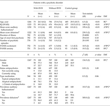
The total study population consisted of 1119 patients with a non-affective psychotic disorder and 586 controls. The group of patients consisted of 852 men (76.1%) and 267 women (23.9%) with a mean age of 27.58 years (s.d. = 7.94). The control group consisted of 269 men (45.9%) and 317 women (54.1%), their mean age was 30.42 years (s.d. = 10.58). Demographic and illness characteristics of the groups are listed in Table 1. On average, the patients were younger and had a lower IQ than the control group; VH patients had a lower IQ than non-VH patients. The mean age of onset first psychosis was similar in both patient groups. For patients, the percentage of men was higher than for controls. The percentage of women was higher in VH patients than in non-VH patients. VH patients showed a higher prevalence of AH (81.3%) and delusions (96.0%) than non-VH patients (28.3% and 61.2%), which in turn showed a higher frequency than healthy controls (0.4% and 2.9%). There were no differences between the patient groups regarding the use or non-use and type of antipsychotic medication.
Demographic and illness characteristics of patients with a non-affective psychotic disorder with and without VH at the present moment, and healthy controls (CASH)

VH, Visual hallucinations; CASH, Comprehensive Assessment of Symptoms and History; RVH, recurrent visual hallucinations; WAIS, Wechsler Adult Intelligence Scale; PANSS, Positive and Negative Syndrome Scale.
a Mean score on CASH items 76–78, at the present moment. Answer possibilities: 0 = none; 1 = questionable; 2 = mild; 3 = moderate; 4 = marked; 5 = severe.
b Mean score CASH items 53–64, at the present moment.
c Prevalence: % that scored on at least one of above-mentioned items ⩾1.
Answer possibilities: 0 = none; 1 = questionable; 2 = mild; 3 = moderate; 4 = marked; 5 = severe.
d Prevalence: % that scored on at least one of above-mentioned items ⩾1.
Diff = significant at a level of 0.05, * = significant at a level of 0.01.
A = patients with RVH v. patients without RVH.
B = patients with RVH v. control group.
C = patients without RVH v. control group.
Prevalence of VH
Lifetime (CAPE)
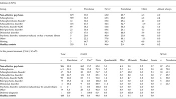
The lifetime prevalence of VH in non-affective psychosis was 37.0%; in schizophrenia 36.5%. Table 2 summarizes VH prevalence rates within the diagnostic subcategories. The highest lifetime prevalence was found in schizoaffective disorder (47.5%). The lifetime prevalence of VH in controls was 3.4%.
The prevalence of VH, lifetime and at the present moment, in non-affective psychosis and healthy controls

VH, Visual hallucinations; CAPE, Community Assessment of Psychic Experiences; NOS, not otherwise specified; CASH, Comprehensive Assessment of Symptoms and History; SCAN, Schedules for Clinical Assessment for Neuropsychiatry 2.1.
Prevalence and frequencies are shown in %.
a Including all subcategories.
b The sum of ‘mild’, ‘moderate’, ‘marked’ and ‘severe’; ‘questionable’ was excluded from analyses.
At the present moment (CASH and SCAN)
The prevalence of VH during at the present moment in non-affective psychosis was 18.9%; the highest prevalence was found in schizophreniform disorder (36.2%). Patients with a substance-induced psychotic disorder or psychosis due to a somatic illness did not report VH at the present moment.
Visual perception and attention
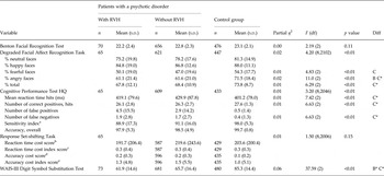
All cognitive test results are listed in Table 3. For the results without AH and delusions as covariates, see online Supplementary Table S1. The results are virtually the same.
Comparison of different aspect of attention and visual information processing between patients with a non-affective psychotic disorder with and without VH at the present moment, and healthy controls (CASH)

VH, Visual hallucinations; CASH, Comprehensive Assessment of Symptoms and History; RVH, recurrent visual hallucinations; WAIS, Wechsler Adult Intelligence Scale.
a [(hits – false positive target)/28] × 100.
b reaction time in the reversal condition – reaction time in the imitation condition
c (reaction time in the reversal condition – reaction time in the imitation condition)/reaction time in the imitation condition
d proportion correct in the imitation condition - proportion correct in the reversal condition
e (proportion correct in the imitation condition – proportion correct in the reversal condition)/proportion correct in the reversal condition
Diff = significant at a level of 0.05, * = significant at a level of 0.01.
A = patients with RVH v. patients without RVH.
B = patients with RVH v. control group.
C = patients without RVH v. control group.
Visual perception
There were no differences in face perception (DFAR and BFRT) between patients with and without VH. Non-VH patients recognized fewer fearful faces and showed a worse overall performance on the DFAR than controls; both patient groups recognized fewer angry faces than controls (VH patients: p < 0.05; non-VH patients: p < 0.01). Patients did not perform significantly different than controls on the BFRT.
Attention
There were no differences in any of the subscores of the tasks assessing attention (CPT-HQ and RST) between both patient groups; specifically, there were no differences on the CPT-HQ in reaction time [mean (s.d.)] [VH patients: 419.1 (79.6); non-VH patients: 429.9 (87.8); p = 0.28], number of hits [VH: 26.1 (2.8); non-VH: 26.3 (2.7); p = 0.28] and number of false alarms [VH: 4.5 (15.5); non-VH 2.9 (14.2); p = 0.32]. Neither were there any significant differences between VH patients and controls. However, non-VH patients responded more slowly than controls on the CPT-HQ [non-VH: 429.9 (87.8) v. controls: 401.2 (78.0); p < 0.01; VH: 419.1 (79.6) v. controls: p = 0.49]. Furthermore, non-VH patients showed fewer hits [26.3 (2.7)] and more false negatives [1.7 (2.7)] than controls [27.6 (1.3) and 0.4 (1.3); both p < 0.01].
There were no significant differences in attentional switching (RST) between the three groups.
Post-hoc analyses
The patient groups showed comparable processing speeds (WAIS-III-DSST). The control group showed faster processing speed than both patient groups (both p < 0.01).
Discussion
This study showed that VH in non-affective psychosis are quite common. We found no VH-related bottom-up impairments or top-down alterations. Both patient groups had worse visual perception and processing speed than controls. Moreover, non-VH patients showed worse attentional performance than controls, whereas VH patients did not. As an earlier study in this same population showed that patients performed worse on this attention task (Meijer et al. Reference Meijer, Simons, Quee and Verweij2012) than controls, one may speculate that this implies a relatively spared attentional performance in VH patients. This seem to be more in accordance with the increased attention hypothesis than the PAD model.
Prevalence
In our large (1119) cohort of patients, 37% reported having experienced VH during their lifetime, and the reported prevalence at the present moment was 18.9%. This is in line with earlier studies, with rates of 14% (Mueser et al. Reference Mueser, Bellack and Brady1990) and 43% (Ndetei & Singh, Reference Ndetei and Singh1983). Only one study involving patients with chronic schizophrenia reported a much higher percentage of VH (72%) (Goodwin & Rosenthal, Reference Goodwin and Rosenthal1971).
It should be noted that CAPE is a self-report questionnaire, whereas CASH and SCAN are interviews. This might also have led to differences in reported prevalence rates, on top of the difference in included period between ‘lifetime’ and ‘at the present moment’.
Visual perception and attention
Our data do not show perceptual and/or attentional differences in psychotic patients with VH as compared to non-VH patients. We found significant differences only between the patient groups and controls. Visual perception and processing speed were equally impaired in both patient groups as compared to controls. In addition, non-VH patients showed worse sustained attention than controls, while the scores of VH patients did not differ significantly from controls.
Based on these results, we conclude that impairments in bottom-up processing are not specifically related to the occurrence of VH in psychosis. This would corroborate the conclusions of a recent review (Waters et al. Reference Waters, Collerton, ffytche, Jardri, Pins, Dudley, Blom, Mosimann, Eperjesi, Ford and Larøi2014). Our data do not show VH-related impaired attention. The results thus seem to be at odds with the PAD model and DLB. However, we should be cautious about rejecting the PAD model, as previous studies used different tasks (McKeith et al. Reference McKeith, Wesnes, Perry and Ferrara2004; Mosimann et al. Reference Mosimann, Mather, Wesnes, O'Brien, Burn and McKeith2004; Koerts et al. Reference Koerts, Borg, Meppelink, Leenders, van Beilen and van Laar2010).
There are similarities between organic disorders and psychosis regarding VH: they are both linked to alterations in the attentional networks (Shine et al. Reference Shine, Halliday, Naismith and Lewis2011; Jardri et al. Reference Jardri, Thomas, Delmaire, Delion and Pins2012). Moreover, in both types the visual cortex and hippocampus seem to play a large role (Waters et al. Reference Waters, Collerton, ffytche, Jardri, Pins, Dudley, Blom, Mosimann, Eperjesi, Ford and Larøi2014).
However, clinical differences between VH in psychosis and PDD/DLB patients, like the subtype of VH (simple v. complex), and associations with hyperarousal and drowsiness, may suggest different pathophysiological mechanisms. The attentional deficits in PDD and DLB seem to be closely related to reduced acetylcholine levels (Fabbrini et al. Reference Fabbrini, Barbanti, Aurilia, Pauletti, Lenzi and Meco2004; McKeith et al. Reference McKeith, Wesnes, Perry and Ferrara2004; Francis & Perry, Reference Francis and Perry2007). The impaired visual perception in PDD is related to hypometabolism of secondary cortices (Meppelink et al. Reference Meppelink, Koerts, Borg, Leenders and van Laar2008), possibly to Lewy bodies depositions in the temporal lobe (Aarsland et al. Reference Aarsland, Ballard, Larsen and McKeith2001), as in DLB (Harding et al. Reference Harding, Broe and Halliday2002). Additional dopaminergic hyperactivity may be involved due to dopamimetic treatment (Fénelon et al. Reference Fénelon, Mahieux, Huon and Ziégler2000). Consequently, the PAD model appears to have a biological substrate in PDD and DLB.
In schizophrenia, on the other hand, hallucinations and delusions are thought to arise from an excess of dopamine subcortically (Abi-Dargham, Reference Abi-Dargham2004). Dopamine has been associated with the integrity of attention-related networks (Dang et al. Reference Dang, O'Neil and Jagust2012); it mediates salience at the level of the mesolimbic pathway (van der Gaag, Reference van der Gaag2006). In patients with paranoid delusions, higher levels of dopamine increase the signal-to-noise ratio, which lead to heightened salience and reduced development of alternative hypotheses (van der Gaag, Reference van der Gaag2006). This would fit very well with the increased attention hypothesis. Our results, however, do not clearly show VH-related increased attention. Nevertheless, non-VH patients showed worse attentional performance than controls (CPT-HQ) and VH patients did not, whereas previous research in the same population showed that patients performed worse on the CPT-HQ (Meijer et al. Reference Meijer, Simons, Quee and Verweij2012). Therefore, our results could indicate a relatively spared attentional performance in VH patients. The less impaired reaction time found in VH patients may be caused by hasty decision making [described by the JTC (Van Dael et al. Reference Van Dael, Versmissen, Janssen, Myin-Germeys, van Os and Krabbendam2006)], in combination with resistance to counter evidence as described by the BADE (Balzan et al. Reference Balzan, Delfabbro, Galletly and Woodward2013).
We analyzed post-hoc whether the indirect difference in reaction time could be explained by differences in processing speed (WAIS-III-DSST), which was not the case. Lower reaction times on auditory and visual tasks in patients with AH as compared to patients without AH have been described previously (Schneider & Wilson, Reference Schneider and Wilson1983). The higher prevalence of AH in VH patients than in non-VH patients confirmed previous findings (Zarroug, Reference Zarroug1975; Mueser et al. Reference Mueser, Bellack and Brady1990; Manford & Andermann, Reference Manford and Andermann1998). This, along with the higher prevalence of delusions in VH patients, suggest a shared underlying mechanism with AH and delusions. It should be emphasized, however, that our results indicate a relatively spared attention. Moreover, the previous finding that VH in psychosis are related to a higher severity of illness (Mueser et al. Reference Mueser, Bellack and Brady1990), could also explain the higher frequency of AH and delusions (and the lower IQ score) in VH patients, instead of increased attention. The co-occurrence of symptoms during disease progression does suggest a similar pathophysiology, but this is not certain.
Cognitive functioning was also analyzed without AH and delusions as covariates (see Supplementary Table S1). The results were virtually the same. There are two possible explanations for this: (1) VH, AH and delusions share a common (not significantly different) pathophysiology; (2) AH and delusions are unrelated to the cognitive functions measured. The latter possibility is quite unlikely (assuming the right tasks were used for attention), as there is evidence for AH-related increased attention (Vercammen et al. Reference Vercammen, de Haan and Aleman2008).
Our study has several limitations. First, only global information on the frequency of VH was acquired. The CAPE does not clearly define frequency ratings, leaving room for subjectivity. A second limitation is the lack of knowledge about whether patients experienced VH during testing, because this may impact the data.
Furthermore, it should be noted that perception and attention can vary over time. We used other tests to assess visual perception and attention than earlier studies to investigate the PAD model, (Mosimann et al. Reference Mosimann, Mather, Wesnes, O'Brien, Burn and McKeith2004; Koerts et al. Reference Koerts, Borg, Meppelink, Leenders, van Beilen and van Laar2010; Gallagher et al. Reference Gallagher, Parkkinen, O'Sullivan, Spratt, Shah, Davey, Bremner, Revesz, Williams, Lees and Schrag2011), which could partially explain our different findings. A point of concern regarding the PAD model is that visual perception and attentional processes referring to this model should be defined more precisely, permitting more reliable comparisons between studies. The tests used in this study might be suboptimal. Actually, the visual perceptual tasks should resemble the ‘hearing a voice in the noise’ task in the auditory domain (Vercammen et al. Reference Vercammen, de Haan and Aleman2008).
The non-impaired reaction times in VH patients might also be explained by better visual characteristics, which the GROUP study did not assess. However, this would have resulted in a higher accuracy, which was not the case. Consequently, this possible explanation is unlikely.
In Table 1, there appear to be 14 VH patients without AH. As mentioned above, VH frequently co-occur with AH (Waters et al. Reference Waters, Allen, Aleman, Fernyhough, Woodward, Badcock, Barkus, Johns, Varese, Menon, Vercammen and Larøi2012). Therefore, this result probably reflects the fact that the AH and VH status are defined on the present moment. This could have biased our results, but it is also possible that the test results reflect the present state.
Finally, this study is somewhat limited by the significantly higher IQ in controls relative to the patients and the general population. Although the controls were randomly selected and the analyses were controlled for IQ, this might have influenced our data. Future studies could address this by matching participants regarding IQ.
In conclusion, the lifetime prevalence of VH is 37% in patients with non-affective psychosis, with a prevalence of 18.9% at the present moment. Our results do not show VH-related bottom-up impairments or direct top-down alterations. Both patient groups showed impaired visual perception and processing speed. Interestingly, non-VH patients showed worse attention than controls, whereas VH patients did not. This might imply a relatively spared attentional performance in VH patients. This would be more compatible with the increased attention hypothesis (as in AH and delusions in psychosis) than with the PAD model in organic disorders. Our finding that AH and delusions occur more frequently in VH patients than in non-VH patients, may support this ‘hallucination-proneness’ in psychosis: an underlying, modality-independent mechanism that evokes hallucinations. Our results may support the current view that VH in psychosis are probably not specifically related to impairments in bottom-up processing, but rather to distorted top-down mechanisms. However, caution is needed when rejecting the PAD model; replication with other tasks would be needed first. Further research, preferably with a ‘noise task’ in both the visual and the auditory domain, as well as using the same tasks as earlier research, is recommended.
Appendix. Genetic Risk and Outcome of Psychosis (GROUP) Investigators
R. Bruggeman1*, W. Cahn2, L. de Haan3, R. S. Kahn2, C. J. Meijer3, I. Myin-Germeys4, J. van Os4,5 and D. Wiersma1
1Department of Psychiatry, University of Groningen, University Medical Center Groningen, Rob Giel Research Center, The Netherlands
2Department of Psychiatry, University Medical Center Utrecht, Rudolf Magnus Institute of Neuroscience, Utrecht, The Netherlands
3Department of Psychiatry, Academic Medical Centre, University of Amsterdam, Amsterdam, The Netherlands
4Maastricht University Medical Centre, South Limburg Mental Health Research and Teaching Network, EURON, Maastricht, The Netherlands
5Department of Psychosis Studies, King's College London, King's Health Partners, Institute of Psychiatry, London, England
Supplementary material
For supplementary material accompanying this paper visit http://dx.doi.org/10.1017/S0033291716000246.
Acknowledgements
We are grateful for the generosity of time and effort by the patients and their families, healthy participants, and all researchers who make this GROUP project possible. The infrastructure for the GROUP study is funded through the Geestkracht programme of the Dutch Health Research Council (ZON-MW, grant number 10-000-1001), and matching funds from participating pharmaceutical companies (Lundbeck, AstraZeneca, Eli Lilly, Janssen Cilag) and universities and mental health care organizations (Amsterdam: Academic Psychiatric Centre of the Academic Medical Centre and the mental health institutions: GGZ Ingeest, Arkin, Dijk en Duin, Rivierduinen, Erasmus Medical Centre, GGZ Noord Holland Noord. Maastricht: Maastricht University Medical Centre and the mental health institutions: GGZ Eindhoven, GGZ Midden-Brabant, GGZ Oost-Brabant, GGZ Noord- Midden Limburg, Mondriaan Zorggroep, Prins Clauscentrum Sittard, RIAGG Roermond, Universitair Centrum Sint-Jozef Kortenberg, CAPRI University of Antwerp, PC Ziekeren Sint-Truiden, PZ Sancta Maria Sint-Truiden, GGZ Overpelt, OPZ Rekem. Groningen: University Medical Center Groningen and the mental health institutions: Lentis, GGZ Friesland, GGZ Drenthe, Dimence, GGNet, Mediant, Yulius and Parnassia psycho-medical centre (The Hague). Utrecht: University Medical Centre Utrecht and the mental health institutions Altrecht, Symfora, Meerkanten, Riagg Amersfoort and Delta.)
Declaration of Interest
None.