Introduction
Cones sensitive to short wavelengths (S) can be distinguished from those sensitive to middle (M) and long (L) wavelengths by subtle details of their synaptic structure. In particular, foveal S-cone terminals (pedicles) are smaller than non-S-cone terminals and have more presynaptic ribbons: 22–23 ribbons for S and 16–20 ribbons for non-S (Herr et al., Reference Herr, Klug, Sterling and Schein2003). By contrast, M- and L-cone terminals have not been distinguished from each other: the volumes of non-S-cone terminals distribute unimodally (Haverkamp et al., Reference Haverkamp, Grünert and Wässle2001; Klug et al., Reference Klug, Herr, Ngo, Sterling and Schein2003), as do the number of synaptic ribbons (Ahnelt et al., Reference Ahnelt, Kolb and Pflug1987, Reference Ahnelt, Keri and Kolb1990; Klug et al., Reference Klug, Herr, Ngo, Sterling and Schein2003). Even immunocytochemical methods, which can mark specifically the opsin in S cones, have not been able to distinguish the two types of non-S-cone opsins (Szél et al., Reference Szél, Diamantstein and Rohlich1988; Lerea et al., Reference Lerea, Bunt-Milam and Hurley1989). To explore the contribution of L and M cones to circuits concerned with color vision, we sought structural features that might distinguish their synaptic terminals.
In the macaque fovea, each non-S-cone terminal contacts one ON and one OFF midget bipolar cell (Polyak, Reference Polyak1941; Boycott & Dowling, Reference Boycott and Dowling1969; Kolb et al., Reference Kolb, Boycott and Dowling1969; Boycott & Hopkins, Reference Boycott and Hopkins1991; Hopkins & Boycott, Reference Hopkins and Boycott1992; Kolb et al., Reference Kolb, Linberg and Fisher1992; Milam et al., 1993; Wässle et al., Reference Wässle, Grünert, Martin and Boycott1994; Klug et al., Reference Klug, Herr, Ngo, Sterling and Schein2003). The ON midget bipolar cell dendrites occupy the central position of the postsynaptic triad, where they are called “central elements” (Missotten, Reference Missotten1965). These contacts, also called “invaginating” (Dowling & Boycott, Reference Dowling and Boycott1966; Boycott & Dowling, Reference Boycott and Dowling1969), have asymmetrically dense regions that are thicker on the presynaptic side (Raviola & Gilula, Reference Raviola and Gilula1975; Vardi et al., Reference Vardi, Morigiwa, Wang, Shi and Sterling1998; Ahmad et al., Reference Ahmad, Klug, Herr, Sterling and Schein2003). The OFF midget bipolar cell dendrites occupy the triad-associated position (Kolb, Reference Kolb1970; Boycott & Hopkins, Reference Boycott and Hopkins1991; Hopkins & Boycott, Reference Hopkins and Boycott1992), and their contacts, called “basal,” harbor one or more symmetrically dense regions (Vardi et al., Reference Vardi, Morigiwa, Wang, Shi and Sterling1998; Ahmad et al., Reference Ahmad, Klug, Herr, Sterling and Schein2003), here called “Outer Densities” in contrast to “Inner Densities” located more proximally on OFF midget bipolar cell dendrites (see below; Herr et al., 2011).
Here, based on reconstruction from high-magnification serial electron micrographs, we report counts of basal contacts and Outer Densities (within those basal contacts) between a non-S cone and its OFF midget bipolar cell. These numbers divide the non-S cones into two distinct populations, possibly providing markers that structurally differentiate L-type cones from M-type cones and L-cone-connected OFF midget bipolar cells from M-cone-connected OFF midget bipolar cells. We also report counts of central elements between non-S cones and ON midget bipolar cells. These numbers are not different for the two populations. From the same material, on the basis of lower magnification material, Calkins (Reference Calkins1994) and Calkins et al. (Reference Calkins, Schein, Tsukamoto and Sterling1994) suggested that counts of contacts at the next synaptic level—between midget bipolar cells and midget ganglion cells—differentiated two populations of non-S circuits, but the current data cast doubt on counts from such low-magnification material.
Materials and methods
The detailed methods are reported in Tsukamoto et al. (Reference Tsukamoto, Masarachia, Schein and Sterling1992), Herr et al. (Reference Herr, Klug, Sterling and Schein2003), and Klug et al. (Reference Klug, Herr, Ngo, Sterling and Schein2003). In brief, we removed a retina from an adult (6 kg), male, Macaca fascicularis monkey that had been anesthetized (10 mg/kg ketamine, followed by 40 mg/kg pentobarbital) and perfused through the aorta for <1 min with saline and then with a phosphate-buffered solution of 2% glutaraldehyde and 2% paraformaldehyde for 10 min, all in accordance with assurance number A3332-01 from the Institutional Animal Care and Use Committee of the Massachusetts Eye and Ear Infirmary. After preparing the retina for electron microscopy, including postfixing in 2% OsO4, dissecting, staining a strip containing the fovea en bloc in 1% aqueous uranyl acetate, and embedding in Epon, we cut a series of 319 consecutive, ultrathin (90 nm), radial (or cross) sections through the center of the fovea, parallel to the horizontal meridian.
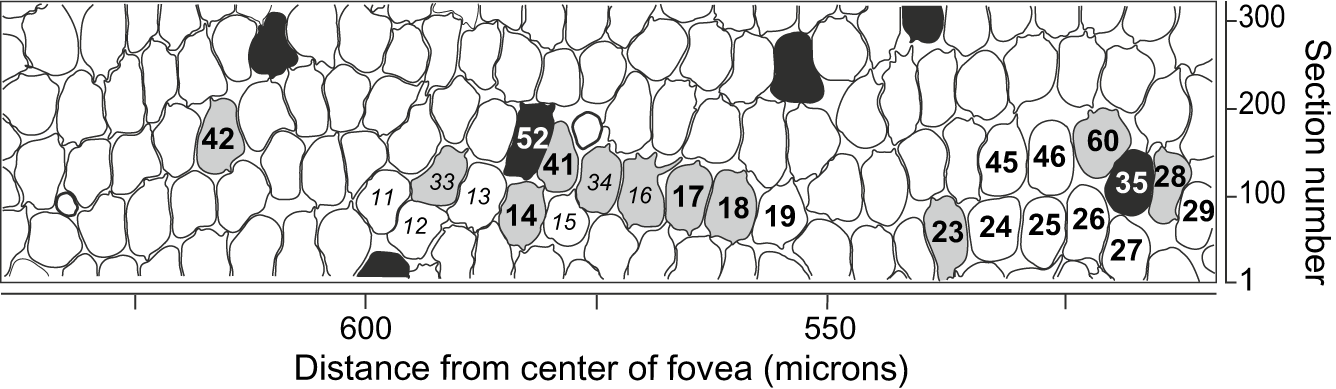
After the sections were mounted and further stained with uranyl acetate and lead citrate, we imaged a region of the retina from 480 to 640 μm nasal to the center of the fovea en montage at 400×, 2000×, and 5000× onto film (negatives) in an electron microscope, a region containing more than 100 cone terminals (Fig. 1). The six black-filled cone terminals in Fig. 1 had been determined to be S-cone terminals based on their postsynaptic connectivity (Herr et al., Reference Herr, Klug, Sterling and Schein2003; Klug et al., Reference Klug, Herr, Ngo, Sterling and Schein2003). The 16 non-S-cone terminals labeled with larger bold numbers in the figure were reimaged at 10,000×. Negatives were printed with 2.5 times or 3.5 times enlargement. Because of lateral displacement of photoreceptor terminals from inner segments (Schein, Reference Schein1988), the center of the imaged region corresponded to ~1 degree of eccentricity in the visual field (Calkins et al., Reference Calkins, Schein, Tsukamoto and Sterling1994). The macaque fovea has a radius of 500 μm, or 2.5 deg, so the photoreceptors that contacted this region were well within the fovea (Polyak, Reference Polyak1941).
Two groups of non-S-cone terminals may be distinguished by differing numbers of basal contacts onto OFF midget bipolar cells and Outer Densities within those contacts. Black-filled outlines are S-cone terminals. Of the 23 numbered non-S-cone terminals, the 12 unfilled ones make more basal contacts onto their OFF midget bipolar cells than the 11 gray-filled ones. The 16 with bold numbers were studied at higher magnification than the 7 with regular numbers. The figure, a tangential view reconstructed from serial vertical sections, is built on the maps of cone terminals presented in Tsukamoto et al. (Reference Tsukamoto, Masarachia, Schein and Sterling1992) and Herr et al. (Reference Herr, Klug, Sterling and Schein2003).

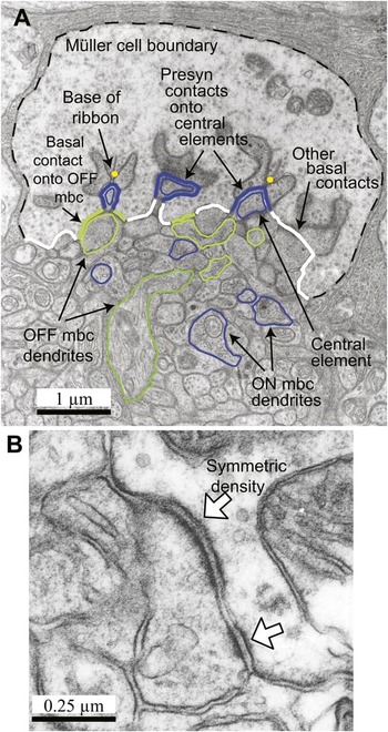
A “hybrid synaptic contour” of the presynaptic face of a cone terminal (Fig. 2A) follows the membrane of the terminal but excludes terminal membrane in apposition to lateral elements supplied by horizontal cells. There, the hybrid synaptic contour detours from the terminal to the regions of apposition between the central elements and the lateral elements, hence the word “hybrid.” For further details, see Herr et al. (Reference Herr, Klug, Sterling and Schein2003, 2011). From digitized tracings of sequential hybrid synaptic contours, we reconstruct the “hybrid synaptic surface” of the presynaptic face of a cone terminal in three dimensions by use of the Montage program (Smith, Reference Smith1987). The hybrid synaptic surface thus shows mainly the presynaptic face of the cone terminal, but it tracks over the central elements supplied by ON bipolar cells instead of following cone terminal membrane over lateral elements. It thus allows visualization of the synaptic clefts within which glutamate neurotransmitter—released at the active zones near the bases of synaptic ribbons—may diffuse to reach regions of the synaptic terminal in contact onto bipolar cell dendrites. It also permits visualization of the basal surface of the cone terminal from a vantage point inside the cone terminal, a vantage point unobscured by the lateral elements supplied by horizontal cells. In Figs. 3–7, each string of yellow spheres marks the base of a synaptic ribbon, and depending on the figure, regions colored blue, green, and red respectively mark central elements, contacts onto an OFF midget bipolar cell, and Outer Densities within those contacts.
A hybrid synaptic contour of a foveal cone terminal (#17) that contacts one OFF midget bipolar cell and one ON midget bipolar cell. (A) Each yellow dot marks the base of a synaptic ribbon in the cone terminal. Thick blue contour elements mark regions of the terminal that are presynaptic to central elements provided by the ON midget bipolar cell (mbc) (thin blue contours). Thick green contour elements mark basal contacts that are presynaptic to the OFF midget bipolar cell (thin green contours). Thick white contour elements mark other basal contacts. The dashed black contour marks the Müller cell boundary of the terminal. Contours such as this one, excluding terminal membrane apposed to lateral elements, are used to assemble the three-dimensional hybrid synaptic surface of the terminal. Original negative imaged at 10,000× magnification. (B) This basal contact region between the cone terminal and a dendrite of its OFF midget bipolar cell contains two dense regions with symmetrically thickened pre- and postsynaptic membranes (open arrows). The interior of the cone terminal is to the right of the OFF midget bipolar cell dendrite. Original negative imaged at 10,000× magnification.

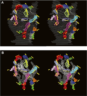
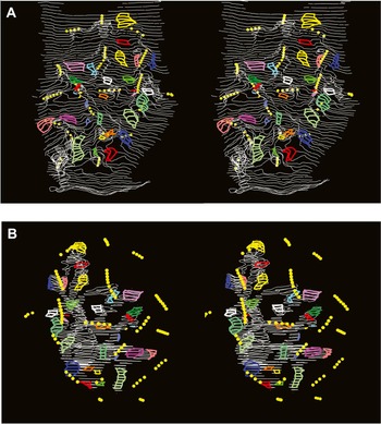
Cone terminal #17, with 19 synaptic ribbons, makes 16 invaginating contacts onto its ON midget bipolar cell. Stereo depth is described for cross fusers. (A). The three-dimensional reconstruction of the hybrid presynaptic surface of the terminal of non-S cone #17 is shown in stereo from a vantage point inside the cone terminal, so the bases of the synaptic ribbons (strings of yellow spheres) are closest to the reader, and the invaginations appear behind as bulges that rise toward the reader. Colored patches show the parts of the presynaptic surface contacting 16 central elements at the tips and distal sides of dendrites provided by the ON midget bipolar cell. (B) The corresponding surface of the dendritic arbor of the ON midget bipolar cell postsynaptic to cone terminal #17 is shown in stereo from the same vantage point, inside the cone terminal. Colored patches are postsynaptic to the cone terminal and correspond to the patches in part (A). To assist mental alignment (of A and B), this part also shows in front the bases of the synaptic ribbons (strings of yellow spheres) from inside the cone terminal.

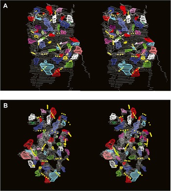
Cone terminal #25, with 17 synaptic ribbons, makes 16 invaginating contacts onto its ON midget bipolar cell. The depth relationships in this stereo figure are the same as described in the legend of Fig. 3.

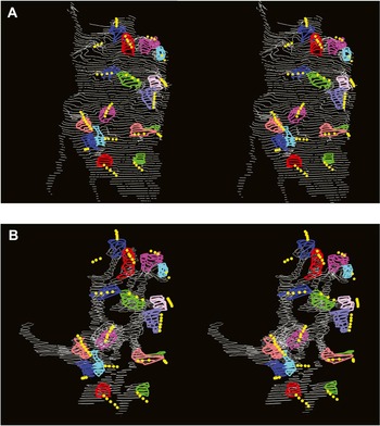
Cone terminal #17 makes 30 basal contacts onto its OFF midget bipolar cell. Colored regions mark contacts between (A) the presynaptic surface of terminal #17 and (B) the postsynaptic surface of its OFF midget bipolar cell. Viewed with cross fusion, the stereo view of the presynaptic surface of the cone terminal in part (A) is shown from the vantage point of the bipolar cell. The surface is rotated in this way so that the presynaptic contacts, which are on the basal surface of the terminal, may be seen unimpeded by the invaginations, here pointing away from the reader. The basal surface is thus closest to the reader, the invaginations farther away, and the synaptic ribbons still farther away. The stereo view of the OFF midget bipolar cell in part (B) is shown from the same vantage point as in the prior figures, namely, from inside the cone terminal. We add the synaptic ribbons from the interior of the cone terminal to help the reader to mentally align the pre- and postsynaptic surfaces.

Cone terminal #25 makes 49 basal contacts onto its OFF midget bipolar cell. The depth relationships in stereo parts (A and B) are as the same as described in the legend of Fig. 5.

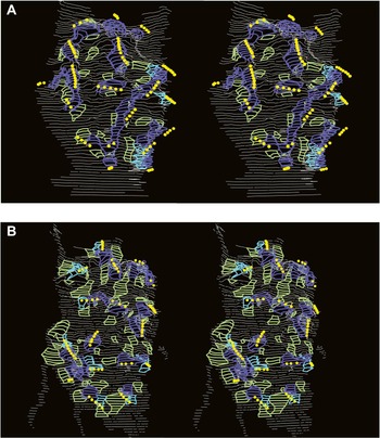
Nearly all the basal contacts from the cone onto the OFF midget bipolar cell are in the triad-associated position, adjacent to the contacts onto central elements. (A) The presynaptic face of cone terminal #17 makes 30 basal contacts (green) with its OFF midget bipolar cell. It also contacts 16 central elements (blue) provided by its ON midget bipolar cell and 3 central elements (cyan) provided by ON diffuse bipolar cells. (B) The presynaptic face of cone terminal #25 makes 49 basal contacts (green) onto its OFF midget bipolar cell. It also contacts 16 central elements (blue) provided by its ON midget bipolar cell and 8 central elements (cyan) provided by ON diffuse bipolar cells.

We used the ContourFitter program (Meyers et al., Reference Meyers, Skinner and Sloan1992) to tile the wire-frame models of hybrid surfaces with triangles, the edges of which were ~0.03 μm long within the plane of a section, the distance between digitized points. To visualize the surfaces, we used Geomview (http://www.geomview.org/) and rendered them with the Blue Moon Rendering Tools (http://en.wikipedia.org/wiki/Blue_Moon_Rendering_Tools). We also measured surface areas from the tiled surfaces. With SPSS 15.0 software, we ran Shapiro–Wilk tests for normality of distributions, Mann–Whitney U nonparametric tests to determine if distributions were significantly different, and two types of cluster analyses (K-means and hierarchical clustering with the between-group linkage procedure).
Data from images
We scanned electron micrograph prints for Fig. 2 with an Agfa scanner at a resolution of 1200 dpi. Except for linear adjustment for brightness and contrast in Adobe Photoshop 7.0, we did no retouching or other image manipulation.
Results
Contacts onto ON midget bipolar cells
We initially reconstructed the complete dendritic arbors of 23 ON midget bipolar cells, each contacted by one non-S-cone terminal, from 2.5× enlargements of 2000× negatives. Later, we reconstructed eight of them from 3.5× enlargements of 5000× negatives and 2.5× enlargements of 10,000× negatives (Fig. 1). Their invaginating dendrites provide central elements of synaptic triads, typically one central element per triad in foveal non-S cones (Herr et al., Reference Herr, Klug, Sterling and Schein2003).
Viewed with cross fusion, stereo Figs. 3A and 4A show the hybrid synaptic surfaces of the presynaptic terminals of two non-S cones, #17 and #25, from a vantage point inside each cone terminal. From inside, each synaptic ribbon—its base marked by a string of yellow spheres—is closest to the viewer, and the invaginations due to postsynaptic central elements appear behind the yellow spheres as hills that bulge toward the viewer. Stereo Figs. 3B and 4B show the corresponding surfaces of the dendritic arbor of the ON midget bipolar cells postsynaptic to these two terminals from the same vantage point, inside the cone terminal. To help readers mentally align the presynaptic surfaces (Figs. 3A and 4A) and the postsynaptic surfaces (Figs. 3B and 4B), the latter figures also display the synaptic ribbons located within the interior of the cone terminal.
Each of stereo Figs. 3 and 4 shows in three dimensions the regions of contact between the presynaptic hybrid surface of a cone terminal and the postsynaptic surface of its ON midget bipolar cell. Correspondingly colored pre- and postsynaptic regions mark the invaginating central elements, aligned beneath synaptic ribbons. However, as can be seen in Fig. 2A, it is the sides of a central element that actually appose the cone terminal. There, the membranes are asymmetrically dense, thicker presynaptically, marking the site of mGluR6 receptors (Nomura et al., Reference Nomura, Shigemoto, Nakamura, Okamoto, Mizuno and Nakanishi1994; Vardi et al., Reference Vardi, Morigiwa, Wang, Shi and Sterling1998, Reference Vardi, Duvoisin, Wu and Sterling2000; Ahmad et al., Reference Ahmad, Klug, Herr, Sterling and Schein2003). By contrast, the tip of a central element, a strip within each colored region underneath the synaptic ribbon, is not directly apposed to the cone terminal and does not harbor glutamate receptors.
Cone terminal #17 had 19 synaptic ribbons and was presynaptic to 16 central elements provided by the dendrites of its ON midget bipolar cell. (Some ribbons contacted only central elements provided by the dendrites of ON diffuse bipolar cells.) Cone terminal #25 had 17 synaptic ribbons and was also presynaptic to 16 central elements provided by its ON midget bipolar cell (Figs. 4A and 4B).
Contacts onto OFF midget bipolar cells
We also initially reconstructed the complete dendritic arbors of the OFF midget bipolar cells for the same 23 terminals and identified their basal contacts, and later, we reconstructed 16 of them from 3.5× enlargements of 5000× negatives and 2.5× enlargements of 10,000× negatives (Fig. 1). Each of stereo Figs. 5 and 6 shows in three dimensions the regions of contact between the presynaptic hybrid surface of a cone terminal (Figs. 5A and 6A) and the corresponding postsynaptic surface of its OFF midget bipolar cell (Figs. 5B and 6B). Unlike the prior figures, viewed with cross fusion, Figs. 5A and 6A show the hybrid synaptic surfaces of the presynaptic terminals of non-S cones #17 and #25 from a vantage point inside the bipolar cell. We rotate the presynaptic surfaces in this way to see the basal contacts of the presynaptic terminals unimpeded by invaginations. As in the prior figures, however, stereo Figs. 3B and 4B show the corresponding postsynaptic surfaces of the dendritic arbor of the two OFF midget bipolar cells from a vantage point inside the cone terminal. To align pre- and postsynaptic surfaces, readers can mentally rotate one or the other.
Cone terminal #17 was presynaptic to its OFF midget bipolar cell at 30 basal contacts (Figs. 5A and 5B). The average surface area of basal contacts was 0.15 ± 0.11 μm2 (mean ± s.d.; N = 30), giving a total surface area of basal contact of 4.4 μm2. Some contacts had one region with symmetrically thickened membranes, but many contacts had more than one dense region (Fig. 2B). Overall, the 30 basal contacts contained 41 of these Outer Densities (21 contacts with 1, 7 with 2, and 2 with 3). The average surface area of these Outer Densities was 0.08 ± 0.05 μm2 (mean ± s.d.; N = 41), giving a total surface area of Outer Density of 3.2 μm2.
Cone terminal #25 was presynaptic to its OFF midget bipolar cell at 49 basal contacts (Fig. 6A and 6B). The average area of each basal contact was 0.20 ± 0.15 μm2 (N = 49), giving a total surface area of basal contact of 9.6 μm2. These contacts contained 68 Outer Densities (32 contacts with 1, 12 with 2, and 4 with 3) with an average area of 0.09 ± 0.06 μm2 (N = 68), giving a total surface area of Outer Density of 6.3 μm2. The numbers of contacts and of Outer Densities for cone terminal #25 were notably greater (1.6× and 1.7X) than those for terminal #17. The total surface areas of basal contact and of outer density were larger by an even greater factor (2.2× and 2.0×).
Of 649 basal contacts made by the 16 cone terminals onto OFF midget bipolar cells, 436 (67%) had one Outer Density, 166 (26%) had two, 37 (6%) had three, 8 (1%) had four, 1 had five, and 1 had seven. Sixteen contacts (2.5%) showed no Outer Density; these probably had dense regions that were invisible because of the plane of section. Thus, all or almost all basal contacts had at least one Outer Density, and each harbored an average of 1.4.
Basal contacts in the triad-associated position
In cross section, basal contacts onto OFF midget bipolar cells often—but not always—appear adjacent to contacts onto central elements (e.g., Fig. 2A), a position that has been called “triad associated” (Kolb, Reference Kolb1970; Boycott & Hopkins, Reference Boycott and Hopkins1991; Hopkins & Boycott, Reference Hopkins and Boycott1992, Reference Hopkins and Boycott1997). Our reconstructions permitted us to investigate this relationship at a unique level of completeness and detail. For cone terminals #17 and #25, stereo Fig. 7A and 7B shows the 30 and 49 basal contacts (green), respectively, together with all the regions presynaptic to central elements provided by ON midget (blue) and ON diffuse (cyan) bipolar cells.
All but one of the basal contacts between cone terminal #17 and its OFF midget bipolar cell were in the characteristic triad-associated position (Fig. 7A). Most of the basal contacts appeared to be associated with just one of the 19 synaptic ribbons, but several in the lower left region of the figure appeared to be associated with more than one. Conversely, some ribbons appeared to be associated with no basal contacts onto the OFF midget bipolar cell, whereas some ribbons appeared to be associated with one, two, and even as many as five.
Similarly, all but one of the basal contacts between cone terminal #25 and its OFF midget bipolar cell were in the characteristic triad-associated position (Fig. 7B). Nearly all the basal contacts appeared to be associated with just one synaptic ribbon, and very few appeared to be associated with more than one. Conversely, several of the 17 ribbons appeared to be associated with one basal contact, about half of the 17 ribbons appeared to be associated with two, and several appeared to be associated with more than two, even up to five.
Two populations of non-S cones
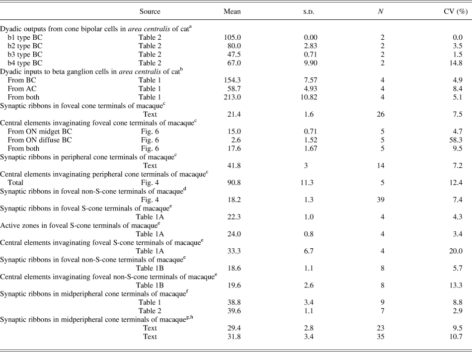
Counts from electron microscope reconstructions or fluorescence microscopy after single labeling of a homogeneous population of objects (e.g., synaptic ribbons, invaginating central elements, basal contacts, receptor patches) generally follow the “10% rule,” that is, they have standard deviations that are typically ~10% (or less) of the mean, except where counts are very low (Table 1) (e.g., Cohen & Sterling, Reference Cohen and Sterling1990, Reference Cohen and Sterling1992; Chun et al., Reference Chun, Grünert, Martin and Wässle1996; Hopkins & Boycott, Reference Hopkins and Boycott1997; Haverkamp et al., Reference Haverkamp, Grünert and Wässle2001; Herr et al., Reference Herr, Klug, Sterling and Schein2003; Klug et al., Reference Klug, Herr, Ngo, Sterling and Schein2003). Because cone terminals #17 and #25 differed so greatly from each other in their numbers of basal contacts and Outer Densities onto OFF midget bipolar cells, we reconstructed the dendritic arbors of the OFF midget bipolar cells postsynaptic to 14 more non-S-cone terminals from high-magnification photomicrographs, as we had done for cone terminals #17 and #25, and counted the numbers of basal contacts. As will be noted in a later section, these 16 cone terminals (including #17 and #25) were among 23 non-S-cone terminals that we had previously reconstructed from lower magnification material.
The 10% rule

CV, coefficient of variation; BC, bipolar cells; AC, amacrine cells.
a Cohen and Sterling (Reference Cohen and Sterling1990).
b Cohen and Sterling (Reference Cohen and Sterling1992).
c Chun et al. (Reference Chun, Grünert, Martin and Wässle1996).
d Klug et al. (Reference Klug, Herr, Ngo, Sterling and Schein2003).
e Herr et al. (Reference Herr, Klug, Sterling and Schein2003).
f Hopkins and Boycott (Reference Hopkins and Boycott1997).
g Haverkamp et al. (Reference Haverkamp, Grünert and Wässle2001).
h From light microscope, not electron microscope.
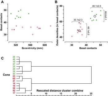
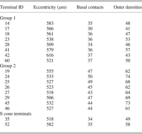
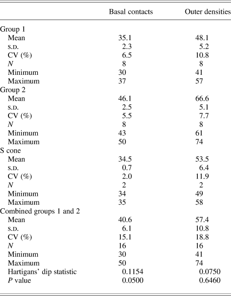
Within the narrow range of eccentricities studied in the fovea (Fig. 1), the numbers of basal contacts were not dependent on eccentricity (Fig. 8A). As shown in Tables 2 and 3 and Fig. 8B, eight terminals made 35.1 ± 2.3 (mean ± s.d.; N = 8) (range 30–37) basal contacts (red-open diamonds in Fig. 8B), whereas the other eight made 46.1 ± 2.5 (N = 8) (range 43–50) (green-filled diamonds in Fig. 8B). The coefficients of variation, 6.5 and 5.5% (Table 3), were each within the 10% rule for counts that characterize a single population (Table 1). By contrast, lumping the data for the 16 cones would have given 40.6 ± 6.1 (N = 16) basal contacts, a coefficient of variation of 15.1%, and a wide range of 30–50 (Table 3). Moreover, Hartigans’ dip statistic was 0.1154 (P = 0.050) (Table 3), consistent with a bimodal distribution of the number of basal contacts (Hartigan & Hartigan, Reference Hartigan and Hartigan1985; Mechler & Ringach, Reference Mechler and Ringach2002). The numbers of basal contacts in the first group of eight were not normally distributed [P = 0.017 given a Shapiro–Wilk statistic of 0.779 with 8 degrees of freedom (df)]. Even though the numbers in the second group were normally distributed (P = 0.503 given a Shapiro–Wilk statistic of 0.929 with df = 8), comparison of the two groups thus required a nonparametric statistic. Based on the nonparametric Mann–Whitney U test, the distributions of the numbers of basal contacts in the two groups of eight were significantly different [Z = −3.378, P (two-tailed) = 3 × 10⁻⁴].
Based on the number of basal contacts between a non-S-cone terminal onto its OFF midget bipolar cell and the number of Outer Densities within those basal contacts, these cones and OFF midget bipolar cell each consist of two clusters. (A) Over this small range of eccentricity, numbers of basal contacts are independent of eccentricity. Green-filled diamonds are members of one cluster [see part (B)]; red-open diamonds are members of the other. The violet-filled squares represent the data for two S cones (#35 and #52). (B) By itself, the number of basal contacts made by a cone terminal onto its OFF midget bipolar cell falls into two groups with no overlap. Likewise, the number of Outer Densities falls into two groups with no overlap. Within each group, the number of Outer Densities is only weakly related to the number of basal contacts: The correlation coefficient R 2 for the left group (#Outer Densities = 1.19 × #basal contacts + 6.33) is 0.28; that for the right group (#Outer Densities = 0.9443 × #basal contacts + 23.07) is 0.22. The pairs of data for the 16 cone-to-OFF bipolar cell circuits reveal two clusters. All the points in each cluster are closer to that cluster’s centroid than to the other cluster’s centroid (open black circles). The pair of data for S cones (violet-filled squares) places them among the cluster with smaller numbers (red-open diamonds). (C) Based on a within-group hierarchical clustering analysis and taking into account the numbers of basal contacts and of Outer Densities, the rescaled dendrogram shows the same two clusters.

Numbers of interactions between a cone terminal and its OFF midget bipolar cell

Statistics for the numbers of interactions between a cone terminal and its OFF midget bipolar cell

CV, coefficient of variation.
Similarly, the numbers of Outer Densities were independent of eccentricity and nonoverlapping (Fig. 8B; Tables 2 and 3). The coefficients of variation were 10.8 and 7.7%, whereas that of the combined groups would have been 18.8%. Based on the Shapiro–Wilk statistic (P = 0.960 and 0.237, given Shapiro–Wilk values of 0.979 and 0.891, each with df = 8), the distributions of the numbers of Outer Densities were both normal. The means were significantly different (unpaired t-test for independent samples: t = 2.54; P = 4.8 × 10⁻⁶); alternatively, based on the nonparametric Mann–Whitney U values, the distributions were significantly different [Z = −3.363, P (two-tailed) = 8 × 10⁻⁴].
The graph of number of Outer Densities against number of basal contacts (Fig. 8B) shows two clusters (Tan et al., Reference Tan, Steinbach and Kumar2006). All the points in the one cluster of points with red-open diamonds are closer (as measured by Euclidian distance) to that cluster’s centroid (35.1, 48.1; black circle) than to the centroid of the other cluster with green-filled diamonds (46.1, 66.6; black circle) and vice versa, consistent with a K-means clustering algorithm with K = 2. (Indeed, consideration of just the number of basal contacts or just the number of Outer Densities produces the same two clusters.) By taking the sum of squares of the “errors” (Euclidian distances from each point to the centroid of its cluster), dividing by the number of points, and taking the square root, we can calculate the “average” distance of a point from its centroid. This distance, 5.3, is smaller than the distance that separates the two centroids, 21.5, by the factor 0.25, a measure of the cohesion of the points in the clusters.
On the basis of the two variables and the squared Euclidian distances between them, the 16 cones were grouped into the same two clusters by hierarchical clustering with the between-group linkage procedure (Fig. 8C). The large (rescaled) distance to join the two clusters, compared to the distance to join the individual cones into each cluster, is strong evidence for two clusters, not one and not three or more. Below, we speculate that the cluster with smaller numbers of basal contacts and Outer Densities represents L cones, whereas the cluster with larger numbers represents M cones, so we color the members of the first cluster red, the members of the second green.
We reported that S-cone terminals #35 and #52 made 34 and 35 basal contacts with their OFF midget bipolar cells and that these contacts harbored 49 and 58 Outer Densities (Klug et al., Reference Klug, Herr, Ngo, Sterling and Schein2003). These pairs of numbers (violet squares in Fig. 8A and 8B) resemble those of the group of non-S cones with smaller numbers (red-open diamonds in Fig. 8A and 8B).
These results encouraged us to reconstruct dendritic arbors of six more non-S-cone ON midget bipolar cells (for a total of eight) from high-magnification material and to count their central elements. Based on the grouping established by the numbers of basal contacts, the first group of four cone terminals was presynaptic to an average of 15.2 ± 2.2 central elements—16, 13, 18, and 14 provided by ON midget bipolar cells #17, #18, #23, and #42. By comparison, the second group of four cone terminals was presynaptic to 16.0 ± 0.0 central elements—16, 16, 16, and 16 provided by ON midget bipolar cells #19, #25, #26, and #46. Clearly, these numbers of central elements did not distinguish the two groups of non-S cones.
Low versus high magnification
As noted above, initially we had randomly chosen 23 non-S-cone terminals and their midget bipolar cells from low-magnification electron photomicrographs (negatives taken at 2000× and enlarged 2.5×). Reconstructing from this material, we found 11 cone terminals making fewer basal contacts (21–23) onto OFF midget cells and 12 cone terminals making more (29–32). Thus, the two types of non-S cones were similarly numerous.
We were concerned that the reconstructions of the dendritic arbors of OFF midget bipolar cells from the low-magnification material might be incomplete, so we randomly selected eight cone terminals of each group from the 23 circuits and reconstructed them and their OFF midget bipolar cells from 5000× negatives enlarged 3.5× and/or from 10,000× negatives enlarged 2.5×. (The larger bold labels in Fig. 1 identify the 16 circuits that were reconstructed from higher magnification material.) As reported above (Fig. 8), the numbers of basal contacts, 30–37 and 43–50, were indeed higher. However, for the same cells reconstructed from both higher magnifications, the counts from 10,000× negatives enlarged 2.5× produced no additional basal contacts or Outer Densities beyond those counted in the 5000× negatives enlarged 3.5×. We conclude that the latter were of sufficiently high magnification to enable us to fully reconstruct the dendritic arbors and to identify all the basal contacts and their Outer Densities. Nonetheless, even the incomplete reconstructions from low-magnification material produced nonoverlapping count distributions and divided the non-S cones into the same two groups.
Discussion
Two groups of non-S cones
Non-S cones in the fovea form two clusters (Fig. 8B and 8C) on the basis of the numbers of basal contacts and symmetric Outer Densities made by their synaptic terminals onto their OFF midget bipolar cells. We hypothesize that that these two groups of non-S cones are L- and M-type cones. If so, these would be the first anatomical differences to be identified between L- and M-type cones. Likewise, these would be the first anatomical differences to be identified between L- and M-type OFF midget bipolar cells.
In human eyes, the ratio of the numbers of L to M cones averages around 2 (Cicerone & Nerger, Reference Cicerone and Nerger1989; Hagstrom et al., Reference Hagstrom, Neitz and Neitz1998; Roorda & Williams, Reference Roorda and Williams1999; Otake & Cicerone, Reference Otake and Cicerone2000; Carroll et al., Reference Carroll, Neitz and Neitz2002; Hofer et al., Reference Hofer, Carroll, Neitz, Neitz and Williams2005), but in macaque monkey eyes, the ratio is closer to 1 (Mollon & Bowmaker, Reference Mollon and Bowmaker1992; Packer et al., Reference Packer, Williams and Bensinger1996; Jacobs & Deegan, Reference Jacobs and Deegan1997; Deeb et al., Reference Deeb, Diller, Williams and Dacey2000; Dobkins et al., Reference Dobkins, Thiele and Albright2000; Roorda et al., Reference Roorda, Mehta, Lennie and Williams2001; Bowmaker et al., Reference Bowmaker, Parry, Mollon, Mollon, Pokorny and Knoblauch2003), as we find here. Thus, in monkey, the relative number in the two groups of non-S cones cannot tell us which group might be the L cones and which might be the M.
However, we have one clue. S cones are responsible not only for the blueness of short-wavelength light, signaled through the blue–yellow color opponent channel, but also for the redness of short-wavelength light, signaled through the red–green color opponent channel (Hering, Reference Hering1878, Reference Hering, Hurvich and Jameson1920; Hurvich & Jameson, Reference Hurvich and Jameson1957; Wooten & Werner, Reference Wooten and Werner1979; Shevell & Humanski, Reference Shevell and Humanski1988; Rodieck, Reference Rodieck, Valberg and Lee1991; Wuerger et al., Reference Wuerger, Atkinson and Cropper2005). Thus, L cones ally with S against M in the psychophysical red–green color opponent channel. Indeed, based on psychophysical findings (Shinomori et al., Reference Shinomori, Spillmann and Werner1999), Klug et al. (Reference Klug, Herr, Ngo, Sterling and Schein2003) suggested that the redness of short-wavelength light is signaled by S-cone OFF midget bipolar cells but not by S-cone ON bipolar cells. Because the numbers of basal contacts and Outer Densities for the S cone-to-OFF midget bipolar cell circuits #35 and #52 (violet-filled squares) lie among the cluster of non-S cones with similarly smaller numbers (red-open diamonds in Fig. 8B), we speculate that the small-numbers of cluster corresponds to the psychophysically allied L cones, whereas the large-numbers cluster (green-filled diamonds in Fig. 8C) corresponds to the antagonist M cones. A greater number of synapses onto these M-cone OFF midget bipolar cells than those driven by L cones might compensate for M cones catching fewer photons on average than L cones. Such compensation could help set the wavelength of unique yellow (Brainard et al., Reference Brainard, Roorda, Yamauchi, Calderone, Metha, Neitz, Neitz, Williams and Jacobs2000).
Further testing
We studied just 16 non-S cones and their interactions with their OFF midget bipolar cells from one monkey fovea at high magnification. Even including the non-S cones studied at lower magnification, bringing the number to 23, the sample was not large—though from the point of view of the effort involved—the number was very large indeed. But, given the fairly narrow count distributions expected for data from electron micrographic reconstructions of one type of cell at one eccentricity and the results of cluster analysis, the data are consistent with two types of non-S cone. Nonetheless, it would be desirable to obtain more of these synaptic counts between cones and their OFF midget bipolar cells from more monkey retinas and at a variety of eccentricities.
In addition, taking advantage of regional characterization of L:M cone ratios (Kuchenbecker et al., Reference Kuchenbecker, Sahay, Tait, Neitz and Neitz2008), our tentative assignment (fewer basal contacts and Outer Densities = L; more = M) could be tested by study of human retina. Indeed, a definitive test of this assignment could be to immunochemically mark and count basal contacts and their Outer Densities in a patch of retina in which the cone array had been imaged previously (Roorda et al., Reference Roorda, Mehta, Lennie and Williams2001; Hofer et al., Reference Hofer, Carroll, Neitz, Neitz and Williams2005).
A promising start was made by Telkes et al. (Reference Telkes, Lee, Jusuf and Grünert2008), who used fluorescence immunocytochemistry to visualize cone synaptic terminals and the dendritic tips of OFF midget bipolar cells in marmoset retina. That study reported a distribution, 18.7 ± 4.0 (mean ± s.d.), in the number of dendritic tips per cone pedicle for central retina (<12 degrees of eccentricity), with a coefficient of variation of 21%. As the authors comment in a different but related context, exact eccentricity-matched samples would be required to establish whether there is a consistent difference. Moreover, the authors also caution there and in Eriköz et al. (Reference Eriköz, Jusuf, Percival and Grünert2008) that their counts are lower than those found in electron microscopic studies that are by their nature of much higher resolution (e.g., Kolb, Reference Kolb1970; Boycott & Hopkins, Reference Boycott and Hopkins1991; Hopkins & Boycott, Reference Hopkins and Boycott1997; Calkins & Sterling, Reference Calkins and Sterling2007).
Outer plexiform layer versus inner plexiform layer
Electron microscopic reconstruction has potential for error, particularly in tracking all the parts of a synaptic terminal or a dendritic arbor. For example, as reported in Results, we generally found more basal contacts onto OFF midget bipolar cell dendrites from 17,500× prints (5000× negatives enlarged 3.5×) than from 5000× prints (2000× negatives enlarged 2.5×). However, we confirmed the counts from 17,500× prints with counts from 25,000× prints (10,000× negatives enlarged 2.5×). Therefore, we have confidence in the counts reported here, all of which are based on reconstructions of the outer plexiform layer from 17,500× or 25,000× images, but we would not have confidence in counts from 5000× images.
From study of midget bipolar terminals in the inner plexiform layer of the same tissue as described here but from prints at just 5000×, Calkins (Reference Calkins1994) and Calkins et al. (Reference Calkins, Schein, Tsukamoto and Sterling1994) reported that counts of synaptic ribbons in ON–OFF pairs of midget bipolar terminals fell cleanly into two groups, one with 32 ± 3 ribbons and the other with 49 ± 3. However, we suspect that many of the bipolar terminals in that study, specifically the ones with fewer ribbons, may have been incompletely reconstructed. It is also possible that the higher ribbon count in some of the bipolar terminals could have been due to mistaken connection to parts of other terminals.
Our suspicion has two grounds. First, while 2000× negatives enlarged 2.5× (5000× prints) are inadequate for complete reconstruction of processes in the outer plexiform layer, as we report here, they should be even more problematic for the finer processes in the inner plexiform layer. Second, a midget bipolar terminal has many lobes, some connected to the main terminal body by very fine processes that are very difficult to follow. And, complete reconstruction of the dendritic arbor of that terminal’s midget ganglion cell, which helps to demarcate the bipolar terminal, includes fine processes that are similarly difficult to follow. As a result, although counting ribbons is not particularly difficult in bipolar terminals, identifying each and every part of a bipolar terminal before counting the ribbons is difficult. Correspondingly, we (Eggers et al., Reference Eggers, Sterling and Schein1999; and unpublished results) reconstructed several midget bipolar terminals and the dendritic arbors of their midget ganglion cells first at 5000× and then again at 17,500×. In the higher magnification material, we found more terminal lobes and consequently more ribbons.
Indeed, of the eight cone-to-OFF midget bipolar circuits for which we here report small numbers of basal contacts and Outer Densities, Calkins (Reference Calkins1994) reported that five (14, 17, 18, 23, and 60) have small numbers of ribbons in their midget bipolar terminals, but two (28 and 41) have large numbers. [One (42) could not be studied because of the slight tilt of the plane of section from vertical in the series.] Of the eight circuits that here we report as having large numbers of basal contacts and Outer Densities, Calkins (Reference Calkins1994) reported that two (19 and 29) have large numbers of ribbons in their midget bipolar terminals, but six (24, 25, 26, 27, 45, and 46) have small numbers. We agree in just 7 of 15 cases. For all these reasons, we no longer have confidence in the counts of synaptic ribbons in midget bipolar terminals from study of the inner plexiform layer at low magnification (5000×) or in a grouping based on those numbers (Calkins, Reference Calkins1994; Calkins et al., Reference Calkins, Schein, Tsukamoto and Sterling1994). We would have confidence in these counts had reconstruction been performed from higher magnification material.
From fluorescence microscopy of common marmoset and macaque monkey retinas, Jusuf et al. (Reference Jusuf, Martin and Grünert2006) counted synaptic ribbons among midget bipolar terminals over a wide range of eccentricity, lumped the counts, found distributions of counts that were very broad and unimodal, and concluded that the bimodal distribution in Calkins et al. (Reference Calkins, Schein, Tsukamoto and Sterling1994) must have been in error. Although we agree with that conclusion, we do not agree with the reasoning: Numbers of synaptic ribbons in both cone terminals and bipolar terminals increase with eccentricity, so lumping counts over eccentricity would smear any underlying bimodal distribution and thus obviate any test of bimodality.
Arrangement of the triad-associated contacts of an OFF midget bipolar cell
Our results resolve a long-standing disagreement. From study of four Golgi-filled OFF midget bipolar cells, Kolb (Reference Kolb1970) suggested that two OFF midget basal contacts were associated with each synaptic ribbon, one on each side. (See also fig. 3.7 of Dowling, Reference Dowling1987.) But, from study of one Golgi-filled OFF midget bipolar cell, Boycott and Hopkins (Reference Boycott and Hopkins1991) found one triad-associated contact near some ribbons and none near others. The two reconstructions in Figs. 7 and 8 show that with rare exception the basal contacts onto OFF midget bipolar cells are indeed adjacent to central elements, that is, in the triad-associated position (Figs. 7 and 8). However, in both of these terminals, an individual synaptic ribbon may be associated with as few as zero basal contacts onto the OFF midget bipolar cell or as many as five. Thus, the simple rule—two triad-associated contacts astride each ribbon onto an OFF midget bipolar cell—does not hold, at least in the sample studied here.
Acknowledgments
We thank Vanessa Preciado for help with figures and the late John (“Jak”) Ribble for help with digitizing; Dr. Yoshihiko Tsukamoto, Patricia Masarachia, and Sally Shrom for preparing the electron microscope material; and Kazuki Uema and Lisa Travis for printing. We also thank Dario Ringach for help with the Hartigans’ dip test. S.S., K.K., I.T.N., and T.M.H. were supported by the NIH grant EY11153 and by a grant from the UCLA Academic Senate; P.S. was supported by the NIH grant EY08124; and S.H. was supported by the National Institute of Mental Health Training grant MH15795-18 and the NIH grant EY11153.