Introduction
The occurrence of microorganisms smaller than 0.2 μm has been questioned based on the minimum requisite size for a complete set of genomic DNA, ribosomes, proteins, cellular membranes, and other intracellular components (Maniloff Reference Maniloff1997). On the other hand, referring to the genomes of Mycoplasma genitalium and other parasitic bacteria, some argued that microbial cells can theoretically be as small as 0.14 μm in diameter (Mushegian & Koonin Reference Mushegian and Koonin1996). The debate regarding the minimal cell size of microorganisms remains unresolved.
Concurrent with this theoretical discussion, research on microorganisms that pass through 0.2 μm-pore-size filters (0.2 μm-passable microorganisms) has been ongoing since the 1970s. For example, it has been demonstrated that bacteria smaller than 0.2 μm in diameter represent between 1% and 10% of all bacteria found in seawater (Hobbie et al. Reference Hobbie, Daley and Jasper1977, Zimmermann Reference Zimmermann1977, Watson et al. Reference Watson, Novitsky, Quinby and Valois1977). In addition, in coastal waters, where bacterial populations are estimated to range between 108 and 109 cells l-1, Fukuba et al. (Reference Fukuba, Elsaied and Naganuma2002) reported that between 100 and 101 viable bacteria l-1 pass through 0.2 μm-pore-size filters.
In 1981, two types of very small bacteria < 0.3 μm in diameter were successfully cultured from seawater (Torrella & Morita Reference Torrella and Morita1981). One displayed an increase in size following incubation, whereas the other did not increase in size even after incubation and was named ultramicrobacteria (UMB). Since then, further isolations and phylogenetic identification of 0.2 μm-passable microorganisms have been performed, and some of these isolates were UMB (MacDonell & Hood Reference MacDonell and Hood1982, Hood & MacDonell Reference Hood and MacDonell1987, Haller et al. Reference Haller, Rolleke, Vybiral, Witte and Velimirov2000, Elsaied et al. Reference Elsaied, Sato and Naganuma2001, Miteva & Brenchley Reference Miteva and Brenchley2005, Dmitriev et al. Reference Dmitriev, Duda, Suzina, Akimov, Vainshtein, Barinova, Abashina, Oleynikov, Esikova and Boronin2007, Geissinger et al. Reference Geissinger, Herlemann, Morschel, Maier and Brune2009). Hahn et al. (Reference Hahn, Lunsdorf, Wu, Schauer, Hofle, Boenigk and Stadler2003, Reference Hahn, Lang, Brandt, Wu and Scheuerl2009) isolated Actinobacteria and Polynucleobacter Cluster (class Betaproteobacteria) with ultramicro cell sizes (< 0.1 μm3), even under eutrophic culture conditions, and called these obligate UMB. It has also been reported that these obligate UMB are cosmopolitan species found in freshwater regions (Hahn Reference Hahn2003, Hahn et al. Reference Hahn, Lunsdorf, Wu, Schauer, Hofle, Boenigk and Stadler2003).
Recent research has investigated community structure and the functional potential of 0.2 μm-passable populations in the environments using culture-independent methods (Miyoshi et al. Reference Miyoshi, Iwatsuki and Naganuma2005, Naganuma et al. Reference Naganuma, Miyoshi and Kimura2007, Nakai et al. Reference Nakai, Abe, Takeyama and Naganuma2011). Miyoshi et al. (Reference Miyoshi, Iwatsuki and Naganuma2005) reported that c. 65% of all 16S rRNA gene phylotypes from 0.1-μm-pore-size filter-captured microorganisms in deep terrestrial aquifers were affiliated with the candidate divisions OD1 and OP11, potentially representing new bacterial phyla. Naganuma et al. (Reference Naganuma, Miyoshi and Kimura2007) detected novel unaffiliated phylotypes only in the 0.2 μm-passable fraction of deep-sea hydrothermal vent samples. Nakai et al. (Reference Nakai, Abe, Takeyama and Naganuma2011) indicated that the metagenome of 0.2 μm-passable microorganisms in deep-sea hydrothermal fluid contained novel genes pertinent to membrane functions.
These findings show that 0.2 μm-passable populations include some phylogenetically and/or functionally important species which need to be isolated and characterized. In addition, our understanding of the biogeography for 0.2 μm-passable bacteria is at present limited to cosmopolitan species found in freshwater (Hahn Reference Hahn2003, Hahn et al. Reference Hahn, Lunsdorf, Wu, Schauer, Hofle, Boenigk and Stadler2003). Here we tested samples taken from widely dispersed sites for 0.2 μm-passable bacteria and investigated phylogenetic relationships in order to preliminarily test the Baas-Becking hypothesis that “everything is everywhere, but, the environment selects” (Baas-Becking Reference Baas-Becking1934), implying that microscopic organisms do not have any large-scale spatial pattern of distribution. Our results provide new information on the phylogeography of 0.2 μm-passable bacteria.
Materials and methods
Sample collection
A total of 31 samples were collected from various environments, including soil, gravel, seawater, and freshwater, in 26 locations around the world, the majority of which were in Japan. Arctic samples were collected in August 2008 from Troll hot springs (79.4°N, 13.4°W) on Spitzenberg Island in the Svalbard archipelago. This site is the northernmost thermal springs documented on land (Jamtveit et al. Reference Jamtveit, Hammer, Andersson, Dysthe, Heldmann and Vogel2006). Antarctic samples were collected in December 2008 from Byers Peninsula (62°4′S, 60°6′W), Livingston Island, South Shetland Islands. Byers Peninsula is one of the largest ice-free areas in the region and serves as an important reference site (ASPA No. 126) within the Antarctic ecosystem (Quesada et al. Reference Quesada, Camacho, Rochera and Velázquez2009). In addition, gravel was collected from the Gobi Desert and eastern edge of the Sahara Desert in November 2008 and December 2008, respectively. Although a significant number of taxa of dust-borne microorganisms have been isolated and characterized in both African and Asian desert systems, only a few papers have reported a detailed identification of culturable bacteria (Chen et al. Reference Chen, Kobayashi, Yamada, Kim, Iwasaka and Shi2011). Samples were collected using sterile gloves and placed in sterile plastic bottles or sample tubes and stored in the dark at room temperature until processing.
Isolation and cultivation of 0.2 μm-passable bacteria
Bacteria were isolated using a method modified from Elsaied et al. (Reference Elsaied, Sato and Naganuma2001). Liquid samples were filtered through three-fold 0.2 μm filters (Advantec, Tokyo, Japan), and concentrated R2A liquid medium (Massa et al. Reference Massa, Caruso, Trovatelli and Tosques1998) was added to the filtrate to bring the final media concentration to 1×. Solid samples, such as soil or pebbles, were suspended in phosphate buffer solution (8 g l-1 NaCl, 1.1 g l-1 Na2HPO4, 0.2 gl-1 KCl, and 0.2 gl-1 KH2PO4), and the resulting suspension was processed in the same manner as the liquid samples. The enrichment cultures were kept in the dark at room temperature for at least three weeks. To confirm the effectiveness on filtration through three-fold 0.2 μm filters, Escherichia coli K12 (IFO 3301) cultures were used as negative growth control. Subsequently, the culture liquids were spread on 1.5% agar medium containing identical ingredients. Single colonies were isolated and streaked on fresh agar plates to purify colony-forming strains. This procedure was repeated at least three times to ensure reliable colony purification.
Phylogenetic characterization
Genomic DNA extraction was performed as described by Aljanabi & Martinez (Reference Aljanabi and Martinez1997). Bacteria-specific 27F (5′-AGAGTTTGATCCTGGCTCAG-3′) and universal 1492R (5′-GGTTACCTTGTTACGACTT-3′) primers were used to amplify near full-length fragments of the 16S rRNA gene (DeLong Reference DeLong1992). Reaction conditions for polymerase chain reaction (PCR) amplification were the same as those used in a previous report (Naganuma et al. Reference Naganuma, Miyoshi and Kimura2007). After confirming the presence of PCR products of the desired fragment size (c. 1500 base pairs) by electrophoresis on 1.5% agarose gel, PCR products were purified using the QIAquick PCR Purification kit (Qiagen, Valencia, CA, USA). All purified PCR products were sequenced using an ABI 3730XL automatic DNA sequencer (Applied Biosystems, Foster City, CA, USA). The 16S rRNA gene sequences obtained were compared against known sequences using a BLASTN search against the DDBJ/EMBL/GenBank nt-database and the Greengenes prokaryotic multiple sequence alignment (prokMSA) database (DeSantis et al. Reference DeSantis, Hugenholtz, Larsen, Rojas, Brodie, Keller, Huber, Dalevi, Hu and Andersen2006). Greengenes is a dedicated full-length 16S rRNA gene database with a curated taxonomy based on de novo tree inference. The obtained sequences and the highly homologous known sequences were aligned using the CLUSTALX multiple alignment program (Larkin et al. Reference Larkin, Blackshields, Brown, Chenna, McGettigan, McWilliam, Valentin, Wallace, Wilm, Lopez, Thompson, Gibson and Higgins2007). Phylogenetic trees were constructed based on sequences after alignment using the neighbour joining (NJ) method in MEGA 4.0 (Kumar et al. Reference Kumar, Tamura, Dudley and Nei2007).
Registration of the nucleic acid sequences
The 16S rRNA gene sequences obtained in this study have been deposited in the DDBJ/EMBL/GenBank database under the serial accession numbers AB539970–AB540022.
Evaluation of cell size under culture conditions using electron microscopy and filtration
The cell morphology of isolates was examined with an ultra-high-resolution field-emission scanning electron microscope (FE-SEM) (S-5200, Hitachi High Technologies, Tokyo, Japan). The cells were fixed in 1:20 dilution of 25% glutaraldehyde solution, and an appropriate number of cells were collected on a 0.1-μm-pore Anopore inorganic membrane (Anodisc) filter (Whatman, Tokyo, Japan) for observation. The membrane is composed of an alumina matrix that is manufactured electrochemically. Filter-captured cells were dehydrated with a series of ethanol washes (50%, 70%, 90%, 95%, 97.5%, 99.5%) and air-dried. In order to prevent electrification and to generate clear images during electron microscope observation, dehydrated cells were coated with a c. 100-Å-thick (0.1 μm) layer of platinum via ion sputtering (E-1030, Hitachi High Technologies, Tokyo, Japan).
We used E. coli K12 (IFO 3301) as control cells. However, changes in cell morphology were unavoidable during sample preparation, therefore cell size under culture conditions was confirmed by passing culture liquids through three-fold 0.2 μm filters (Advantec, Tokyo, Japan). Concentrated R2A liquid medium (Massa et al. Reference Massa, Caruso, Trovatelli and Tosques1998) was added to the filtrate to bring the final media concentration to 1×. Because the pores of the filter used for filtration are created by exposure to neutron radiation, a portion of the pores overlap. As filters contain a small number of pores that are larger than the nominal pore size, we minimized the possibility of larger cells passing through by filtering samples or culture suspensions through three layers of filters. The resulting filtrate was incubated for at least three weeks. To confirm the effectiveness on filtration through three-fold 0.2 μm filters, E. coli K12 (IFO 3301) cultures were used as negative growth control.
Results and discussion
Phylogeny of 0.2 μm-passable bacteria
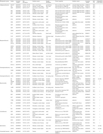
We obtained 53 isolates of 0.2 μm-passable bacteria. The phylogenetic analysis based on 16S rRNA gene sequences classified them into five phyla of Proteobacteria (34 isolates), Bacteroidetes (11), Actinobacteria (5), and Spirochaetes (2), with one unclassified isolate showing less than 85% similarity to known strains and isolates (Table I; BLASTN search results against the Greengenes database are shown in Table SI, which will be found at http://dx.doi.org/10.1017/S0954102012000831).
Phylogenetic affiliation of isolated strains and culturability of the filtrates.

aSimilarity.
bLiquid cultures of each isolate were filtered through three 0.2 μm-pore-size filters. The filtrates were added to R2A medium and observed after three weeks.+= growth was observed, -=no growth was observed.
Of the 34 protobacterial isolates, 15 were affiliated with the class Alphaproteobacteria, 12 with the class Betaproteobacteria, and seven with the class Gammaproteobacteria. No Delta, Epsilon or other proteobacterial isolates, most of the known species of which are microaerobic to anaerobic, were obtained from the collected aerobic samples.
Of the 15 Alphaproteobacterial isolates, seven similar isolates were only remotely related (at 92–93% similarity) to the genus Rhizobium, five were closely related (> 98%) to a Phaeospirillum species/strain, two Antarctic isolates (of which the 16S rRNA gene sequences are 100% identical) were almost identical (99.8%) to an Antarctic Mycoplana strain, and one was almost identical (99.8%) to Afipia felis. In contrast, the Betaproteobacterial isolate pool was dominated by a Hylemonella species/strain (10/12 isolates). Gammaproteobacterial isolates were scattered in five genera of Saccharospirillum, Pseudomonas, Oceaniserpentilla, Reinekea, and Vibrio.
About half of the Bacteroidetes isolates were affiliated with the genera of Flexibacter, Cytophaga, and Gracilimonas, while the other half was loosely related (at < 96% similarities) to two unidentified Bacteroidetes. Isolates of Actinobacteria and Spirochaetes were closely related (> 97%) to previously reported species/strains. The last isolate “Shr3” from the Sahara is totally unrelated (< 85%) to known cultured organisms and thus is of great taxonomic interest, but will be described and discussed elsewhere.
Relative dominance of Proteobacteria and Bacteroidetes is affected by the recovery of isolates belonging to an unnamed group of Rhizobiales and the genus Hylemonella (Proteobacteria) as well as Flexibacter and Cytophaga (Bacteroidetes). To the best of our knowledge this is the first report of 0.2 μm-passable Rhizobiales, although certain Rhizobium species such as R. leguminosarum have shown a negative relationship between cell size and low nutrient conditions (Postma et al. Reference Postma, Vanelsas, Govaert and Vanveen1988). Our results suggest that this relationship may hold for other groups in the cryosphere. The Hylemonella strains are well-studied 0.2 μm-passable fast-growing freshwater bacteria that have slender spirillum-shaped cell morphology (Wang et al. Reference Wang, Hammes, Boon and Egli2007). Flexibacter and Cytophaga species also exhibit 0.2 μm-passable slender filamentous forms (Hood & MacDonell Reference Hood and MacDonell1987, Hahn Reference Hahn2004, Hahn et al. Reference Hahn, Stadler, Wu and Pockl2004) as well as 0.2 μm-passable dwarf cells during their life cycles (Elsaied et al. Reference Elsaied, Sato and Naganuma2001).
Phylogeography of 0.2 μm-passable bacteria
Generally, microorganisms have large population sizes and very short generation times resulting in high dispersal rates. Studies have shown some protist, fungal, and bacterial taxa with cosmopolitan distribution, rather than restricted geographic distribution (reviewed by Green & Bohannan Reference Green and Bohannan2006). The Baas-Becking (or ubiquity) hypothesis is supported by findings that identical bacterial 16S rRNA gene sequences have been detected from lakes in both the Arctic and Antarctic (Pearce et al. Reference Pearce, Cockell, Lindstrom and Tranvik2007). Conversely, there are reports that some microbial taxa have restricted geographic distribution from dispersal limitations, which implies that not all microorganisms can disperse globally (reviewed by Martiny et al. Reference Martiny, Bohannan, Brown, Colwell, Fuhrman, Green, Horner-Devine, Kane, Krumins, Kuske, Morin, Naeem, Øvreås, Reysenbach, Smith and Staley2006).
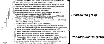
The seven isolates belonging to the order Rhizobiales were mutually closely related (97.2–100% similarity), and formed a separate cluster within the phylogenetic tree (Fig. 1; phylogenies of all isolates and their closest known sequences are presented in Fig. S1, which will be found at http://dx.doi.org/10.1017/S0954102012000831). This cluster of seven isolates included a clone (GQ264001) isolated from a waste site in North America. In addition, the five isolates belonging to the genus Phaeospirillum (class Alphaproteobacteria) collected in the Arctic, Japan, and China were also mutually closely related (97.2–100% similarity). Given that similar isolates were collected from geographically disparate locations, it would appear that these 0.2 μm-passable bacteria are more ubiquitous in terrestrial environments than aquatic environments. To test this conjecture, it will be necessary to isolate and culture a larger sample of 0.2 μm-passable bacteria from a wider range of terrestrial samples. In contrast, the eight novel strains belonging to the phylum Bacteroidetes isolated from the Arctic and Japan were not mutually closely related (75.5–93.0% similarity).
Neighbour-joining phylogenetic tree based on nearly complete 16S rRNA gene sequences from Rhizobiales and Rhodospirillales strains. The percentages of replicate trees in which the associated taxa clustered together in a bootstrap test (1000 replicates) are shown next to the branches. There were a total of 1259 positions in the final dataset. Origins of the isolates and clones are in round brackets, and nucleotide accession numbers are in square brackets. The scale bar indicates 0.02 substitutions per nucleotide site.

The KNC strain isolated from Japanese river water was very closely related (99.7% similarity) to Microbacteriaceae bacterium MWH-VicE1 belonging to the Luna cluster (phylum Actinobacteria) isolated by Hahn et al. (Reference Hahn, Stadler, Wu and Pockl2004). This cluster consists of strains isolated from lakes and ponds in Europe, East Africa, and China, and is known to be an obligate ultramicrobacterial cluster.
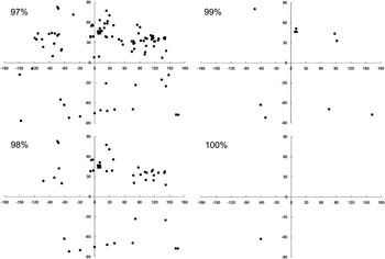
The two Antarctic isolates (S-19 and S-42) belonging to the order Rhizobiales isolated from two samples (beach sand and freshwater algal mat) collected from Maritime Antarctica were found to be closely related (99.8% similarity) to Mycoplana sp. ZS1-12 isolated from sandy intertidal sediments of the Larsemann Hills coast (Princess Elizabeth Land, East Antarctica) (Yu et al. Reference Yu, Li, Zeng and Chen2010). In addition, BLASTN search results showed that the sequence of Antarctic isolate S-19 showed ≥ 97%, ≥ 98%, ≥ 99%, and 100% similarity to 901, 226, 13, and one 16S rRNA gene sequences of known isolates and/or from environmental samples, respectively. The latitudes and longitudes of the sample collection sites corresponding to these sequences are shown in Fig. 2 (accession numbers of all sequences showing ≥ 97% similarity to each of 0.2 μm-passable isolates, together with their sample collection sites and sources, are listed in Table SII (which will be found at http://dx.doi.org/10.1017/S0954102012000831). Sample collection sites corresponding to the 901 sequences with ≥ 97% similarity were geographically disparate and from diverse environmental conditions. On the other hand, samples corresponding to 13 sequences with ≥ 99% similarity included those from cryospheric habitats, such as Ellesmere Island in the Canadian Arctic (one sequence) and Antarctica (five sequences). Sequences closely related (≥ 99%) to the S-19 sequence were also isolated from samples collected at the mid-range latitudes: a biofilm found in the Central Alps in Switzerland (one sequence) and glaciers and snow recovered from China (three sequences). These four sequences were also from cryospheric habitats. One sequence 100% identical to S-19 was the other Antarctic isolate (S-42) obtained in this study. In summary, 10 of the 13 sequences with ≥ 99% similarity to the S-19 sequence (c. 77%) were isolated from samples from cryospheric habitats, such as Arctic, Antarctic, and alpine environments. This implies that such Rhizobiales strains occur in the cryospheric regions, but their abundance and biomass may be scarce depending on the geographic location.
Distribution of the sample collection sites corresponding to the sequences derived from known isolates and environmental samples related to the Antarctic strain S-19. The x- and y-axes represent 0° latitude and longitude respectively. Sequences related to the S-19 sequence were identified from the DDBJ/EMBL/GenBank database, and grouped by level of similarity to it (97%, 98%, 99%, or 100% similarity: E-value = 0). The number of sequences similar to the S-19 sequence increased from one to 901 when the similarity threshold was changed from 100% to 97%. Sequences similar to the S-19 sequence on the database but lacking information on country of isolation source were not included in the graphs.

In clear contrast to the Antarctic isolate described earlier, Spirochaeta strain IZ-19-1 isolated from lake water collected in western Japan was related to only one Chinese coastal strain. No known isolates or environmental sequences were found to be similar to IZ-19-1 as of June 2011 even with a 97% similarity threshold (Fig. S2 & Table SII, which will be found at http://dx.doi.org/10.1017/S0954102012000831). This implies that Spirochaeta strain IZ-19-1 belongs to the Asian endemic group that inhabits a restricted region.
Candidates of new taxa obtained from 0.2 μm-passable fractions
The results of BLASTN searches on the obtained 16S rRNA gene sequences indicate that a total of 18 isolates (seven belonging to the order Rhizobiales (class Alphaproteobacteria), two to the genus Saccharosprillium (class Gammaproteobacteria), eight to the phylum Bacteoidetes, and the isolate “Shr3”) were less than 97% homologous with known strains and isolates, suggesting novel taxa at species, genera, class, or even higher levels (Table I).
This result may arise as faster-growing bacteria were effectively excluded by filtration (Hahn Reference Hahn2004), allowing isolation of slower-growing bacteria that had been previously undetectable. Even a minor species could be recovered by a culture method designed to select for multiplication of minorities (Pedrós-Alió Reference Pedrós-Alió2006). Our design was to exclude a majority of fast-growing heterotrophic bacteria by filtration with triple-serial 0.2 μm-pore filters. More than 99.9% of bacterial cells in natural environments are excluded, leaving only a fraction as small as < 0.1% that would pass the filters (Fukuba et al. Reference Fukuba, Elsaied and Naganuma2002). As a result, the presence of novel microbial lineages that had previously been obscured by faster-growers was revealed.
Cell size of 0.2 μm-passable bacteria under culture conditions
Field-emission SEM images of the cell morphology of NP1 and NP3 (genus Phaeospirillum, class Alphaproteobacteria) and IZ6, GB3 and S-42 (order Rhizobiales, class Alphaproteobacteria) showed that the cells were fat and rod-shaped, with minimum diameters between 0.3 and 0.4 μm. In contrast, cells of GB9 (Phaeospirillum) and GB10 (Rhizobiales) were larger, with diameters between 0.5 and 1.0 μm. Larger cells may occur in culture suspensions despite filtration through 0.2 μm filters by cell polymorphism. In the present study, liquid cultures were filtered twice, and although growth activity was observed in the filtrate of GB10 cultures (Table I), no diminutive cells were detected. It is possible that such bacteria that grow larger than 0.2 μm under culture conditions may also generate cells smaller than 0.2 μm temporarily, which passed through the 0.2 μm filters. For example, Cytophaga species have cyclic morphological change, namely young filament and old coccus, and the coccid cells are known to release sub-0.2 μm cells (Elsaied et al. Reference Elsaied, Sato and Naganuma2001). In the future, detailed observation of cell morphology over the course of all lifecycle stages is needed.
IZ16 and NP9 isolates (Rhizobiales), which were seen on FE-SEM to be relatively large, did not grow after secondary filtration, and it is possible that the cells are small in the environment from which the samples were collected. This effect may result from cells entering dormancy and/or entering a viable but non-culturable (VBNC) state in the environment. Cells in these states are typically of reduced size (e.g. Hood & MacDonell Reference Hood and MacDonell1987), presumably to adapt to low organic concentrations by increasing the surface-to-volume ratio or a starvation-survival strategy. Alternatively, even though these isolates may form sub-0.2 μm cells at some point of their life cycle, the number of diminutive cells might have been negligible, and the cells were not identified in the filtrate. In genus Afipia (class Alphaproteobacteria), the isolated IZ17 cells were small with minimum diameters between 0.2 and 0.3 μm.
Cells of the IZ19-2 isolate (genus Hylemonella, class Betaproteobacteria), the IZ4 isolate (related to an unidentified species belonging to the class Betaproteobacteria), and the ShrSW isolate (genus Oceaniserpentilla, class Gammaproteobacteria) were relatively small and rod-like, with minimum diameters between 0.2 and 0.3 μm. Cells of the KE isolate (genus Vibrio, class Gammaproteobacteria) were extremely small, with a minimum diameter of c. 0.15 μm.
Cells of the KIP, KIY, and KOr isolates belonging to the CFB group (phylum Bacteroidetes) were relatively long and rod-like, with maximum diameters between 1.5 and 3.0 μm. No obvious sub-0.2 μm cells were observed among these isolates. Although it is possible that cell morphology is altered by fixation and dehydration, the minimum diameters of the cells were between 0.2 and 0.3 μm. These cells are likely to pass through the 0.2 μm pores, not because of their diminutive cell size, but because of their long rod shape. This will need to be validated by other experiments such as detailed observation of the filtrates. Eight of the 11 isolates belonging to the phylum Bacteroidetes grew in the twice-filtered culture.
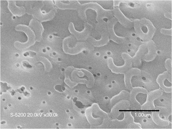
The isolated KNC cells were selenoid and closely related to the obligate UMB belonging to the Luna cluster (phylum Actinobacteria) (Fig. 3). Other members of the Luna cluster have also been reported to form selenoid or crescent-shaped cells (Hahn et al. Reference Hahn, Lunsdorf, Wu, Schauer, Hofle, Boenigk and Stadler2003). The name Luna cluster is derived from this crescent-shaped cell morphology, which may be associated with flexibility and thus 0.2 μm-passability of the cells.
Scanning electron micrographs of strain KNC (phylum Actinobacteria) that exhibit typical solenoid or crescent-shaped cell morphology. Scale bars = 1 μm, pore sizes = 0.1 μm in diameter.

Conclusions
Phylogeographic analysis of 0.2 μm-passable bacteria in this study demonstrated that some isolates were related closely to geographically very distant strains, while others were found only in a restricted region. Thus, the isolates identified in this study showed diverse distribution patterns. In future, we will consider 0.2 μm-passable bacteria showing characteristic distributions as reference microorganisms to test the Baas-Becking hypothesis. However, the possibility exists that the presence of bacteria in the environment occurred from inadvertent selectivity and/or sampling effort. To validate our results, we would use isolation and phylogenetic identification techniques on the bacteria of the 0.2 μm-filtrable fraction in the cryosphere and other environments. Further studies would require additional techniques such as multi-locus sequence typing and phenotypic characterization.
Acknowledgements
We are grateful to Drs Synnøve Elvevold and Kim Holmén at the Norwegian Polar Institute and Dr Yoshihide Ohta at University of Oslo, who helped us immensely in conducting the investigation at Svalbard in the Arctic. We are grateful to the crew of the RV Toyoshio-maru, Hiroshima University, and the cruise participants for their on-board assistance. We also acknowledge the Natural Science Centre for Basic Research and Development of Hiroshima University for FE-SEM analysis. This work was supported by a Grant-in-Aid for Scientific Research (No. 18255005) and Research Fellowships for Young Scientists (No. 11J30005) from the Japan Society for the Promotion of Science and a grant from the Institute for Fermentation, Osaka, Japan. Antarctic samples were collected during the Spanish program LIMNOPOLAR International Polar Year 2007–08 campaign with the support of the Spanish Polar Programme, Ministerio de Ciencia e Innovación (POL2006-06635/ANT). This article was published thanks to the financial support given by the Ministerio de Ciencia e Innovación (Spain) with the grant ref. CTM2011-12973-E. The constructive comments of the reviewers are also gratefully acknowledged.
Supplemental material
Two supplemental figures and two supplemental tables will be found at http://dx.doi.org/10.1017/S0954102012000831