Case report
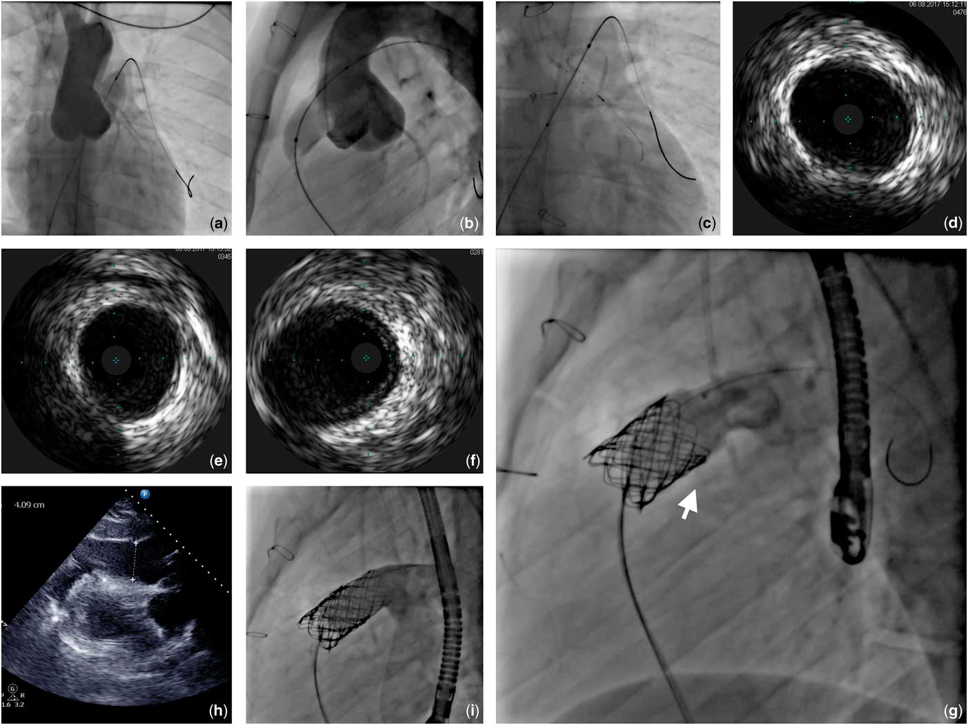
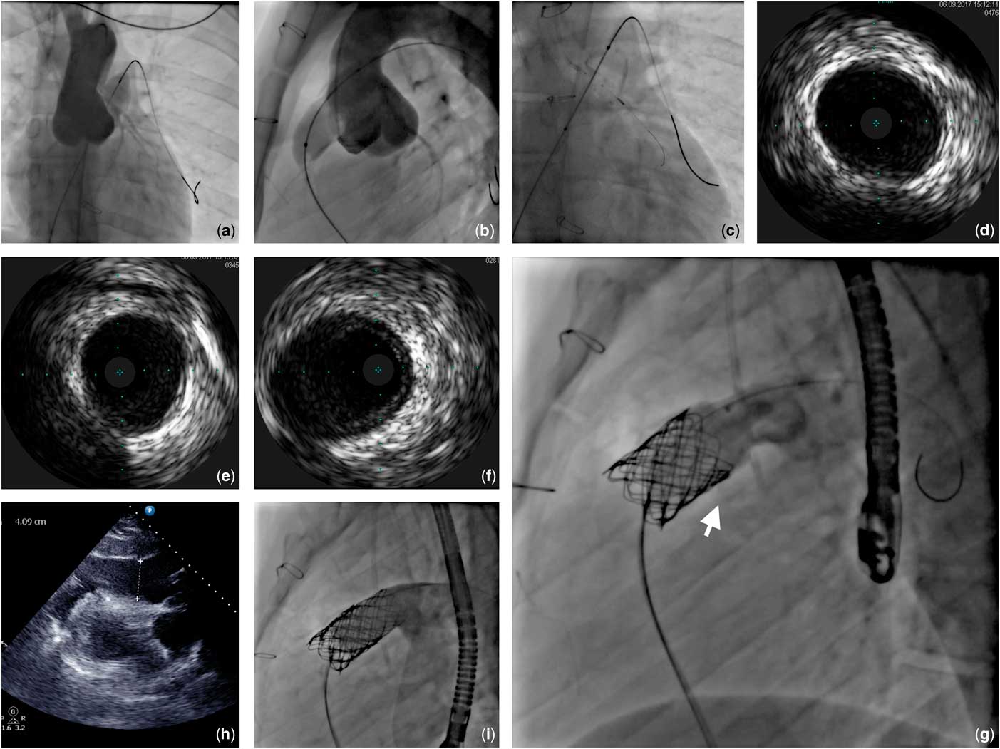
Approximately 5% of patients undergoing right ventricle outflow tract stenting are at risk of coronary compression.Reference Gillespie and McElhinney 1 Exclusion of potential constriction is recommended and potential oversight may be fatal.Reference Eicken, Ewert and Hager 2 At times, the suggested method – balloon inflation in right ventricle outflow tract with simultaneous aortography or selective angiography – can be inconclusive or falsely negative.Reference Biermann, Schönebeck, Rebel, Weil and Dodge-Khatami 3 We present a case of 27-year-old male post Ross operation qualified for percutaneous pulmonary valve implantation due to pulmonary homograft degeneration with the pressure gradient across the pulmonary valve was 45 mmHg, increasing dyspnoea, and exercise tolerance worsening. The procedure was performed under general anaesthesia via venous and arterial femoral access. A multipurpose catheter was used to measure pressures in the right ventricle, main pulmonary artery, and left pulmonary artery. Subsequently, a 22 mm Z-MED balloon (B. Braun, Inc., Bethlehem, PA, United States of America) was introduced to right ventricle outflow tract over a Lunderquist guidewire (Cook) and inflated with simultaneous left coronary artery angiography. Neither of the views that were obtained, posteroanterior or lateral provided a definite exclusion of coronary compression (Fig 1a and 1b); therefore, a decision to perform intravascular ultrasound was made (Fig 1c). A manual pullback of ultrasound probe demonstrated an oval-shaped left main stem with pulsatile shape changes (Fig 1d) and a normal left anterior descending artery. Another pullback was performed with a balloon inflated in right ventricle outflow tract showing a similar size and silhouette of the left main stem (Fig 1e) and no compression of the left anterior descending artery. Given that no difference was appreciated, a 45 mm covered stent (CP Stent™; NuMED Inc., Hopkinton, NY, United States of America) was crimped on a 22 mm balloon-in-balloon (NuMED Inc.), implanted and post-dilated with a 22 mm Mullins-X balloon (NuMED, Inc.). Final investigation with intravascular ultrasound confirmed an unchanged left main stem (Fig 1f) and left anterior descending artery conformation. Subsequently, a Melody transcatheter pulmonary valve (Medtronic, Minneapolis, MN, United States of America) crimped on a 22 mm balloon-in-balloon was delivered into the right ventricle outflow tract and deployed. No pericardial effusion was present in trans-oesophageal echocardiography and the procedure was concluded (Fig 1g). Two hours later, the patient complained of mild chest pain. In transthoracic echocardiography up to 4 cm of contained fluid localised in the posterior part of pericardial sack was found (Fig 1h). Retrospective analysis of final angiogram revealed a possibility of subtle extravasation at the distal part of the homograft (Fig 1g, arrow). A decision was made to perform a hybrid procedure consisting of additional covered stent implantation followed by pericardial drainage and implantation of another pulmonary valve. Using the same implantation technique without repeating coronary compression exclusion, a 45 mm covered stent on a 22 mm balloon-in-balloon was deployed distally, covering approximately 50% of the previously implanted scaffold. In the next step, pericardial drainage was established through substernal incision and 300 ml of fluid was evacuated. Ultimately, a second valve on a 22 mm balloon-in-balloon was implanted (Fig 1i). Follow-up transthoracic echocardiography showed no pericardial effusion and correct bioprosthesis function. Within 48 hours the drainage was removed and 6 days after the procedure the patient was discharged.
(a,b) Conventional views not providing definite exclusion of the coronary compression. (c) Intravascular ultrasound probe placed in the left coronary artery and balloon placed in the right ventricle outflow tract. (d) Initial intravascular ultrasound pullback demonstrating an oval-shaped left main stem. (e) Intravascular ultrasound pullback with simultaneous balloon inflation in the right ventricle outflow tract demonstrating similar silhouette of the left main stem. (f) Unchanged left main stem conformation after covered stent implantation. (g) Final effect of the first procedure. Arrow pointing at a possible extravasation point at the distal, posterior part of the homograft. (h) Effusion in the pericardium. (i) Final effect after second pulmonary valve implantation.
Discussion
It is estimated that 4–5% of patients undergoing right ventricle outflow tract stenting are at risk of coronary compression.Reference Gillespie and McElhinney 1 Serious complications are rare, but have been described;Reference Biermann, Schönebeck, Rebel, Weil and Dodge-Khatami 3 , Reference Mauri, Frigiola and Butera 4 therefore cautious exclusion of potential coronary constriction is recommended in every patient undergoing right ventricle outflow tract stenting. Pre-procedural assessment with cardiac MRI and CT can be helpful in assessing the static distance between the outflow tract and the coronary tree. These modalities, however, are not capable of predicting the change in conformation of the right ventricle outflow tract after stenting, as well as stent’s impact on the adjacent structures. Therefore, the routine protocol is to perform a balloon inflation in the right ventricle outflow tract with simultaneous aortography or selective angiography. This method, however, can render inconclusive or falsely negative results.Reference Biermann, Schönebeck, Rebel, Weil and Dodge-Khatami 3 In our case, initial routine imaging was unconvincing, therefore a decision to use intravascular ultrasound was undertaken. Intravascular ultrasound is an established method to assess coronary arteries in the coronary artery disease setting. It has been implemented in the pulmonary valve implantation framework as well, but for a different purpose. Ferrari et al reported using intravascular ultrasound for valve positioning and post-implantation control.Reference Ferrari, Niclauss, Berdajs and von Segesser 5 In our case, manual pullback of ultrasound probe in the left coronary artery before and during balloon inflation in the right ventricle outflow tract demonstrated a stable and unchanged shape and size of the left coronary artery, therefore excluding potential compression. Application of the intravascular ultrasound requires use of the guiding catheter as well as introduction of a coronary guidewire to the distal part of the artery, which in rare cases may lead to a mechanical injury of the vessel. In typical patients referred to right ventricle outflow tract stenting, however, such complication seems less likely than in patients with coronary artery disease, owing to lower age and theoretically less advanced atherosclerosis. Potential difficulties with interpretation of the study arise from the anatomical differences in the subset of patients post-surgical reimplantation of the coronary arteries including possible stretching of the coronaries during the heart cycle. The strength of the technique comes from the fact that the entire length of the left anterior descending artery and the left main stem may be studied with one pullback of the ultrasound probe before and during the balloon inflation. Such investigation potentially requires less contrast than aortography with automatic injector and eliminates the need for repeated injections due to inconclusive image, overlapping arteries, etc. Nonetheless, other potential alternatives for coronary compression exclusion have been reported in the literature, such as a three-dimensional rotational angiography,Reference Pockett, Moore and El-Said 6 as well as placing a coronary wire in the left coronary artery to serve as a landmark during a balloon inflation in the right ventricle outflow tract.Reference Góreczny, Eicken and Ewert 7
Homograft rupture is a serious, potentially lethal complication of right ventricle outflow tract interventions. In our case, the rupture happened after covered stent implantation, most likely following stent post-dilatation or pulmonary valve implantation. Signs of contrast extravasation in the angiography were subtle and therefore not appreciated during the initial angiographic assessment of the final result. Furthermore, no symptoms of cardiac tamponade were present and no pericardial effusion was detected in the transthoracic echocardiography performed at the end of the procedure. Nonetheless, after the onset of symptoms and appearance of effusion in the pericardium, percutaneous attempt at sealing the rupture with additional covered stent accompanied by surgical drainage was deemed necessary and proved to be effective.
Conclusions
Coronary compression exclusion is crucial in patients undergoing right ventricle outflow tract stenting. In case of doubtful or inconclusive images obtained from aortography or selective angiography during balloon inflation in right ventricle outflow tract, intravascular ultrasound might be a feasible and useful technique. Signs of homograft rupture may be subtle, whereas symptoms of cardiac tamponade delayed. Meticulous assessment of final angiography in search for slightest potential extravasation along with careful postoperative monitoring is essential. In selected patients percutaneous treatment of homograft rupture is achievable and beneficial.
Acknowledgement
The authors would like to thank Professor Tomasz Moszura, Doctor Piotr Scisło and Doctor Paweł Czub for their help, support and guidance during the procedure.
Financial Support
This study did not receive any funding.
Conflicts of interest
ZH is a proctor for Medtronic.
Ethical Standards
All procedures performed in studies involving human participants were in accordance with the ethical standards of the institutional and/or national research committee and with the 1964 Helsinki Declaration and its later amendments or comparable ethical standards.
Informed Consent
Informed consent was obtained from the patient described in the case report.