Introduction
Endoscopic ear surgery is an evolving science in the field of otology; it provides a minimally invasive transcanal approach to the middle ear. Training in endoscopic ear surgery is an essential skill set. Surgeons can achieve this training utilising various methods, such as cadaveric temporal bone dissection (the ‘gold standard’ method), animal models, mannequins, synthetic temporal bone models and virtual reality simulators.Reference Okhovat, Milner and Iyer1
There are many pros and cons associated with each method. Cadaveric dissection is one of the best approaches. However, cadavers are expensive and difficult to procure because of local regulations. Trainees can opt for the formalin-preserved temporal bone specimens for dissection. Nevertheless, the tissue quality is low and the strong fumes can make the surgical exercise cumbersome for trainees. Virtual simulators are highly expensive and are lacking in tactile sensation.Reference Zaidi, Khan and Parab2
Animal models are inexpensive and are an excellent alternative to human cadavers, but the anatomical orientation is incongruent with human cadavers. The external auditory canal of an animal model is narrow and bent, making tympanomeatal flap elevation more complicated than in a human ear canal. In addition, it is difficult to manoeuvre the instruments while operating using such models. Animal models are recommended for residents’ training if they can overcome these difficulties.Reference Anschuetz, Bonali, Ghirelli, Mattioli, Villari and Caversaccio3
All of these training methods have been impossible during the coronavirus disease 2019 lockdown period, as the materials are not often readily available. A trainee or a new surgeon should be practising surgical techniques continuously in order to obtain consistent outcomes, which will eventually result in them being a good surgeon. This is the idea behind the innovative mode of training in endoscopic ear surgery using a papaya petiole.
Endoscopic ear surgery training using the papaya petiole is simple and affordable, it simulates the experience of real surgical procedures, and the equipment is readily available. A surgical trainee's cardinal difficulty is holding the endoscope and manipulating the instruments within the external ear canal, under conditions of limited depth perception. The challenge in endoscopic ear surgery is to successfully elevate the tympanomeatal flap. The Narendrakumar papaya petiole model method makes it easy to master this skill. This model is easy to set up and is cost-efficient.
Materials and methods
Equipment requirements
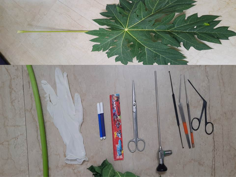
Requirements for this endoscopic ear surgery model include: a papaya petiole, a surgical glove, rubber bands, artificial clay, otological instruments, and an endoscope with a camera system (Figure 1).
Equipment requirements for the endoscopic ear surgery training model using a papaya petiole.

Setting-up procedure
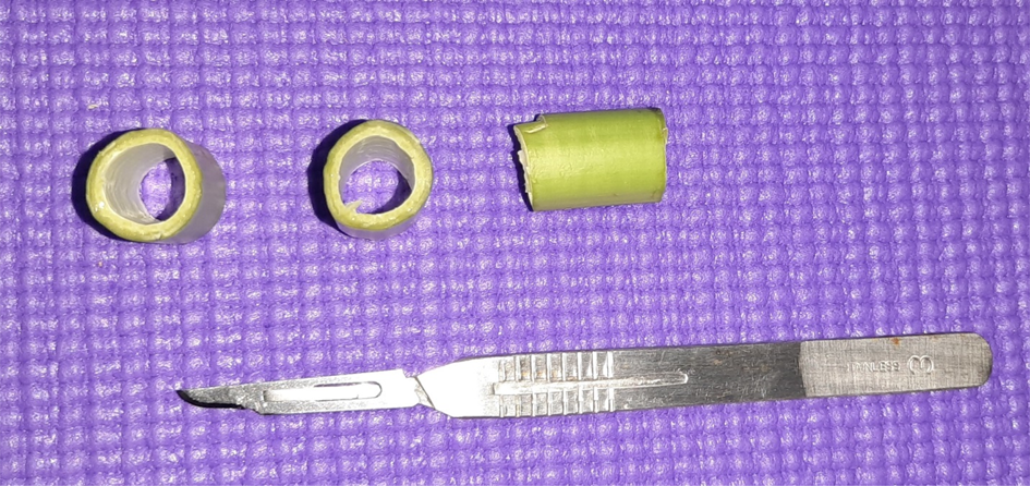
The method of using these materials to set up the model is not very complicated. Firstly, the papaya petiole is cut from the papaya tree and its leaves, which are then discarded, leaving only the petiole for the procedure. The approximate length of the human external ear canal is 24 mm, which can be used as an average measurement to cut the papaya petiole into multiple pieces, each with the same length (Figure 2).
The papaya petiole is cut into small pieces.

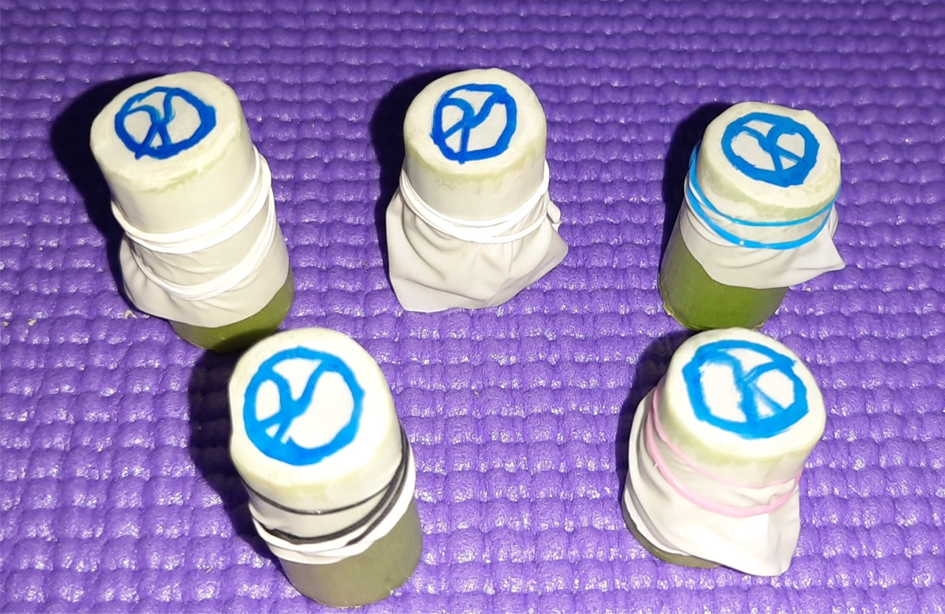
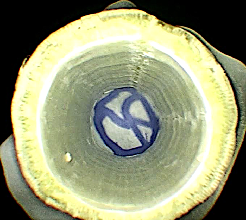
Having the papaya petiole ready for practice, we use the tip of the surgical gloves cut with a length of 2 cm to enclose one side of the papaya petiole. Then, wrapping it with a rubber band, the glove tip is held, making sure that it is pulled taut. The tympanic membrane structure can be drawn using a regular marker over the glove surface for orientation and best practice (Figure 3).
The tip of a surgical glove is secured around the papaya petiole, with a ‘tympanic membrane’ drawn for orientation.

Once the basic form has been created, the petiole tube is stabilised using the artificial clay (the same clay used by children for creative play) moulded in its exact shape.
Finally, the endoscope is inserted through the petiole's open end to enable an experience similar to that of a real endoscopic ear surgery procedure.
Surgical exercises
The following exercises, conducted after creating the artificial ear structure using the papaya petiole, allow manipulation of the otological instruments to fine-tune surgical skills.
Exercise one
Using a myringotomy knife, an incision is made over the anteroinferior quadrant of the tympanic membrane. The surgical glove tip in the model gives the surgeon a similar texture to the actual membrane, making it a more realistic exercise. The grommet is then inserted in a sliding oblique manner, placing it where the incision was made (Figure 4).
Endoscopic picture showing grommet insertion in the papaya petiole model.

Exercise two
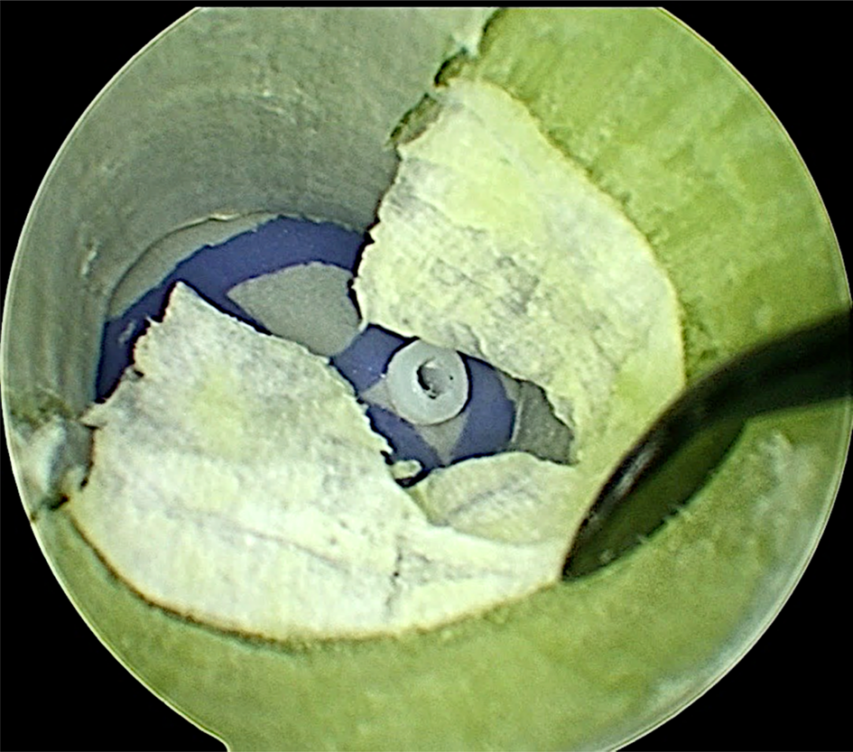
Using Plester and Rosen knives, an incision is made over the papaya petiole's inner surface, which represents an external canal wall. Elevation of the tympanomeatal flap, the first and most challenging step of endoscopic ear surgery, can be made easier by practising using this model. The petiole gives the same delicate, papery thin layer as the flap (Figure 5).
Endoscopic picture showing tympanomeatal flap elevation training in the papaya petiole model.

Exercise three
The previously inserted grommet can be removed.
Exercise four
We can improvise the model setup to practise foreign body removal by using tiny items of different textures (e.g. chalk, a peanut, a pin) placed inside the petiole model. Removal of these items can be practised using an endoscope and other preferred instruments. Repeating this step can also improve hand–eye co-ordination, which will ultimately aid handling of the endoscope.
Exercise five
Instead of surgical glove, a tissue paper with a punched-out hole can be used to replicate the tympanic membrane. This can enable learning and practice at freshening the edges of a tympanic membrane perforation.
Discussion
Surgical models and simulators are valuable learning tools to practise and improve surgical techniques. Various studies have shown that such models and simulators can dramatically increase residents’ learning more than real-time experience. Factors deciding the suitability of a model or simulator for surgical training purposes include easy availability, low cost and concordance with real life.Reference Dedmon, Kozin and Lee4
The popularity of the minimally invasive endoscopic approach to ear surgery is on the rise. ENT trainees seeking advanced training in this technique are increasing exponentially. At present, the hours spent by trainees and residents participating in endoscopic ear surgery are limited, preventing them from acquiring sufficient training and relevant endoscopic skills.Reference Okhovat, Milner and Iyer1 Although the principles and steps of ear surgery are similar to those of endoscopic and microscopic approaches, the skills required for hand–eye co-ordination and single-hand manipulation of instruments are different. For instance, the endoscopic approach requires proper use of the endoscope, prompting every trainee to be versatile in using it. Though the learning curve varies, the level of expertise will correlate with the surgeon's experience and comfort in using an endoscope.Reference Mijovic and Lea5
The increasing use of endoscopy in otology means that ENT trainees are becoming more exposed to endoscopic ear surgery during their residency, but their exposure to surgery is varied.Reference Ng, Ong and Ngo6 The endoscope provides a panoramic, high-definition and wide-angle view of the surgical field. However, there are significant technical challenges that surgeons must overcome when adopting it for surgical procedures. Given the monocular nature of the endoscope, surgeons have to operate under conditions of limited depth perception, which can make precise instrumentation very difficult, especially with a background of minimal endoscopic experience.Reference Dedmon, Xie, O Connell, Dillon, Wellborn and Bennett7
This article discusses how an interested otologist can begin experimenting with endoscopy, and elaborates on the steps needed to improve endoscopic ear surgery skills. In endoscopic ear surgery, the approach, primary dissection and reconstruction are performed under direct endoscopic visualisation. The endoscope is used as the main modality of visualisation, rather than as an adjunct at the end of the case. Advances in instrumentation may improve the precision of endoscopic ear surgery and make such surgery technically easier.Reference Golub8
Papaya, a material that can be used for training, is readily available in tropical and subtropical areas. Other alternative hollow-stemmed plants with petioles include sow thistle, horsetail, Joe Pye weed, Japanese knotweed, Himalayan balsam, cow parsley and rakkyō. Although trainees can use these plants for practising endoscopic ear surgery skills, they do not provide the close to real-life experience of the papaya petiole when practising tympanomeatal flap elevation.
Conclusion
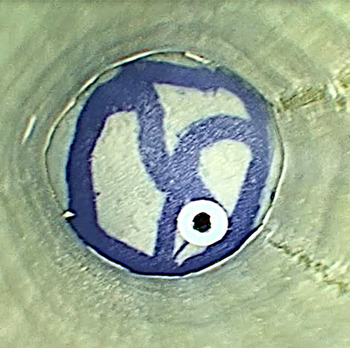
The main contribution of this article is that it suggests a means to train residents to improve their surgical techniques in a simple way, using materials from daily life and the environment around us. I have designed this innovative dissection model using the Narendrakumar papaya petiole technique (Figure 6 and Appendix 1 (a short video, available on The Journal of Laryngology & Otology website)), which can be used to facilitate the acquisition of the most challenging aspects of transcanal endoscopic ear surgery, including one-handed dissection with limited depth perception, and the use of angled endoscopes and instruments. Such techniques ultimately help to improve a surgeon's surgical skills, even during unprecedented and difficult times like those currently experienced, using readily available and cost-efficient raw materials. Surgical skills training can improve endoscopic ear surgery techniques by allowing repetitive practice in a safe environment. Such training enhances dexterity and provides experience in performing the surgical procedure, improving the likelihood of future successful outcomes.Reference Anschuetz, Bonali, Ghirelli, Mattioli, Villari and Caversaccio3
Screenshot (taken from Appendix 1) showing an endoscopic view of the papaya petiole endoscopic ear surgery model.

Acknowledgements
I would like to acknowledge the assistance provided by Dr S Geethalakshmi for final proof reading, and Ms N Sai Snehha who aided in the editing of this article.
Competing interests
None declared
Appendix 1. Training in endoscopic ear surgery using the papaya petiole.
A short video demonstrating training in endoscopic ear surgery using the Narendrakumar papaya petiole technique is available online at The Journal of Laryngology & Otology website, at https://www.youtube.com/watch?v=lkMwpIEhuEE&t=5s