Introduction
The African variegated grasshopper Zonocerus variegatus (Linnaeus) (Orthoptera: Pyrgomorphidae) is a common insect pest in the forest regions of West and Equatorial Africa. The grasshopper is a polyphagous insect with its different life stages showing a preference for different food plants (Chapman et al., Reference Chapman, Page and McCaffery1986; Chiffaud and Mestre, Reference Chiffaud and Mestre1990; Le Gall, Reference Le Gall2000; Kekeunou et al., Reference Kekeunou, Weise, Messi and Tamo2006).
Gregarious stages of Z. variegatus survive on weeds such as Eupatorium odoratum, Ageratum conyzoides, Talinum triangulare, Lantana camara, Aspilia latifolia, Aspilia africana, Solanum torvum and Tridax procumbens (Toye, Reference Toye1982). Adult Z. variegatus show a preference for cassava (Manihot esculenta), while the early instars (gregarious stages) preferentially feed on Siam weed (Chromolaena odorata) (Idowu and Sonde, Reference Idowu and Sonde2004).
The grasshopper is able to sequester toxic chemicals in the plants it consumes, such as the alkaloids present in C. odorata (Modder, Reference Modder1983) and cyanogenic glucosides in cassava (Bernay et al., Reference Bernay, Chapman, Cook, Mcveigh and Page1975; Idowu and Modder, Reference Idowu and Modder1998). The exact mechanisms of this detoxification are not well understood, and relatively few studies have been carried out on Z. variegatus physiology. Balogun (Reference Balogun1972) and Modder (Reference Modder1977) reported the presence of glycosidase enzymes in the gut homogenate of Z. variegatus, but the origin and functions of these enzymes have not been investigated in detail.
Idowu and Edema (Reference Idowu and Edema2003) reported that gut sections of Z. variegatus (fore-, mid- and hindgut) harboured a variety of micro-organisms, mainly bacteria, yeast and moulds, and that the microbial load was highest in adults, followed by 6th, 4th and 3rd instars in that order.
Several studies have shown that diverse insects and other organisms harbour symbiotic micro-organisms in their gut cavity (Ohkuma, Reference Ohkuma2003). The gut of the adult locust Schistocerca gregaria has a wide range of bacterial types, with the predominant flora in the midgut and hindgut regions being members of the family Enterobacteriaceae and motile Streptococci (Hunt and Charnley, Reference Hunt and Charnley1981). While most of the gut microbes are commensals or parasites, some are known to play substantial biological roles for their hosts. For example, gut microbes are needed for cellulose digestion in termites (Ohkuma, Reference Ohkuma2003).
The objective of the study reported here was to investigate the enzymatic profile of microflora present in the gut sections of Z. variegatus, with a view to understanding the mechanisms of digestion in this insect, whose gregarious stages can cause widespread damage to important field crops, including cassava.
Materials and methods
Sampling
Grasshoppers were collected from wild and farmed cassava within the University of Agriculture, Abeokuta campus, between February and June 2006. Collections were made daily with sweep nets at about 06.30–07.00 h. The insects were maintained in an insectary in a wire mesh cage measuring 47 × 30 × 30 cm, under ambient temperature and relative humidity, with good ventilation. A total of 20 insects were collected per instar, from which five each were randomly picked as samples from 3rd, 4th, 5th and 6th instars and adult stage.
Dissection
A dissecting kit and board were surface sterilized with 70% ethanol before use. The insects were killed by breaking their cervical membrane to sever the ventral nerve cord (Idowu and Edema, Reference Idowu and Edema2003). Each grasshopper was surface sterilized by swabbing with a tincture of iodine followed by 70% ethanol. The body cavity was opened by a ventral longitudinal incision. The gut was partitioned in situ to demarcate the three gut regions: the fore, mid- and hindgut. The gut contents of the various regions were emptied into labelled Petri dishes and the gut walls washed thoroughly with distilled water to release any adhering material. The content of each gut section was then mixed thoroughly with 1 ml sterile distilled water. For each gut section, three such isolations were made to serve as replicates.
Microbiological analysis
A 1 ml sample of each gut isolate was placed in 9 ml sterile distilled water and tenfold serial dilutions were made. A 0.5 ml aliquot of each diluted sample was inoculated using the pour-plate technique into a potato dextrose agar (PDA) medium for enumeration of fungi, and a nutrient agar (NA) medium for enumeration of bacteria. PDA plates were incubated at 30 °C for up to 3 days, while NA plates were incubated at 37 °C for 24–48 h.
Isolation and characterization of gut microflora
Colonies that emerged from the PDA and NA media were examined for cultural characteristics. Pure cultures were then obtained by repeated subculturing, and maintained on agar slants of respective media, for further analysis. Characterization was by microscopic examination for gram reaction, cell morphology, motility, pigmentation and sporulation. Catalase and oxidase activities, nitrate reduction, patterns of sugar utilization, as well as urea and starch hydrolysis were also examined.
Bacterial and yeast isolates were identified based on their cultural, morphological and biochemical characteristics (Kreger-van Rij, Reference Kreger-van Rij1984; Sneath et al., Reference Sneath, Mair, Sharpe and Holt1986). The bacterial isolates were further examined using API identification kits (API Systems, Biomerieux, France): API 20E strips for enteric bacteria and other gram-negative rods were used, primarily to distinguish between Klebsiella and Enterobacter and between Enterobacter and Escherichia coli (Sneath et al., Reference Sneath, Mair, Sharpe and Holt1986). Mould isolates were identified to the level of genus from their micro-morphological features, as well as the colour and nature of their sporulating structures and conidia (Barnett and Hunter, Reference Barnett and Hunter1972).
Determination of extracellular enzymatic activity
The isolates were screened for production of the enzymes amylase, esterase, lipase, protease, cellulase and linamarase.
Amylase activity
Cultures were screened for their ability to hydrolyse starch on a medium containing Sabouraud dextrose agar and 2% soluble starch in Petri dishes. Following incubation at 25 °C for 2–5 days to promote cell growth, the Petri dishes were flooded with iodine solution. A clear zone around the colony in an otherwise blue medium indicated starch hydrolysis (amylase production) (Yarrow, Reference Yarrow, Kurtzman and Fell1998).
Esterase activity
The ability to hydrolyse esters was tested on a medium containing: 10 ml polyoxyethylene sorbitan monooleate (Tween 80), 5 g NaCl, 0.1 g CaCl2·2H2O, 10 g peptone and 20 g/l agar 20, pH 6.83. Esterase activity was indicated by a visible precipitation (opaque halo) around the colony.
Lipase activity
Samples were tested on a medium containing 5 ml Tween 80 and 1 g bromocresol green at pH 6.0 (Atlas and Parks, Reference Atlas and Parks1993). A clear yellow zone around the colony in an otherwise deep blue medium indicated lipase activity.
Protease activity
Extracellular protease production was determined on a yeast extract peptone glucose (YEPG) medium containing 20 g/l casein, pH 6.5; YEPG agar: agar 5 g (Strauss et al., Reference Strauss, Jolly, Lambrechts and Van Rensburg2001). A clear zone around the colony indicated protease activity.
Cellulase activity
Cellulase production was detected on a YEPG medium (10 g yeast extract, 10 g peptone, 20 g glucose, 15 g/l agar and 5 g/l carboxymethyl cellulose) (Strauss et al., Reference Strauss, Jolly, Lambrechts and Van Rensburg2001). Following cell growth, the presence of extracellular cellulase was detected by the Congo Red method.
Linamarase activity
The sodium picrate method was used to estimate the cyanide-degrading capacity of the isolated micro-organisms. Picrate paper was prepared by dipping a strip of filter paper (7.0 × 1.0 cm) into a sodium picrate solution, a mixture of equal volumes of 5% Na2CO3 and saturated picric acid solution (Idowu and Modder, Reference Idowu and Modder1998). A solution of cassava extract containing cyanide was obtained from the cassava-processing farm at the University of Agriculture, Abeokuta, and 4 ml of the solution was dispensed into a McCartney bottle, with a drop of toluene added to facilitate the release of cyanide. After 30–60 min, the colour of the paper changed from yellow to brown, indicating the presence of cyanide, released in gaseous form. The procedure was then repeated with each of the microbial isolates introduced into different McCartney bottles containing the same amount (4 ml) of cassava extract solution. Bacterial isolates were incubated at 37 °C and fungal isolates at 30 °C for 5–7 days to allow degradation of the cyanide in the solutions by any isolate-producing linamarase. Following incubation, a drop of toluene was added to each of the samples to facilitate the release of cyanide gas (if any). The picrate paper was suspended in each solution and corked. After 30–60 min, the various samples were observed. A change in colour from yellow to brown indicated the presence of linamarase. Tap water was used in place of cassava extract solution in the control experiment.
Statistical analyses
The data generated were subjected to statistical analyses using SPSS 11.0 for Windows. Means that were statistically different at a 95% confidence level were separated using the Student-Newman-Keuls test.
Results
Total viable counts
The total viable counts of bacteria and fungi in Z. variegatus gut regions are presented in Table 1. The highest density of colony-forming units (cfu/ml) of bacteria was found in the hindguts of adults, while the lowest was found in the hindgut region of 3rd instars. Total counts in the various gut regions were significantly different (P ≥ 0.05).
Total viable counts (106 cfu/ml) of bacteria and fungi in Zonocerus variegatus gut regions by developmental stage

Values are means of three replicates ± standard deviation. Means in rows or columns followed by different letters are significantly different at P < 0.05 (Student-Newman-Keuls test).
Characterization of microflora
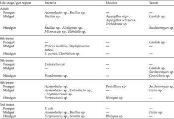
Yeasts, moulds and bacteria were isolated from the different gut regions of Z. variegatus (Table 2). We identified four genera of yeasts: Candida, Saccharomyces, Geotrichum and Pichia, and four of moulds: Aspergillus (two species), Trichoderma, Penicillium and Rhizopus. Among the bacterial isolates, the gram-positive non-motile cocci were identified as Staphylococcus and Streptococcus. The gram-negative motile and non-motile rods identified were members of the family Enterobacteriaceae, from the genera Proteus, Alcaligenes, Escherichia, Enterobacter, Pseudomonas and Serratia.
Distribution of microflora from Zonocerus variegatus gut regions

Distribution of microflora
Table 2 also shows the distribution of yeast, mould and bacterial isolates by gut region. Among the yeast isolates, Saccharomyces sp. was the most widely distributed in the gut regions of the different developmental stages of Z. variegatus examined. This was followed by Candida and Pichia sp., while Geotrichum was the least commonly isolated from the gut regions. There was no distinct trend in the distribution of moulds, but none was isolated from the gut sections of 5th and 6th instars.
Extracellular enzyme production by isolates
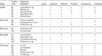
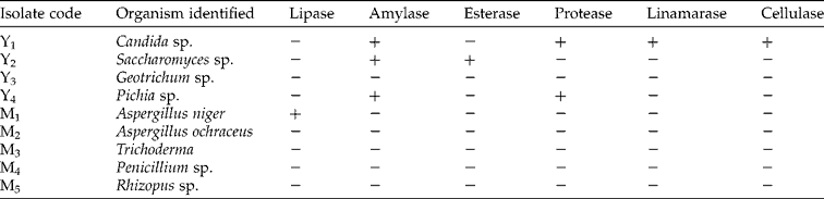
Tables 3 and 4 show the extracellular enzyme activity profile of the microbial isolates. Only the mould Aspergillus sp. was able to degrade lipids. Amylase activity was observed only in bacterial and yeast isolates. Esterase and protease activities were detected in most of the bacterial isolates across virtually all the developmental stages. A number of the bacterial isolates showed linamarase and cellulase activities.
Enzymatic activity profile of bacterial isolates from the gut regions of Zonocerus variegatus

− , negative, i.e. organism isolated at the stage did not produce the enzyme; +, positive, i.e. organism isolated at the stage produced the enzyme.
Enzymatic activity profile of yeast (Y) and mould (M) isolates from Zonocerus variegatus
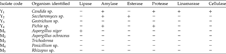
− , did not produce the enzyme; +, produced the enzyme.
Discussion
Zonocerus variegatus is a polyphagous grasshopper (Chapman et al., Reference Chapman, Page and McCaffery1986; Kekeunou et al., Reference Kekeunou, Weise, Messi and Tamo2006), which appears to require a mixed diet for proper development (Idowu and Sonde, Reference Idowu and Sonde2004). Some of its host plants contain secondary compounds, such as cellulose and cyanogenic glucosides, which the grasshopper digests using extracellular enzymes.
The current study has shown that the gut of Z. variegatus harbours micro-organisms, including bacteria and moulds, which are capable of synthesizing enzymes that assist the insect in degrading secondary constituents present in its diet. Studies on cockroaches, termites and locusts have shown that the digestion of dietary cellulose was mediated by micro-organism-derived enzymes (Ohkuma, Reference Ohkuma2003; Delalibera et al., Reference Delalibera, Handelsman and Raffa2005).
Although most of the moulds isolated in this study were not able to degrade the substrates used, it is possible that they play other roles beside the production of enzymes. There are reports that inhabiting symbionts play physiological roles such as the provision of essential nutrients for the host insects (Baumann et al., Reference Baumann, Moran, Baumann and Dworkin2000; Akman et al., Reference Akman, Yamashita, Watanabe, Hattori, Sakai and Ishikawa2002). For instance, Martins (Reference Martins1991) reported that some of these micro-organisms are rich sources of proteins, lipids and carbohydrates. Endocellular symbionts live inside specialized insect cells and provide essential nutrients for their hosts, which in turn provide a suitable habitat for them (Gross, Reference Gross2006).
There are divergent views as to the origin of the microbes in the guts of arthropods. While some researchers believe that they are endogenous, vertically transmitted to the next generation, others argue that they are exogenous, ingested along with food into the gut of the animals (Hunt and Charnley, Reference Hunt and Charnley1981). In agreement with earlier reports (Idowu and Edema, Reference Idowu and Edema2003), the present study shows that the composition of microbial flora in the gut of Zonocerus reflects its environment, indicating that the microbes are ingested by the insect along with its food. The closely related locust species S. gregaria also derives its relatively simple microbiota from ingested food plants, which thereafter multiply within its gut (Dillon et al., Reference Dillon, Venard and Charnley2000). In support of the exogenous origin of microbes in the gut of insects, changes in the diet of the cricket (Acheta domesticus) resulted in the emergence of a new microbial community structure (Gross, Reference Gross2006). Recently, Ademolu (Reference Ademolu2008) has found that the gut of newly hatched 1st instars of Zonocerus was sterile of microbes, meaning that no micro-organisms were vertically transmitted.
Our study provides insight into the physiological mechanisms by which Z. variegatus is able to live successfully on food plants that are rich in potentially toxic secondary compounds. These compounds are processed by enzymes derived from micro-organisms ingested with the insect's diet. Some degree of specialization exists among microbes living in the gut of insects; some are responsible for enzyme production while others serve as sources of nutrients.
Evidently, gut microbiota of Z. variegatus constitute critical metabolic resources for the grasshopper. The transformation of plant secondary compounds by microbial-mediated enzymes has led to the grasshopper's successful adaptation to diverse plant species, particularly cassava, leading to the insect's current status as a major agricultural pest in Nigeria and surrounding regions.