Introduction
Distinct population of stem cells that are located in the male mammalian testes, are responsible for genetic information transmission to the next generation with differentiation of spermatogonia into sperm during spermatogenesis (Bristol-Gould et al., Reference Bristol-Gould2006, Pacchiarotti et al., Reference Pacchiarotti2010). Type A single (As) spermatogonia (known as Spermatogonial stem cell, SSC) starts spermatogenesis. SSCs are located on the basement membrane of the seminiferous tubules and have two fates include of self-renewal and undergo spermatogenesis. Different types of cells produced during spermatogenesis such as A paired (Ap), A aligned (Aal), A1, A2, A3, A4, intermediate and B generally consist of two groups, undifferentiated spermatogonia (As, Ap and Aal) and differentiated spermatogonia (A1, A2, A3, A4, intermediate and B) (Barroca et al., Reference Barroca2009, Schrans-Stassen et al., Reference Schrans-Stassen1999a). All of the spermatogenesis stages are controlled by stem cell factor (SCF) signalling pathway (Mei et al., Reference Mei, Wang and Wu2015). Kit is known as an SCF that is activated after binding to kitl (expressed in the sertoli cells). Kit/kitl system is crucial for the survival, migration, proliferation and anti-apoptosis of Primordial Germ Cells (Gu et al., Reference Gu2009, Ohta et al., Reference Ohta2000). The expression and function of CD-117 receptor are important for proliferation, migration and differentiation and consequently male reproductive potential (Prabhu et al., Reference Prabhu2006). Dym M et al. described that CD-117 messenger RNA is present in differentiated spermatogonia, intermediate and type B but later studies proven that it was also present in undifferentiated spermatogonia (Dym et al., Reference Dym1995, Sorrentino et al., Reference Sorrentino, Giorgi, Geremia, Besmer and Rossi1991). Furthermore, Packer Al et al. has been showed that CD-117 antibody ACK2 injection into male mice caused releasing number of differentiated spermatogonia whereas undifferentiated spermatogonia were not affected (Packer et al., Reference Packer, Besmer and Bachvarova1995). Schrans-Stassen BH et al. described that clear signal was seen in type A spermatogonia and leydig cell but not in the sertoli cells via CD-117 antisense probe hybridization (Schrans-Stassen et al., Reference Schrans-Stassen1999a). The localization of CD-117 expression and response cells to express, is important to find functions of CD-117 as a specific marker (2).
In this way, we now have studied CD-117 expression and localization by Fluidigm real-time RT polymerase chain reaction, immunocytochemistry, immunohistochemistry and flow cytometry. Expression of CD-117 confirmed by in-vitro characterization of GSCs. The results clearly indicate CD-117 express in testicular germ cells which are during proliferation and differentiation. Our investigation will help the future advance studies related to in vitro differentiation and any other kind of experiments that different germ cells’ characterization is required.
Material and methods
In the present study, animal experiments were approved by the Ethical Committee of Amol University of Special Modern Technologies (Ir.ausmt.rec.1398.03.07).
Digestion and culture of testicular cells
In this experimental study, testis cells from C57BL/6 mouse strain were placed in a enzymatic digestion solution which was contain DNAse (0.5 mg/ml) (Sigma Aldrich), Dispase (0.5 mg/ml) and Collagenase IV (0.5 mg/ml) (Sigma Aldrich) in Hank’s Balanced Salt Solution (HBSS buffer) (PAA, USA). After filtering of digested testicular cells through a cell strainer, these cells were cultured in GSCs culture media which was contain 1% L-glutamine (PAA, USA), 1% N2-supplement (Invitrogen, USA), StemPro-34 medium, 5µg/ml bovine serum albumine (BSA) (Sigma Aldrich, USA),6mg/ml D+ glucose (Sigma Aldrich, USA), 1% penicillin/streptomycin (PAA, USA), 30 ng/ml oestradiol (Sigma Aldrich, USA), 1% non-essential amino acids (PAA, USA), 0,1% ß-mercaptoethanol (Invitrogen, USA), 10 ng/ml FGF (Sigma Aldrich, USA), 60 ng/ml progesterone (Sigma Aldrich, USA), 100 U/ml human LIF (Millipore), 8 ng/ml GDNF (Sigma Aldrich, USA), 1% MEM vitamins (PAA, USA), 30 µg/ml pyruvic acid (Sigma Aldrich, USA), 20 ng/ml epidermal growth factor (Sigma Aldrich, USA), 1% ES cell qualified FBS, 1 µl/ml DL-lactic acid (Sigma Aldrich, USA) and 100 µg/ml ascorbic acid (Sigma Aldrich, USA) at 37°C and 5% CO2 in air. This method was according to one-step enzymatic digestion protocol (12, 15).
Cell viability
The testicular cells were planted onto culture plates (100-mm) and cultured for 4 days and were trypsinized for viability determination. The trypan blue solution was added on day 4, and incubated for 5 min at room temperature, To test the proliferate effect of growth factor. Finally, the ratio of viable/dead cells was counted and determined.
Gene expression analysis on the Fluidigm Biomark system
The level of expression of the CD-117 gene, SSCs and TSC cells were examined by Fluidigm BioMark system. SSCs and TSC cells were picked up with a micromanipulator technique, lysed with solution of lysis buffer which was containing 9 μl RT-PreAmp Master Mix (5.0 μl Cells Direct 2× Reaction Mix (Invitrogen, USA), 0.2 μl RT/Taq Superscript III (Invitrogen, USA), 2.5 μl 0.2× assay pool and 1.3 μl TE buffer. We then examined the amount of amplified product of RNA-targeted copies with TaqMan Fluidigm real-time PCR on the Fluidigm BioMark system. Samples were analyzed in two technical repeats. The Ct values were calculated using GenEx software and Excel (Azizi et al., Reference Azizi2016, Reference Azizi, Niazi Tabar and Skutella2021 Conrad et al., Reference Conrad, Azizi and Skutella2018, Conrad et al., Reference Conrad2016).
Immuno-cyto-fluorescenece staining
Isolated cells from testis were fixed with 4% paraformaldehyde, permeabilized with 0.1% Triton X-100 /PBS, blocked with 1% BSA/PBS and incubated with primary antibody CD-117 (Santa Cruz, 130-091-729). The process was continued with overnight incubation (usually ∼16hs) of fluorochrome species-specific secondary antibody at 4°C. The labelled cells were identified by simple nuclear counterstain with 0.2 µg/ml 4′, 6-diamidino-2-phenylindole (DAPI) dye. Antibodies-labelled positive cells were considered by a confocal laser scanning microscope Zeiss LSM 700, and images of cells were obtained using a Zeiss LSM-TPMT camera (Azizi et al., Reference Azizi2016, Azizi et al., Reference Azizi2010, Bojnordi et al., Reference Bojnordi2017).
Immuno-histo-fluorescenece staining
Testicular tissue picked up after de-capsulation of tunica albuginea, washed with PBS, and fixed in 4% paraformaldehyde. Tissue dehydrated during tissue processing and surrounded in Paraplast Plus. Then, tissue was cut with a microtome (usually around 8–10 µm thickness). Sections from testis tissue were mounted on Hydrophilic Plus slides and stored at room temperature until used. During immuno-histo-fluorescenece staining process, slides were washed by xylene and slowly replacing water through a series of decreasing concentrations of ethanol. Before staining, antigen retrieval was done by heat-induced epitope retrieval methods at 95°C for 20 min and non-specific binding site in the tissue sections was blocked with 10% serum/0.3 % Triton in PBS. The characterization of immuno-histo-fluorescenece and immuno-cyto-fluorescenece staining for these sections was followed as past study (Azizi et al., Reference Azizi2016, Niazi Tabar et al., Reference Niazi Tabar, Azizi, Hashemi Karoii and Skutella2022).
Flow cytometric analysis
After viability determining of cells by trypan blue staining, cells were re-suspended in PBS/FBS staining buffer and incubated with cell surface primary antibodies CD-117 which was conjugated with fluorochrome (CD-117-APC, 130-091-729) fluoresce-activated cell sorting (FACS) for 1h. Samples were subjected to a wash procedure and flow cytometric analysis was performed with a BD-FACS Calibur Flow Cytometer.
Statistical analysis
The comparison of expression of CD-117 in neonate GSCs, adult GSCs and TSCs groups was analyzed by using the independent samples t test and ANOVA. Statistical analysis was performed using IBM SPSS Statistics for Windows, Version 25.0 (IBM Corp., Armonk, NY, USA) (P < 0.001).
Results
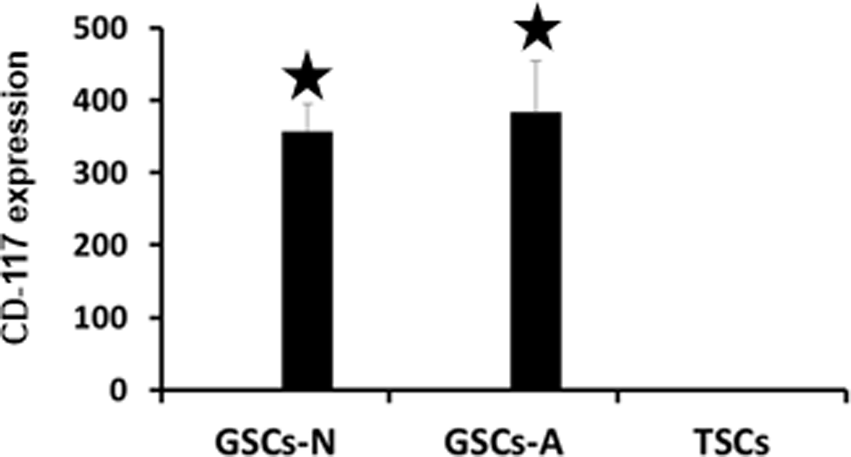
In this experimental research, we study the localization of CD-117 germ cells marker in the seminiferous tubule of mouse testis sections via immuno-histo-fluorescenece analysis. The immuno-histo-fluorescenece results for the testis section showed the positive expression of CD-117 protein in the seminiferous tubules but in different localization. Immuno-histo-fluorescenece analysis indicated that the cytoplasmic and surface expression of CD-117 receptor was translated in cells which located above the basal compartment of the testicular lumen until differentiated part of the seminiferous tubule (fig 1). As immuno-histo-fluorescenece images analysis, CD-117 was completely negative in spermatogonia which located in the basal compartment and spermatid during differentiation to sperms in the luminal location (Fig 1; B1, B2). In addition, merge of blue DAPI (to staining all the nucleus present in seminiferous tubule) and CD-117 (as main marker staining) images proved the strong expression of CD-117 between basal lamina to luminal section of seminiferous tubules of mice (Fig 1; C1, C2). High magnification of confocal microscopy result demonstrated that CD-117 marker was negative in the basal compartment of the testicular lumen and interstitial tissue cells of seminiferous tubule. Analysis of the immunocytochemistry images revealed the cytoplasmic and surface expression of CD-117 in generated germ stem cells (GSCs) while testicular stem cells (TSCs) were negative (fig 2). To study in-vitro analysis of CD-117 protein expression, neonate and adult GSCs were isolated from testis after enzyme digestion and viability determination, then isolated cells cultivated in mentioned growth factors. Isolated GSCs were considered as in our previous study (Azizi et al., Reference Azizi2016). Expression of CD-117 mRNA by Fluidigm real-time RT-PCR indicated significant expression (P < 0.001) of CD-117 in neonate and adult GSCs in comparison to TSCs. Also, we observed that the expression of CD-117 was not different between neonate and adult GSCs (fig 3). Flow cytometry results for surface expression of CD-117 protein between GSCs cells and testis cells demonstrated about 10% of adult testicular cells express CD-117 which was not significant with GSCs cells (fig 4A).
Immuno-histo-fluorescenece analysis of CD-117 in testis section. Immuno-histo-fluorescenece analysis for expression of CD-117 in the testis. CD-117 protein was expressed after the base membrane and differentiated part of testis (arrow). Negative expression in basal compartment and after differentiation part (arrowhead). CD-117 expression (A1, A2). CD-117 expression merge with DAPI (B1, B2) (Scale bar = 50 μm).

Immuno-cyto-fluorescenece analysis of CD-117 in GSCs. Immuno-cyto-fluorescenece analysis for expression of CD-117 in the GSCs. Immuno-cyto-fluorescenece analysis showed that final colonies from expansion of our extract cells, express CD-117. Blue DAPI (C1, C2); CD-117 expression (B1, B2); Merge images (A1, A2) (Scale bar = 50 μm).

mRNA expression of CD-117 gene. Fluidigm Real-time PCR analysis. Significant expression of CD-117 in the neonate GSCs and adult GSCs in compare to TSCs.

Characterization of GSCs. Flow cytometry analysis for expression of CD-117. A: CD-117 expression in testis and GSCs. B: Histogram obtained from the FACS analysis of CD-117-APC (antibody-conjugated with fluorochrome) in stem cells compartment.

Discussion
Confocal microscopic characterization for testis section demonstrated localization of CD-117 germ cells marker in the cells which localized above the basal compartment of the testicular lumen. It seems CD-117 germ cells marker express in proliferation and differentiation position of spermatogonial cells in testis. In-vivo results revealed negative expression of CD-117 in the basement membrane and interstitial tissue compartments. Schrans-Stassen BH et al. has been reported that a strong signal was observed in A differentiated spermatogonia whereas any signal couldn’t be detected in A undifferentiated (Schrans-Stassen et al., Reference Schrans-Stassen1999a). It has long been believed that differentiated spermatogonia are irreversible which mean CD-117+ cells can’t reverse to CD-117- cells but Kai T et al. reported that in Drosophila Melanogaster, functional spermatogonia stem cells that are from convert of differentiated spermatogonia, can restore germinal stages (1992). Barroca et al. reported that purified differentiated spermatogonia when transplanted into the r-irradiated germ cell-depleted adult mice testes and in presence of fibroblast growth factor2 (FGF2) and glial cell line-derived neurotrophic factor (GDNF) (Barroca et al., Reference Barroca2009, Zhang et al., Reference Zhang2011). Similarly to CD-117, other developmental stage-specific biomarkers of male germ cells are the main parts of reproductive biology researches including PGP9.5, sall4, glial cell-derived neurotrophic factor receptor alpha-1 (GFRa-1) and UTF1.furtheremore, lee et al. has been shown that PGP 9.5 expressed in undifferentiated spermatogonia but not in differentiated spermatogonia (Lee et al., Reference Lee2013). Park et al. has been reported that in postpubertal porcine testes, sall4 was detected in CD-117-positive spermatogonia (Park et al., Reference Park2017). Similarly, has been proven that in the prepubertal porcine, UTF1 was detected in most of the PGP 9.5 positive spermatogonia (Lee et al., Reference Lee2014, Roshan et al., Reference Roshan, Azizi, Majelan and Tabar2024) . An immuno-histo-fluorescenece analysis reported that c-kit is negative in A single and paired (As, Ap) and Aal are negative during proliferation period, at which time to differentiated into A1 spermatogonia; another important remarkable report was that the c-kit positive spermatogonia pattern during proliferation development vitamin A-induced differentiation seems to be necessary (Schrans-Stassen et al., Reference Schrans-Stassen1999b). Interestingly, a striking parallel can be seen with haematopoiesis in which the pluripotent stem cells are c-kit low expression (Doi et al., Reference Doi1997). It has been reported that a strong c-kit mRNA and protein expression was detected in leydig cells in normal testis of mice whereas no signal was observed in sertoli cells that proved that no c-kit protein can be formed in sertoli cells (22). Many researches in the recent decade, revealed that CD-117 has a key role in ratio maintaining between differentiation and self-renewal of spermatogonial stem cells. Alternation in this ratio also entails stem cell depletion resulting in SCO (sertoli cell only syndrome) (Morimoto et al., Reference Morimoto2009, Tabar et al., Reference Tabar, Sojoudi, Henduei and Azizi2022).
Conclusion
The study of our data confirmed the CD-117 receptor gene expressed above the basement membrane of the seminiferous tubule of testis and also the differentiated part. It appears CD-117 marker express during proliferation and differentiation of spermatogonial cells. Therefore, it would be interesting to identify in vitro proliferation and differentiation of germ line stem cells in testis by up- or down-regulation of CD-117 marker.
Acknowledgements
I acknowledge the generous support of the China Scholarship Council (CSC) in funding my studies and research, which has been instrumental in facilitating my academic progress and international collaboration.
Funding
This research was supported by the China Scholarship Council (CSC), University of Heidelberg, Institute for Anatomy and Cell Biology III, Department of Neuroanatomy, Germany and the Amol University of Special Modern Technologies, Islamic Republic of Iran.
Competing interests
There is no conflict of interest in this study.
Ethical standards
In the present study, animal experiments were approved by the Ethical Committee of Amol University of Special Modern Technologies (Ir.ausmt.rec.1398.03.07).