Introduction
A detection technique for various biomarkers in blood or urine enables us to make an at-home diagnosis simply, rapidly, and inexpensively. In particular, semiconductor-based biosensors such as ion-sensitive field-effect transistors (ISFETs) and microelectromechanical-system (MEMS)-based biosensors are expected to be developed for multi-marker detection using a single chip. ISFETs detect the charge of molecules immobilized on a channel for ion,[Reference Esashi and Matsuo1, Reference Sawada, Mimura, Tomita, Nakanishi, Tanabe, Ishida and Ando2] DNA,[Reference Sakata, Kamahori and Miyahara3] or protein[Reference Ohno, Maehashi, Inoue and Matsumoto4] detection. Although an ISFET-based biosensor can realize a large-scale integration for the parallel processing of multiple biomarkers, the detection area is limited by several nanometers from the substrate surface owing to the Debye length, and therefore an ISFET-based biosensor cannot be applied to the detection of charged macromolecules.[Reference Sakata and Miyahara5] For the detection of biomacromolecules such as proteins, a MEMS-based biosensor that uses nanomechanical deflection caused by molecular binding to an antibody fixed on the sensing area is applied.[Reference Raorane, Lim, Chen, Craik and Majumdar6] The static deformation is proportional to the surface stress caused by the Coulomb repulsive force between adsorbed molecules.[Reference Watari, Galbraith, Lang, Sousa, Hegner, Gerber, Horton and McKendry7] Based on the sensing method of the stress-induced nanomechanical deflection, MEMS surface stress sensors have been used for various targets such as DNA,[Reference Fritz, Baller, Lang, Rothuizen, Vettiger, Meyer, Guntherodt, Gerber and Gimzewski8] RNA,[Reference Zhang, Lang, Huber, Bietsch, Grange, Certa, McKendry, Guntherodt, Hegner and Gerber9] proteins,[Reference Wu, Datar, Hansen, Thundat, Cote and Majumdar10] viruses,[Reference Gorelkin, Erofeev, Kiselev, Kolesov, Dubrovin and Yaminsky11] and different chemicals.[Reference Berger, Delamarche, Lang, Gerber, Gimzewski, Meyer and Guntherodt12] Surface stress-based biosensors unconstrained by molecular size can possibly allow the establishment of multi-biomarker detection using different antibodies immobilized on individual sensor pixels. In addition, surface stress occurs not only with molecular adsorption but also with the structural changes to the biomacromolecules,[Reference Ghatkesar, Lang, Gerber, Hegner and Braun13, Reference Buchapudi, Xu, Ataian, Ji and Schulte14] and therefore, a surface stress-based sensor can be used to conduct both quantitative and qualitative analyses of adsorbed molecules.
Conventional MEMS surface stress sensors have adopted capacitive[Reference Satyanarayana, McCormick and Majumdar15] or piezoresistive[Reference Wee, Kang, Park, Kang, Yoon, Park and Kim16, Reference Yoshikawa, Akiyama, Gautsch, Vettiger and Rohrer17] methods for an electrical readout transduced from the nanomechanical deflection. However, the limit of detection of the MEMS surface stress sensor is less than that of other biosensors.[Reference Arlett, Myers and Roukes18] To solve this problem, we recently developed a surface stress-based biosensor that uses a MEMS Fabry–Perot interferometer (FPI) with a silicon photodiode.[Reference Takahashi, Oyama, Misawa, Okumura, Ishida and Sawada19] Using the drastic change in transmittance caused by membrane deformation of several tens of nanometers, the electrical readout signal is obtained as a photocurrent with a high signal-transducing efficiency. In addition, the proposed sensor can utilize the signal processing technique of a complementary metal–oxide–semiconductor image sensor.[Reference Maruyama, Hizawa, Takahashi and Sawada20] The integrated FPI sensor array enabled us to develop a label-free multi-biosensing platform with high sensitivity.
In terms of the sensitivity of surface stress sensors, the suspended membrane of the FPI requires a soft material with a low Young's modulus, which offers improved sensitivity with molecular adsorption. Capacitive sensors and piezoresistive sensors use a semiconductor or metal for the deformable membrane because these materials allow a change in capacitance or resistance associated with membrane deformation for signal transduction, respectively. In contrast, optical interferometric sensors can accommodate organic materials for a deformable membrane. The previously fabricated FPI sensor used parylene-C with a Young's modulus of 2.8 GPa as the deformable membrane, and was shown to be able to detect antigens at a concentration of 10 ng/mL.[Reference Takahashi, Hizawa, Misawa, Taki, Sawada and Takahashi21] In this regard, an elastomer seems to be the most promising material for a stress sensor because of the low Young's modulus of 1–100 MPa, which is two to three orders of magnitude smaller than that of plastic materials, such as polymethylmethacrylate, polystyrene, and poly (para-xylylene) (parylene).
We previously developed an elastomer sheet hundreds of nanometers thick using a polystyrene-polybutadiene-polystyrene (SBS) triblock copolymer (referred to as an SBS nanosheet),[Reference Sato, Murata, Fujie and Takeoka22] and demonstrated unique physical properties such as flexibility, stretchability, a low Young's modulus, and physical adhesiveness, reported in other types of nanosheets (e.g., polysaccharides).[Reference Fujie, Matsutani, Kinoshita, Okamura, Saito and Takeoka23, Reference Fujie24] In addition, the freestanding SBS nanosheet also achieves a large elastic deformation of 38% strain with cyclic straining.[Reference Kumagai, Sato, Takeoka, Sawada, Fujie and Takahashi25] Moreover, the freestanding SBS nanosheet can be attached to a substrate using a dry transfer technique without vacuum or high-temperature processes owing to the physical adhesiveness of the SBS nanosheet.[Reference Takahashi, Fujie, Sato, Takeoka and Sawada26]
In this paper, we propose an elastomer-based MEMS FPI transducer fabricated using the dry transfer of an elastomer nanosheet to cover the microcavities of a silicon substrate. We demonstrated a spectrum shift caused by nanomechanical deflection of the dry-transferred nanosheet in a liquid environment. The surface stress sensitivity of the FPI transducer associated with molecular adsorption was also evaluated using the surface-functionalized elastomer nanosheet.
Materials and methods
Materials
Polysaccharide electrolytes, chitosan (M w 88,000), and sodium alginate (Na alginate, M w 106,000) were purchased from Nacalai Tesque, Inc. (Kyoto, Japan) (see Supplementary Material Fig. S1). Polystyrene-block-polybutadiene-block-polystyrene (SBS; M w 140,000) was purchased from Sigma-Aldrich Japan (Tokyo, Japan). Poly(vinylalcohol) (PVA, M w 22,000, 86.5–89% hydrolyzed) was purchased from Kanto Chemical Co., Inc. (Tokyo, Japan). All organic solvents (reagent grade) were purchased from Kanto Chemical Co., Inc. (Tokyo, Japan) and used without purification. Silicon wafers (SiO2 substrates) were purchased from KST World Co. (Fukui, Japan), and cut into an appropriate size (typically 2 cm × 2 cm), and immersed in a mixture of sulfuric-acid/hydrogen-peroxide (3/1 v/v) at 120 °C for 15 min before being extensively rinsed with deionized (DI) water (resistivity of 18 MΩ cm).
Preparation of SBS nanosheets and layer-by-layer deposition
Freestanding SBS nanosheets were prepared by following our previous report (see Supplementary Material Fig. S2).[Reference Sato, Murata, Fujie and Takeoka22] In brief, a polyethylene terephthalate (PET) film was used as a base film. Next, an aqueous solution of PVA (20 mg/mL) was coated onto the PET film as a sacrificial layer using a microgravure coating method (gravure speed of 30 rpm, line speed of 1.3 m/min, drying temperature of 100 °C). Subsequently, the SBS polymer dissolved in tetrahydrofuran was coated onto the PVA/PET film (rotation speed of 30 rpm, line speed of 1.3 m/min, drying temperature of 80 °C).
Next, for electrostatic immobilization of the analytical molecules, 5.5-layer pairs of chitosan and sodium alginate were formed on the SBS layer using a spin-coating assisted layer-by-layer (LbL) method. First, the SBS/PVA/PET film was attached to a PDMS slab for spin coating after the surface was treated with plasma cleaner (30 mA, 1 min). Then, 1 mg/mL of chitosan and 1 mg/mL of Na alginate dissolved in 1% (v/v) acetic acid and DI water, respectively, were alternatively spin coated onto the SBS layer through a dual water-rinsing process (rotation speed of 4500 rpm, rotation time of 15 s for each spin-coating step). In the resulting film, the chitosan-layer-terminated LbL-SBS nanosheet was finally detached from the PET film using a paper-tape frame. After dissolving the sacrificial PVA layer in DI water, freestanding LbL-SBS nanosheets supported by the paper-tape frame were collected in air.
Atomic force microscopy (AFM) observation
The surface morphologies of the SBS and LbL-SBS nanosheets were analyzed using an atomic force microscope (AFM, Innova SPM, Bruker Co., Ltd., MA, USA), equipped with a piezoscanner (maximum scan size of 50 µm × 50 µm). Tapping mode tips (RTESPA-CP, Silicon AFM Probes, Bruker, USA) were used for the surface scanning, with a resonance frequency within the range of 290–330 kHz. The AFM tapping phase images were filtered and analyzed using NanoScope Analysis software (Bruker).
X-ray photoelectron spectroscopy (XPS) analysis
For a chemical analysis of the SBS and LbL-SBS nanosheets, XPS was carried out using a JPS-9010TR with a monochromatic Al KR x-ray source (1486.6 eV) incident at 90° relative to the axis of a hemispherical energy analyzer. Photoelectrons were collected at a takeoff angle of 90° from the surface, and using an analyzer spot diameter of 1.1 mm. The survey spectra were collected from 0 to 1100 eV, and the high-resolution spectra were obtained for C1s, O1s, and N1s. All spectra were collected at room temperature with a base pressure of less than 1 × 10⁻⁸ Pa. Electron binding energies were calibrated with respect to the C1s line at 284 eV.
Bulge test
Mechanical properties of SBS and LbL-SBS nanosheets were evaluated by a bulge test. Ultimate tensile strength and elastic modulus of the nanosheets were determined using following equations:
 $$\sigma = \displaystyle{{P \times a^2} \over {4 \times h \times d}}$$
 $$\varepsilon = \displaystyle{{2 \times d^2} \over {3 \times a^2}}$$
 $$E = \displaystyle{\sigma \over \varepsilon} $$
where σ, P, a, h, d, ε, and E represent the tensile stress, applied pressure, radius of the circular hole of the steel plate, thickness, deflection, tensile strain, and elastic modulus of the thin film, respectively.
Characterization of LbL-SBS nanosheets
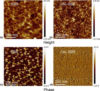
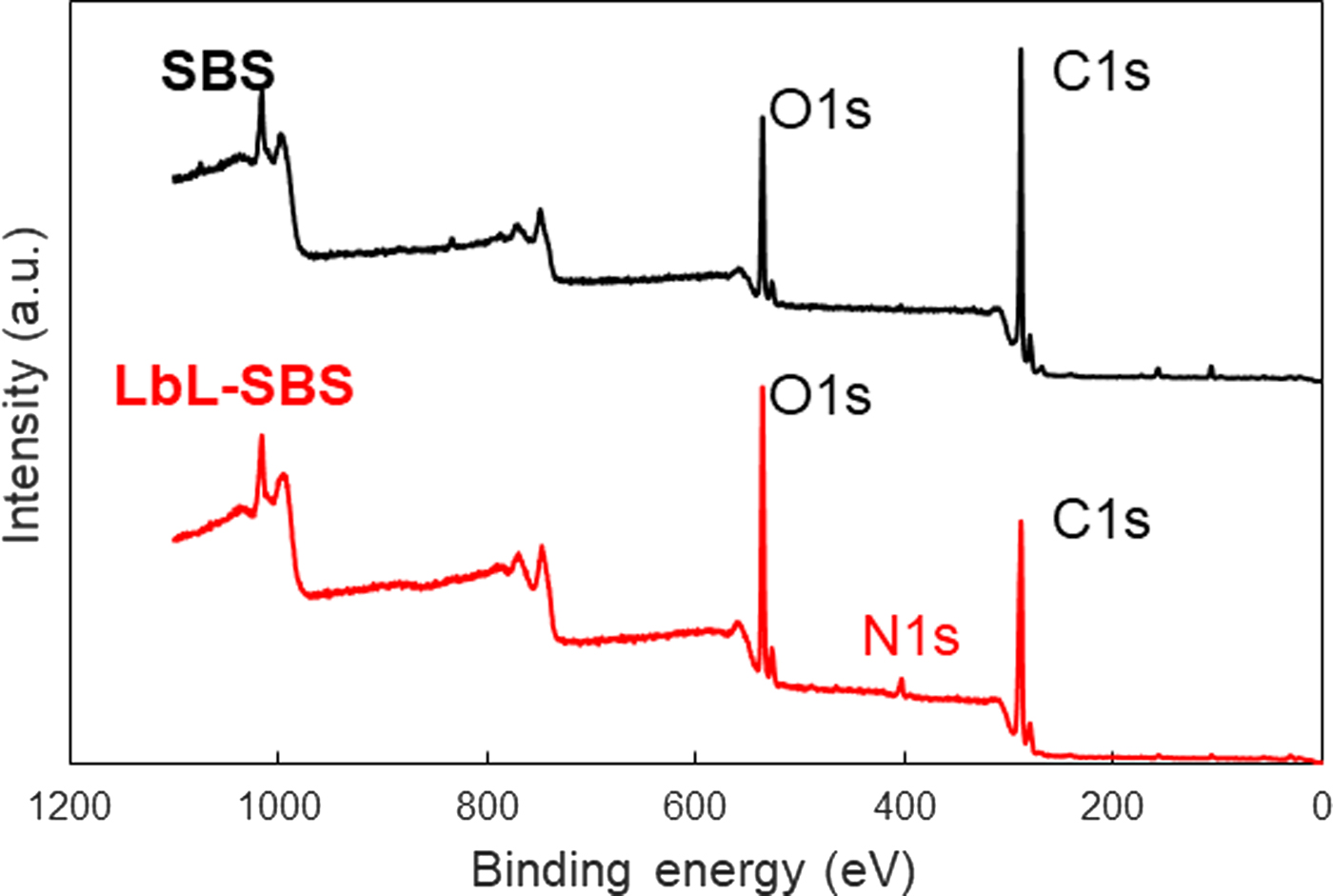
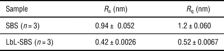
We obtained 5.5-layer pairs of chitosan/Na alginate on an SBS nanosheet using the LbL method. The thickness of the pristine SBS nanosheet was 477 ± 16 nm, whereas that of the LbL-SBS nanosheet was 493 ± 13 nm. Therefore, the subtracted thickness of the 5.5-LbL film was 16 ± 3 nm, which showed good agreement with a previous result.[Reference Fujie, Matsutani, Kinoshita, Okamura, Saito and Takeoka23] The AFM surface morphology of the LbL-SBS nanosheet was relatively smooth, compared with that of the pristine SBS nanosheet (Fig. 1). Notably, the tapping-phase images reveal a phase-separated structure of the SBS nanosheet, which was hindered after the LbL deposition. Correspondingly, the surface roughness (R q) of the LbL-SBS nanosheet (0.52 ± 0.0067 nm) became half that of the pristine SBS nanosheet (1.2 ± 0.060 nm), as shown in Table I. The LbL formation on the SBS nanosheet was also confirmed through an XPS analysis, which showed the presence of a new N1s peak at 400 eV (Fig. 2).
AFM characterization of LbL-SBS nanosheets. Top panels: height images of (a) SBS and (b) LbL-SBS. Bottom panels: phase images of (c) SBS and (d) LbL-SBS.

XPS spectra of SBS and LbL-SBS nanosheets. The N1s peak attributed to amine groups of chitosan was found for LbL-SBS.

Surface roughness of SBS and LbL-SBS nanosheets

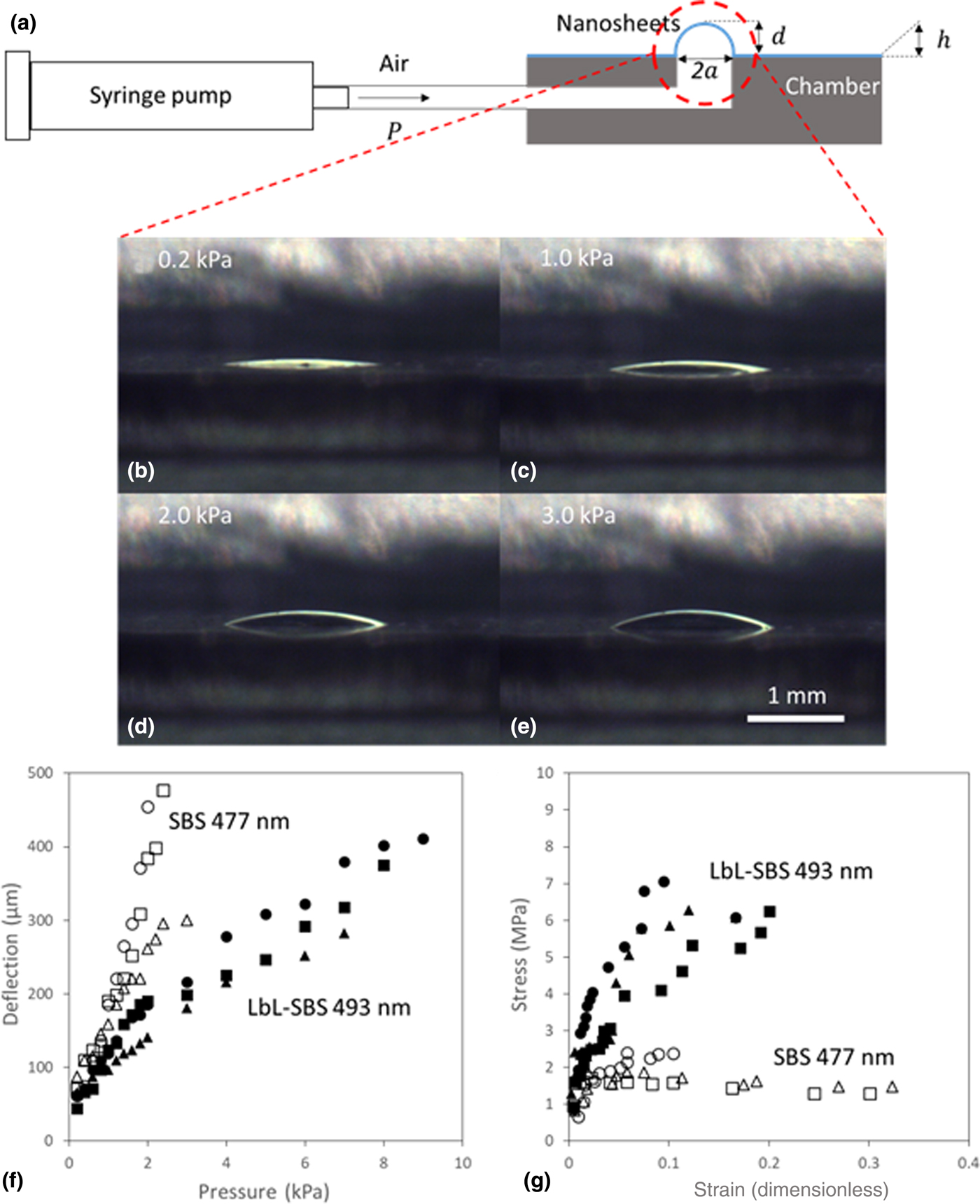
The mechanical influence of the LbL film on the SBS nanosheet was also evaluated using a bulge test (Fig. 3a). Both nanosheets were deflected by the applied air pressure in a step-wise manner (Fig. 3b). According to the stress–strain curve based on the deflection, the elastic modulus of the LbL-SBS nanosheet (0.125 ± 0.033 GPa) was 4.5 times higher than that of the SBS nanosheet (0.028 ± 0.011 GPa) (Fig. 3c). Because the elastic modulus of the chitosan/Na alginate nanosheets was around 1 GPa,[Reference Fujie, Matsutani, Kinoshita, Okamura, Saito and Takeoka23] the chitosan/Na alginate layer increased the elastic modulus of the LbL-SBS nanosheet. However, the elastic modulus of the LbL-SBS nanosheet was still 22.4 times smaller than that of a typical diaphragm, such as parylene-C [https://vsiparylene.com/parylene-advantages/properties/ (accessed September 25, 2018)].
(a) Schematic representation of an apparatus for bulge test. (b–e) Cross-sectional images of deflected LbL-SBS nanosheets, sequentially compressed using external pressure of 0.2–3.0 kPa, respectively. (f) Pressure-deflection and (g) stress–strain curves of SBS nanosheets with thickness of 477 nm, and LbL-SBS nanosheets with thickness of 493 nm [i.e., n = 3 for SBS (blank symbol) and LbL-SBS (filled symbol), respectively].

Device design
Design of optical interferometric transducer
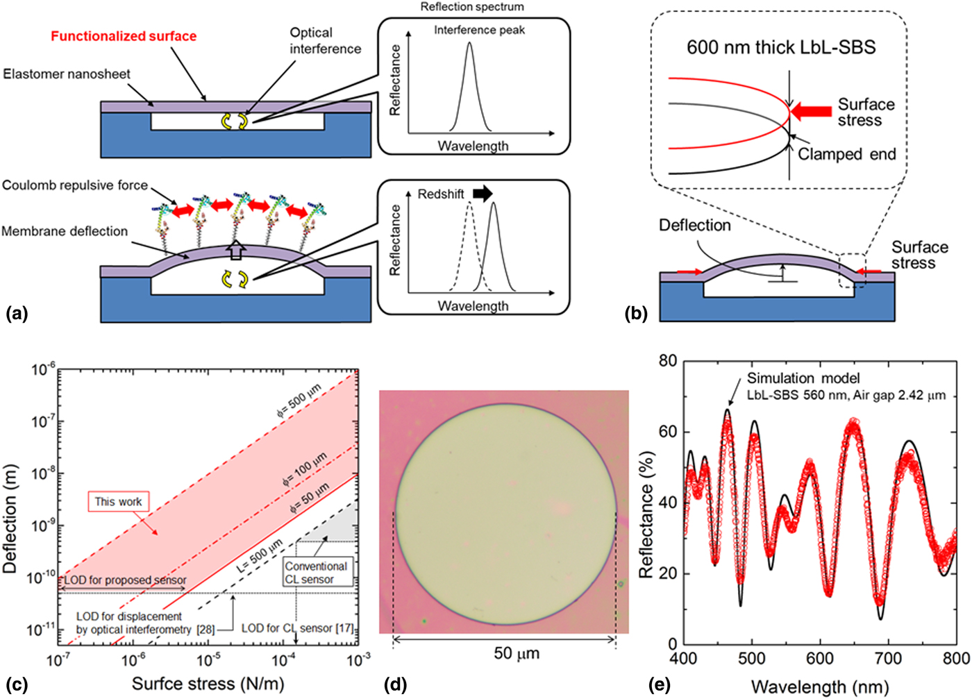
Figure 4(a) shows a schematic illustration of a MEMS optical interferometry-based surface stress transducer constructed using a suspended membrane over a silicon substrate with an air gap. The suspended membrane and the substrate act as two reflecting mirrors. An optical length configured by the air gap and the suspended membrane determines the optical interference. The target molecules are specifically bound using pre-immobilized receptors on the membrane such as an antigen–antibody reaction. The SBS nanosheet is utilized as the suspended membrane to increase the stress sensitivity owing to a low Young's modulus. When charged biomolecules are bound on the elastomer nanosheet, the membrane deforms into the upper direction by electrostatic repulsive force of the adsorbed molecules,[Reference Fritz27] resulting in a redshift of the optical interference peak wavelength. To obtain an electrical signal, a photodetector can be implemented under the FPI for measuring the transmission light intensity. When a single wavelength light such as an LD or LED is illuminated onto the FPI, the intensity of the incident light on the photodetector is drastically changed. Therefore, we can obtain the electrical readout signal as a photocurrent. A drawback of a highly sensitive label-free biosensor is a trade-off relationship between the sensitivity and false positive responses, and the proposed surface stress sensor responds to temperature change, pressure change, etc. To avoid the false positive responses, a reference sensor without receptor molecules is required, resulting in discrimination with positive and false positive responses.
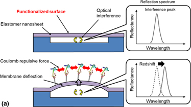
(a) Schematic image of the surface stress sensor based on a MEMS FPI. The FPI consists of a suspended thin film and an air gap, which determine the interference peaks of the reflection spectrum. The peak position is red shifted in association with the membrane deflection caused by Coulomb repulsive force adsorbed molecules on functionalized surface. (b) Mechanical deflection of elastomer nanosheet induced by surface stress simulated using FEM. The sensing membrane was designed to have a diameter of 50–500 µm and a thickness of 600 nm. (c) Typical membrane deflection as a function of the surface stress is plotted for comparing proposed transducer and conventional cantilever (CL) sensor. Limit of detection (LOD) of surface stress is improved by two to three orders of magnitude compared to the CL sensor. (d) Optical micrograph of the developed FPI surface stress sensor. (e) Typical reflection spectrum of the FPI with the dry-transferred LbL-SBS nanosheet. The suspended LbL-SBS nanosheet was 560-nm thick and 50 µm in diameter.

Mechanical simulation
Compared to a previously reported surface stress transducer using a piezoresistive method, the proposed transducer used a soft material with a Young's modulus of 0.01–1 GPa, which is expected to improve the stress sensitivity. To estimate the sensitivity using an LbL-SBS nanosheet, a mechanical deflection of the suspended membrane associated with the surface stress was calculated by applying the finite element method (FEM) using COMSOL. The suspended membrane was designed to have a diameter of 50–500 µm and a thickness of 600 nm. The physical parameters of the LbL-SBS nanosheet were a Young's modulus of 125 MPa, Poison's ratio of 0.49, and density of 0.95 g/cm3. Figure 4(b) shows the calculated deformation with isotropic compressive surface stress applied at the periphery of the membrane.[Reference Yoshikawa, Akiyama, Gautsch, Vettiger and Rohrer17] The result indicates that the LbL-SBS membrane with a diameter of 500 µm is deflected upward, reaching 947 nm with 1 mN/m. The typical deflection curve as a function of the surface stress is shown in Fig. 4(c). For quantitative comparison of transducers, we also calculated the deflection of a conventional cantilever type. The minimum detection limit of the surface stress of the conventional cantilever transducer have been reported to be 0.1–1 mN/m,[Reference Yoshikawa, Akiyama, Gautsch, Vettiger and Rohrer17] which agrees well with our calculation result with 500-μm-long, 100-μm-wide, and 1-μm-thick. With the minimum detectable displacement of 50 pm using the optical interferometry method,[Reference Schmidt, Werther, Furstenau, Matthias and Melz28] the detection limit of the surface stress was obtained to be 0.1 µN/m or less, which is three orders of magnitude greater than a conventional surface stress transducer. From the point of view of the MEMS structure, the stiffness of the clamped membrane is much higher than that of a cantilever and bridge type. However, it is indicated that an elastomer nanosheet with an edge-clamped circular diaphragm decreases the total stiffness owing to the low Young's modulus and thin small thickness.
Fabrication
An elastomer-based MEMS FPI transducer was fabricated using an LbL-SBS nanosheet (see Supplementary material Fig. S3). Based on the mechanical simulation, we prepared LbL-SBS nanosheets for the device fabrication with the actual thickness of 560 nm. To form a suspended membrane with a microcavity structure, a Si wafer with shallow trenches was prepared using reactive ion etching. For an improvement of the adhesion between the SBS nanosheet and Si substrate, a silane-coupling agent was coated onto the Si wafer, followed by a transferring of the nanosheet onto the Si wafer. An LbL-SBS nanosheet was developed on a PET film with a pre-laminated PVA sacrificial layer, as mentioned above. The LbL-SBS nanosheet was released from the PET film by dissolving the PVA layer in hot water at 60 °C. After drying, the released LbL-SBS nanosheet was transferred onto the Si substrate under dry conditions at room temperature. The high adhesiveness of the elastomer nanosheet provides permanent bonding without a vacuum process or high-temperature bonding.
Optical property
Figure 4(d) shows an optical microscope image of the developed freestanding LbL-SBS nanosheet with a diameter of 50 µm. The reflection spectrum of the elastomer-based FPI was measured using microscopic spectroscopy. A spectrometer (Ocean Optics, USB2000) was attached to a reflection microscope (Olympus, BX51) using an optical fiber with a core diameter of 100 µm. The magnification of the objective lens was 20×, and the measurement spot diameter was 5 µm. Figure 4(e) shows a typical reflection spectrum of the FPI with the dry-transferred LbL-SBS nanosheet. Eight interference peaks were observed in the reflection spectrum from the diaphragm within the visible region. A fitting curve using a refractive index of 1.5 with a thickness of 560 nm and a gap of 2.42 µm showed good agreement with the experimental value.
Experimental results of MEMS interferometer
Evaluation of membrane deflection in liquid
For an evaluation of the sensitivity of the proposed transducer, an anti-bovine serum albumin antibody (anti-BSA) was used as a model molecule for the driving force to deform the membrane because the surface stress sensor can obtain a deformation of the membrane owing to an adsorption of any molecule. We prepared phosphate buffer saline (PBS) (pH 7.4) for a negative control, and 10 µg/mL anti-BSA dissolved in PBS for the surface stress sensing. For comparison of the molecular adsorption capacity of a transducer surface, both transducers with Na alginate and chitosan coated on the top of the surface were prepared. Antibody molecules are expected to be adsorbed by the electrostatic interaction on the surface of the chitosan, whereas the molecular adsorption capacity of Na alginate surface is expected to be low. The reflection spectrum from the FPI transducer in the solution was measured using the same microscopy setup as mentioned above.
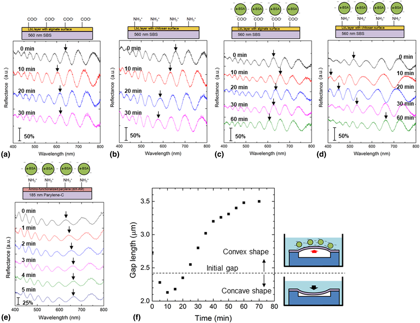
Figures 5(a) and 5(b) show typical reflection spectra of the FPI with a Na alginate surface and a chitosan surface in a PBS solution corresponding to the treatment time, respectively. All spectral shifts shown in Fig. 5 were recorded as moving picture (see Supplementary Video S1). The movie shows the spectral shift in the first 10 min of Fig. 5(a) at 50× speeds. The arrows in Fig. 5 indicate the position where a certain peak has shifted. Although the peak positions of the reflection spectra were found to redshift immediately after being inserted into the PBS which is considered to be physical influence due to surface tension of PBS, the spectra of both devices were found to blue shift after 30 min of immersion in the PBS, indicating that the membrane was deformed into a lower direction. It is thought that air in the microcavity gradually leaked owing to the gas permeability of the elastomer nanosheet and no molecular adsorption on the membrane in PBS. In addition, the transient response of the membrane when the membrane comes in contact with the liquid can be relaxed by adding surfactants to reduce surface tension.
Spectrum shift of elastomer surface stress sensor by displacement of LbL-SBS nanosheet in a liquid. Typical reflection spectra of the FPI with (a) Na alginate surface and (b) chitosan surface in PBS solution corresponding to treatment time. Typical spectrum shift of the interferometric sensor with (c) Na alginate surface and (d) chitosan surface immersed in 10 µg/mL anti-BSA solution. (e) Typical spectrum shift of the parylene-C-based interferometric sensor immersed in a 10 µg/mL anti-BSA solution. (f) The time course of the membrane position is plotted. Although the shape of the membrane for the first 20 min showed a concave shape, the membrane position gradually increased and maintained a convex shape through the immobilization of antibody molecules.

Figures 5(c) and 5(d) show typical spectrum shifts of the FPI transducer immersed in a 10 µg/mL anti-BSA solution. The transducer with the Na alginate surface behaved similarly as the spectral shift in PBS, whereas the FPI transducer with the chitosan surface demonstrated a redshift after observing the blueshift within the first 10 min. It should be noted that the motion of the spectral shift continued to redshift after changing from a blueshift. The elastomer membrane will deform into the upper direction in a liquid because the peak position has shifted toward the long wavelength side as compared with the initial state. The tenth order interference peak, indicated by an arrow, was found to move by 260 nm within 50 min. We assumed that the membrane was deformed by the surface stress induced through the molecular adsorption because the amino group of the chitosan layer can electrostatically adsorb the anti-BSA antibody. The difference in response between the chitosan and alginate surfaces can also be explained by the electrostatic interaction because anti-BSA with an isoelectric point of 4.8–5.2,[Reference Ge, Kojio, Takahara and Kajiyama29] and a carboxyl group of alginate, are negatively charged whereas the amino-group of chitosan is positively charged at pH 7.4. Therefore, a freestanding elastomer membrane with a chitosan-functionalized surface using the LbL method demonstrated a surface stress response by adsorbing the anti-BSA antibody.
For a quantitative comparison, we measured the nanomechanical deflection using a previously developed parylene-C-based surface stress sensor with the same molecular concentration. The sensing membrane was formed using 20-nm thick amino-functionalized parylene (diX-AM) and 185-nm thick parylene-C with a diameter of 50 µm. The tenth order interference peak was found to redshift by a maximum of 37 nm, and saturation in 5 min, as shown in Fig. 5(e). Despite the membrane thickness of parylene-based FPI, which is as thin as one-third of an LbL-SBS nanosheet, the SBS-based FPI exhibits an improvement of both the maximum deflection and the deflection speed owing to the elastic property of SBS.
The cavity length can be calculated through a rigorous coupled-wave analysis using experimentally obtained reflection spectra. Curve fitting was performed for the reflection spectra for 10 and 60 min treatment using the anti-BSA antibody PBS solution, which are the points furthermost to the left and right of spectrum peak shift (see Supplementary Material Fig. S4). A slight attenuation of the light intensity was found, whereas we obtained the agreed-upon fitting curves with interference peak positions. The membrane at 10 min moved down with 0.3 µm displacement compared to the initial gap length measured in air. Meanwhile, the gap length at 60 min was 3.48 µm, which means that the membrane deformed into the upper direction. From the calculation results, the time course of the membrane position was plotted, as shown in Fig. 5(f). The dotted line shows the initial flat state of the membrane. Whereas the shape of the membrane for the first 20 min concave, the membrane position gradually increased and maintained the convex shape through the immobilization of the molecules. This result suggests that the chitosan-functionalized membrane effectively adsorbed the biomolecules through an electrostatic interaction, resulting in a membrane deflection caused by a change in surface stress. In addition, the response of the membrane indicated the saturation of molecular adsorption to the freestanding membrane. Note, however, that the deformed membrane gradually changes back to the flat position, which is considered to be caused by pressure reduction due to air leakage. To measure concentration of target molecule, it is possible to evaluate by comparing the initial velocity of deflection response after adding an analyte. It is expected that highly sensitive surface stress sensing is associated with the molecular adsorption owing to the use of an elastomer nanosheet.
Conclusions
We developed an elastomer-based MEMS FPI using a dry transfer technique to form a microcavity. The fabricated FPI comprises a freestanding LbL-SBS nanosheet with a thickness of 560 nm provided a sufficient optical property such as reflectance and peak-to-valley ratio for optical biosensing. For molecular immobilization on the proposed transducer, a chitosan-functionalized elastomer nanosheet was used for the suspended membrane. We demonstrated the spectrum shift associated with the nanomechanical deflection of the dry-transferred freestanding LbL-SBS nanosheet in a liquid environment. The nanomechanical deflection caused by molecular adsorption was enhanced using an elastomer nanosheet for the deformable membrane, which enables the detection of biomarkers with high sensitivity by freely modifying the membrane surface, leading to a universal biosensing platform. The detection capability of the surface stress biosensor depends on the sensitivity of the transducer as well as the bio-interface such as the density of a cross linker and affinity of a receptor molecule. The elastomer transducer with improved surface stress sensitivity is expected to detect lower concentration of target molecules by optimization of the bio-interface on the elastomer transducer.
Supplementary material
The supplementary material for this article can be found at https://doi.org/10.1557/mrc.2019.11
Acknowledgments
This work was supported in part by a Grant-in-Aid for Young Scientists (A) (26709025), Grant-in-Aid for Scientific Research (B) (18H03539), and Grant-in-Aid for Scientific Research on Innovative Areas (18H05469) from the Japan Society for the Promotion of Science, and by the Precursory Research for Embryonic Science and Technology (PRESTO) (JPMJPR1526 and JPMJPR152A) from the Japan Science and Technology Agency.