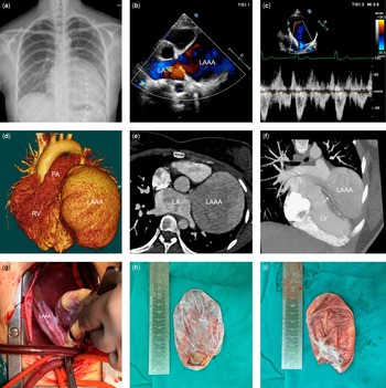
A 19-year-old girl was admitted to our hospital because of significant cardiomegaly in her chest X-ray during general medical examination. Her chest X-ray showed a spherical heart, and the cardiothoracic ratio was 0.6–0.7 (Fig 1a). Her physical examination was unremarkable except for heart enlargement on both sides. The patient had no symptoms associated with this lesion. Electrocardiography showed sinus rhythm and normal P wave. Transthoracic echocardiography demonstrated a 135×100×80-mm saccule-shaped outpouching aneurysm from the base of the left atrial appendage. Bidirectional blood flow with low velocity was detected between the left atrium and the aneurysm via a 17×15-mm neck (Fig 1b and c; Supplementary video 1). No mural thrombi and other structural anomalies were detected by echocardiography. CT displayed a giant left atrial appendage aneurysm, which was located posteriorly and to the left (Fig 1d; Supplementary video 2). The left atrium and the saccule-shaped aneurysm had synchronous enhancement appearances (Fig 1e). The dilatant left atrial appendage aneurysm compressed the banana-shaped left ventricle, whereas the left coronary artery just ran between the left atrial appendage aneurysm and the extruded left ventricle (Fig 1f). The patient received surgical intervention under cardiopulmonary bypass via median sternotomy. During the operation, a pouch-like appendage was exposed when pushing the heart right side (Fig 1g). We resected the left atrial appendage aneurysm, and closed the defect with a continuous suture. The collapsed left atrial appendage aneurysm was about 100 mm in length, and a fibrous mass was found inside near the neck (Fig 1h and i). The patient made an uneventful postoperative recovery and was discharged on day 7.
( a ) Chest X-ray demonstrating severe cardiomegaly with a spherical shape. ( b and c ) Transthoracic echocardiography demonstrating a saccule-shaped outpouching aneurysm from the base of the left atrial appendage and bidirectional shunting with low velocity between the left atrium and the aneurysm via a 17×15-mm neck (asterisk). ( d ) CT with three-dimensional reconstruction showing a giant left atrial appendage aneurysm (LAAA) located posteriorly and to the left. ( e ) CT in two dimensions showing synchronous enhancement appearances in the left atrium and the LAAA. ( f ) CT in two dimensions showing dilatant LAAA compressing the banana-shaped left ventricle, as well as the left coronary artery (white arrow) running between the LAAA and the extruded left ventricle. ( g ) Intraoperative images showing the gross appearance of the LAAA when pushing the heart right side. ( h and i ) Pathological gross images showing the size of the LAAA and the fibrous mass found inside near the neck. LA=left atrium; LP=left posterior; LV=left ventricle; PA=pulmonary artery; RV=right ventricle.

Discussion
Congenital left atrial appendage aneurysm is a very rare cardiac anomaly, characterised by a localised outpouching or diffuse enlargement of the left atrial appendage.Reference Aryal, Hakim and Ghimire 1 The left atrial appendage aneurysms grow in size over several years and tend to become symptomatic, with palpitations, dyspnoea, and thromboembolic events occurring most frequently;Reference Aryal, Hakim and Ghimire 1 however, it is still controversial whether these symptoms have a direct relationship with left atrial appendage aneurysms or are simply manifestations of other underlying cardiac disorders. To our knowledge, this is the largest congenital left atrial appendage aneurysm reported without any symptom.
Acknowledgement
None.
Financial Support
This research received no specific grant from any funding agency or from commercial or not-for-profit sectors.
Conflicts of Interest
None.
Ethical Standards
Informed consent was obtained from the patient. The authors assert that all procedures contributing to this work comply with the ethical standards of China and with the Helsinki Declaration of 1975, as revised in 2008, and has been approved by the Ethics Committee of West China Hospital.
Supplementary material
To view supplementary material for this article, please visit https://doi.org/10.1017/S1047951116002791