Introduction
The genus Iheringascaris was established by Pereira (Reference Pereira1935) with I. iheringascaris as type species based on the characters of a distinctly plicated cuticle devoid of spines, cuticular alae, equal spicules, two lateral doubled papillae on dorsal lip, subventral lip with amphid, adjacent mediolateral doubled papilla and single lateral papilla, absence of dentigerous ridges, presence of interlabia, excretory pore opening near level of nerve ring, absence of gubernaculum, caudal papillae in lateral row as well as regular pre- and post-anal papillae, and a pre-equatorial vulva. Mozgovoi (Reference Mozgovoi1950) studied the taxonomy of Ascaridata where the genus Iheringascaris was invalidated as a synonym.
Ascaris inquies was described by Linton (1901) from Rachycentron canadum (L.) and cobia (Rachycentridae). Later on, Rasheed (Reference Rasheed1965) redescribed the species and placed it under the genus Thynnascaris (Dollfus, Reference Dollfus1933). The genus Iheringascaris was considered a junior synonym to Thynnascaris by Anderson et al. (Reference Anderson, Chabaud and Willmott1974), and later I. inquies (Linton) was accommodated as a new combination by Deardorff & Overstreet (Reference Deardorff and Overstreet1981) with I. iheringascaris as its synonym. These authors also proposed transfer of Neogoezia elacateiae (Khan & Begum, Reference Khan and Begum1971) as a junior synonym of I. inquies. They also proposed a key to species of Iheringascaris to differentiate it from the species of Hysterothylacium Ward & Magath (Reference Ward and Magath1917) on the basis of deep transverse annulations in cuticle that were supposedly absent in the latter genus.
Although Akther et al. (Reference Akther, Alam, D'Silva, Bhuiyan, Bristow and Berland2004) have reported Goezia bangladeshi from fish of the River Ganges in neighbouring Bangladesh, the first report of Thynnascaris was published by Kalyankar (Reference Kalyankar1971) from the Arabian Sea at Goa, India. This, however, was later transferred to Iheringascaris, as I. inquies. The first report of these parasites from Indian fish was published as larvae of I. inquies by Kalyankar (Reference Kalyankar1972) from the gills of sea-crabs in Ratnagiri. This identification was later questioned by Deardorff & Overstreet (Reference Deardorff and Overstreet1981). Bruce & Cannon (Reference Bruce and Cannon1989) were not convinced about the fragility of evidence for differentiation between Iheringascaris and Hysterothylacium. However, they upheld the revival of the former genus by Deardorff & Overstreet (Reference Deardorff and Overstreet1981) on the basis of the presence of distinct cuticular annulations. Their work also validated the presence of a bilateral excretory system in the former as against a unilateral excretory system in the latter, as the essential characters to separate the two genera from each other. Deardorff & Overstreet (Reference Deardorff and Overstreet1980) transferred several species of genus Thynnascaris to Hysterothylacium and retained the separate identity of the genus Iheringascaris.
Bruce & Cannon (Reference Bruce and Cannon1989) enumerated issues related to the nomenclature problems associated with ascaridoid nematodes due to inadequate descriptions or poorer state of specimens of ascaridoid nematodes studied by Yamaguti (Reference Yamaguti1961) and Fujita (Reference Fujita1940). Moravec et al. (Reference Moravec, Nagasawa and Urawa1985) reported 11 species of Contracaecum to be synonymous with H. aduncum. Soota (Reference Soota1983) placed 11 species as species inquirenda and expressed reservations about the validity of an additional 29 species of ascaridoid roundworms. Bruce & Cannon (Reference Bruce and Cannon1990) later proposed a key to uphold the validity of genus Iheringascaris using the characters of clearly defined posterior margin of interlabia and a bilateral excretory system. The characteristics of the worms reported here, especially with respect to the details of the unilateral excretory system and marked cuticular striations on the body would, therefore, be critical to validate genera segregation in the taxonomy of ascaridoid nematodes.
A combination of morphological and molecular strategies has been advocated recently for accurate identification of nematodes (e.g. Paggi et al., Reference Paggi, Mattiucci and D'Amelio2001; Shamsi et al., Reference Shamsi, Norman, Gasser and Beveridge2009a, Reference Shamsi, Norman, Gasser and Beveridgeb). Several reports envision that the rRNA gene can be useful for taxonomic differentiation among helminth parasites. However, the conservation status of genes in the ribosomal region does not provide an impeccable conclusion as regards the exact identification of some helminth parasites. Hence, the need for more conserved genes has surfaced, and the mitochondrial genes are now being considered for deriving a consolidated phylogenetic structure of helminth parasites (Hebert et al., Reference Hebert, Cywinska, Ball and de Waard2003; Santamaria et al., Reference Santamaria, Lanave, Vicario and Saccone2007).
The present study, aims at describing a new species of Iheringascaris morphometrically and its molecular characterization based on gene sequences of 18S ribosomal DNA and cytochrome c oxidase subunit I (coi) mitochondrial DNA.
Materials and methods
Collection and examination of fish for parasites
A total of 219 specimens of worms were collected from the silver whiting, Sillago sihama (n = 255), spotted catfish, Arius maculatus (n = 246), and whale shark, Rhinchodon typus (n = 657), at Dona Paula Beach, Goa off the Central West coast of India. Adult nematodes were washed thoroughly in physiological saline. A small piece of the mid-body of each individual nematode was removed with a scalpel and preserved in 75% ethanol for molecular analysis. The remainder of the roundworms were processed for morphological examination after Malhotra (Reference Malhotra1986). Some specimens were fixed in 70% ethanol for scanning electron microscopy (SEM).
Drawings were made with the aid of a drawing tube and measurements were made directly with an eyepiece micrometer. Measurements of roundworms were recorded in millimetres and expressed as range, followed by mean+SE in parentheses, unless otherwise stated. Photomicrographs were taken using Biovis Image Analysis software (http://www.motic.com). The SEM examination was conducted on specimens that were rehydrated and the head, tail and body parts post fixed in osmium tetroxide in 1 m sodium phosphate buffer (3 h), then passed through graded alcohols, followed by two steps of amyl acetate. SEM analysis was conducted at SIF-AIIMS, Department of Anatomy, All India Institute of Medical Sciences, New Delhi. Adult nematodes were identified to species based on the available keys and descriptions (Anderson et al., Reference Anderson, Chabaud and Willmott1974; Deardorff & Overstreet, Reference Deardorff and Overstreet1981; Gibson, Reference Gibson, Stone, Platt and Khalil1983; Sprent, Reference Sprent, Stone, Platt and Khalil1983; Bruce & Cannon, Reference Bruce and Cannon1989, Reference Bruce and Cannon1990). Specimens have been deposited in collections of the Zoological Survey of India, Dehradun, India.
Molecular analyses
The total genomic DNA was isolated by using the standard phenol–chloroform procedure (Sambrook et al., Reference Sambrook, Fritsch and Maniatis1989), followed by an overnight digestion with proteinase-K at 37°C, alcohol precipitation and washing with 70% ethanol or the DNeasy Tissue kit (Qiagen Inc.; www.qiagen.com). The pellet of DNA was suspended with 50 μl of Tris-EDTA (TE) buffer. The concentration of isolated DNA was estimated using a UV spectrophotometer. The DNA was diluted to get a final concentration of 50 ng/μl.
The mtDNA cytochrome c oxidase subunit I (coi) gene was amplified in 50 μl volume with 2 μl (50 mm) MgCl2, 0.25 μl (2.5 mm) of each deoxynucleotide triphosphate (dNTP), 0.5 μl (10 pm) of each primer, 0.6 U of Taq polymerase and 50 ng of genomic DNA. The primers used for the amplification of the coi gene were: forward primer LCO1490, 5′-GGTCAACAAATCATAAAGATATTGG-3′; reverse primer HCO2198, 5′-TAAACTTCAGGGTGACCAAAAAATCA-3′ (Folmer et al., Reference Folmer, Black, Hoeh, Lutz and Vrijenhoek1994). The thermal regime consisted of an initial denaturation at 94°C for 1 min, followed by 30 cycles at 94°C for 1 min, 48°C for 1 min, 72°C for 2 min, and a post-amplification extension for 7 min at 72°C.
The small subunit ribosomal RNA (18S rRNA) gene was also amplified in 50 μl volume with 5 μl of 10 × Taq polymerase buffer, 2 μl of MgCl2 (25 mm), 0.25 μl of each dNTP (0.05 mm), 0.5 μl of each primer (0.01 mm), 0.6 U of Taq polymerase and 2 μl of genomic DNA. The primers used for the amplification of the 18S rRNA gene were: Nem18SF, 5′-CGCGAATRGCTCATTACAACAGC-3′; and Nem18SR, 5′-GGGCGGTATCTGATCGCC-3′ (Floyd et al., Reference Floyd, Rogers, Lambshead and Smith2005). The thermal regime consisted of an initial denaturation step of 5 min at 94°C followed by 35 cycles of 30 s at 94°C, 30 s at 54°C and 1 min at 72°C followed, in turn, by a final extension of 10 min at 72°C.
The polymerase chain reaction (PCR) products were run on 1.2% agarose gel and visualized by ethidium bromide staining. The purification of products in the range of 50–75 ng/μl was done using MiniElute PCR purification Kit (Qiagen Inc.). Then they were dissolved in distilled water and amplified using the BigDye Terminator Cycle Sequencing Kit (Applied Biosystems Inc., Foster City, California, USA). The purification of products was done again using Centri-Sep spin columns (Princeton Separations, Adelphia, New Jersey, USA) and sequenced by ABI 3730 autosequencer (Applied Biosystems).
The DNA sequences were aligned for phylogenetic analysis using the Clustal W computer program (Thompson et al., Reference Thompson, Gibson, Plewniak, Jeanmougin and Higgens1997) and DNA sequences were edited in BioEdit (Hall, Reference Hall1999). The evolutionary distances were computed by Kimura's two parameter method (Kimura, Reference Kimura1980). The neighbour-joining tree (Saitou & Nei, Reference Saitou and Nei1987) was constructed using MEGA version 4.0 (Tamura et al., Reference Tamura, Dudley, Nei and Kumar2007). The tree was evaluated using the bootstrap test (Felsenstein, Reference Felsenstein1985) based on 1000 replications.
The trees were outgroup-rooted using the nucleotide sequences from Ascaridia (GenBank accession no.: EF180058). Partial coi sequences of specimens in the present study were compared for homology with sequences present in the GenBank of the National Center for Biotechnology Information (NCBI) by using BLAST (Basic Local Alignment Search Tool) (Altschul et al., Reference Altschul, Gish, Miller, Myers and Lipman1990).
Results
A detailed examination of the worms collected predominantly from silver whiting, S. sihama, as well as from Indian sharks, R. typus and spotted catfish, A. maculatus, showed that they can be divided into two groups. Those from R. typus were identified as I. inquies (n = 372) and those from S. sihama and A. maculatus were placed into a new species named herein as Iheringascaris goai n. sp.
The genetic characterization of newly proposed species, I. goai n. sp. using 18S rRNA and coi sequence analyses validated morphological differentiation of characters of these nematodes from I. inquies that was collected from sharks and A. maculatus in the same area of study in India.
Morphology: Iheringascaris goai n.sp
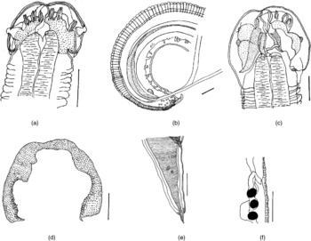
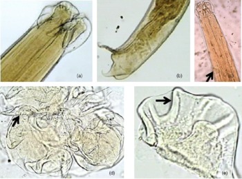
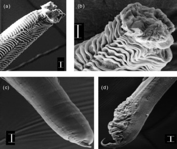
Description. Worms medium-sized with stout body (male, fig. 1a and b). Body enclosed in a cuticular envelope with prominent cephalic flanges (fig. 1a and c). The head distinctly demarcated from the rest of the body. Head with three lips, each with marked indentations. The dorsal lip as well as the two subventral lips with a pair of cephalic papillae each. Interlabia present (fig. 1d). The alae run from immediately posterior to subventral lips to the tail (figs 1e and ). Excretory pore opens latero-ventrally (fig. 2c), located at almost one-third of the distance along the total length of the oesophagus, from anterior extremity. The dentigerous ridges on each of the lips on the head, distinctly marked on the inner edge (fig. 2d), and each lip has filamentous outgrowths distally (fig. 2e). Buccal cavity with a triangular opening, bearing a chitinized rim. Interlabia bifurcated at the base, not distally (fig. 3a), without deep interlabial grooves. The hind extremity of the oesophagus terminates into a smaller bulbar ventriculus. Nerve ring located at a distance of 0.18–0.20 (0.188 ± 0.004) from the anterior extremity. The intricate cuticularized valvular configuration is distinct at the junction of the oesophagus and intestine. The body reaches greatest width at about the anterior third of the body length. Cuticular striations are prominent (figs 3a and b), particularly in the anterior to mid region of the body, but annulations become inconspicuous in the hind region of the body (fig. 3c). Caudal alae narrower (fig. 3d). Rectal glands, two pairs pre-anal and one pair post-anal and smaller, present in both sexes of worms. Two long, sub-equal spicules, 15–17% of the length of body of worms. The wavy cuticular margins of folds on spicules were characteristically typical. The caudal papillae comprise a lateral row along with a medial post-anal row (fig. 3c) as they approach the extremity. The caudal end of the male is strongly curved ventrally, terminating in a bluntly pointed tip at the extremity, devoid of minute spines. Excretory vesicle is elongated pyriform, enveloped in a double-layered sac. Rectal glands oval.
Iheringascaris goai n. sp.: (a) male head; (b) male tail extremity with spicules; (c) female head; (d) interlabia; (e) tail extremity in a female; (f) female post-equatorial vulva. Scale bars: 0.05 mm (a, c, d), 0.1 mm (b, e, f).

Iheringascaris goai n. sp.: (a) male head ( × 400); (b) posterior end of male showing curved tail extremity and spicule ( × 100); (c) anterior end of female showing location of excretory vesicle ( × 100); (d) transverse section of oral end of female showing dentigerous ridges on inner margin of lips; (e) transverse section of oral end of female showing filamentous indentations on lips. (See online at http://journals.cambridge.org/jhl for a colour version of this figure.)

Scanning electron micrographs of Iheringascaris goai n. sp.: (a) head with dorsal lip and ventral interlabium; (b) cephalic extremity with deep cuticular striations on anterior end; (c) tail extremity in a male with caudal papillae; (d) tail, lateral view with alae (scale bars: 10 μm).

Type. Holotype ZSI 1 ♂ ZSI/NRS/IV/N/392 and 1 ♀ ZSI/NRS/IV/N/393; paratypes AU 5 ♂ PNLS 116 and 5 ♀ worms- PNLS 117.
Type host. Arius maculatus (Siluroidea: Tachysuridae).
Other host. Sillago sihama (Percoidea: Sillaginidae).
Localization in host. Small intestine.
Type locality. Arabian Sea (coastal areas of Dona Paula beach), Goa, Central West coast of India.
Etymology. The new species has been named after its marine habitat in the coastal areas of Goa in Central West Coast of India.
Morphology: Iheringascaris inquies (Linton, Reference Linton1901)
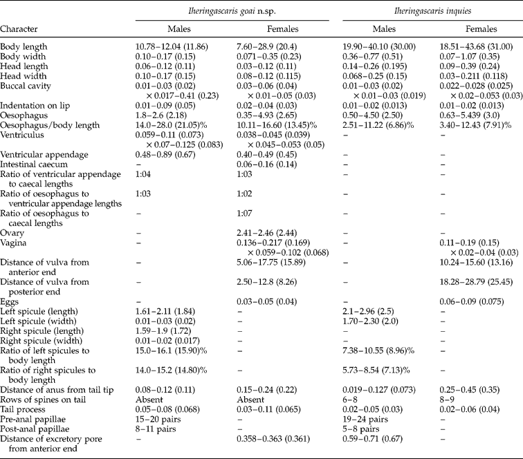
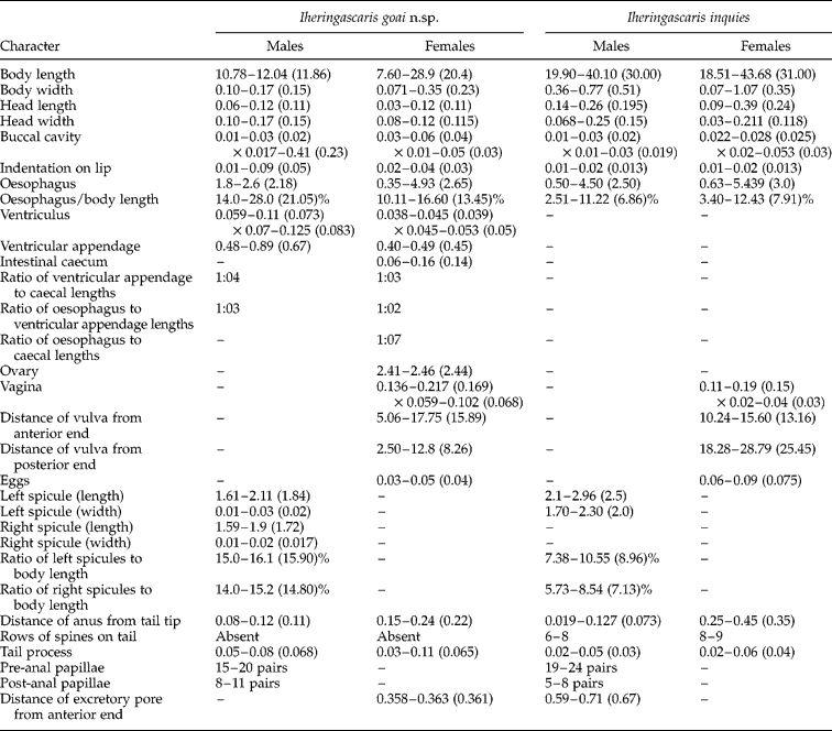
Specimens in this study were similar in morphology of taxonomically important features with those described by Bruce & Cannon (Reference Bruce and Cannon1989). The summary of measurements of specimens of I. inquies is presented in table 1.
Morphometric measurements of I. goai n. sp. and I. inquies in the present study.
Material examined. 6♂, 5♀ (ZSI/NRS/IV/N/394; ZSI/NRS/IV/N/395; PNLS118; PNLS119).
Host. Rhincodon typus (Orectolobidae, Laminiformes).
Localization in host. Small intestine.
Locality. Arabian Sea (coastal areas of Dona Paula beach), Goa, central west coast of India.
Gene sequence analyses
18S rRNA gene analysis
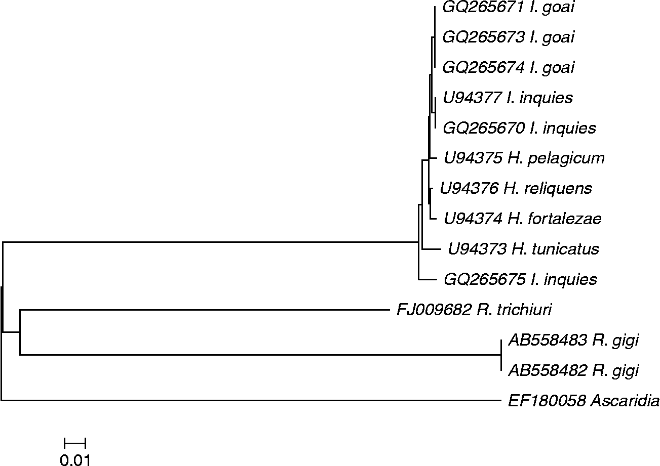
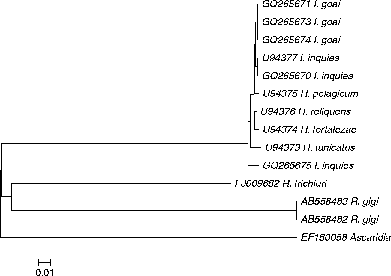
The read length of all 18S rDNA sequences was 864 bp, but a few insertions and deletions were also detected. The average K2P distance of individuals within I. goai n. sp. based on 18S gene sequence, was 0.0145, as compared with an average distance of 0.0117 and 0.0058 of the genera Hysterothylacium and Ascaris, respectively (fig. 4). On an average, therefore, there was 1.2 times more variation among the individuals of I. goai as compared to different species of Hysterothylacium and 2.5 times more variation as compared to different species of Ascaris. It may be noteworthy that there were 808 (93.5%) conserved domains and 17 parsimony informative sites within the 864 bp long 18S region of the same organism.
Neighbour-joining tree based on nucleotide 18S rDNA sequences and reference species. Nucleotide 18S rDNA sequence data are as described in the text with GenBank accession numbers. Bootstrap values based on 1000 replicates were used (values not shown). Scale bar represents an interval of the Kimura two-parameter (K2P) model.
Analysis of the GC content
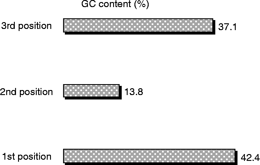
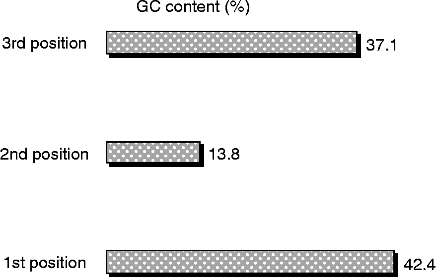
The overall GC content of I. goai n. sp., based on coi gene composition, was found to be 48% (fig. 5). This is consistent with the GC values of all other specimens taken into consideration for this study. Since 18S rDNA is a non-protein-coding gene, the GC content was of little significance in inferring the phylogenetics of this organism.
Distribution of the GC content based on the codon position in the sequence.
A neighbour-joining tree was generated using the Kimura 2 Parameter to calculate pairwise distances (fig. 4). The three specimens, GQ265674, GQ265673 and GQ265671 form a separate clade which does not show any significant similarity to any other organism listed in the GenBank. These may, therefore, be ascribed a separate entity, I. goai n. sp.
coi Gene analyses
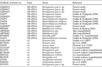
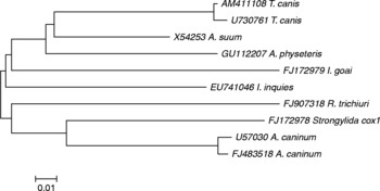
The coi representative sequence of the three specimens studied using 18S rDNA gene (table 2) could be readily distinguished based on DNA barcoding since the results obtained by coi gene complemented those obtained by 18S rDNA gene sequence analyses (fig. 6). Exact matches between different sequences of I. goai n. sp. could not be made due to non-availability of coi sequences of this genus in GenBank. However, analysis based on comparison with other, related genera is presented here.
Representative groups of 18S and cytochrome c oxidase (coi) gene sequence selected for the study.

Neighbour-joining tree based on nucleotide coi gene sequences and reference species. Nucleotide coi sequence data are as described in the text with GenBank accession numbers. Bootstrap values based on 1000 replicates were used (values not shown).

Discussion
Worms of the new species had distinct cuticular striations on the body, a unilateral excretory system with the pore opening near the surface of the body, and a post-equatorial vulva, suggesting thereby that these did not belong to Hysterothylacium. Alignment of the 18S and coi gene sequences showed the existence of two distinct genotypes (figs 4 and 6), with differences in the morphology of the two species, the newer one named here as I. goai n. sp. A morphological comparison of worms of the new species with those of I. inquies is also included (table 1).
The morphological comparison of the new species with specimens of I. inquies collected from similar hosts, viz. sharks, R. typus as well as A. maculatus from the same habitat, i.e. Arabian Sea at Goa, revealed significant variations. In the new species, males possessed a smaller body compared to I. inquies, shorter head, smaller oesophagus (14–28% of body length versus 11%), narrower intestine and a greater number of post-anal papillae. In general terms, worms of the new species were significantly smaller in size than I. inquies. In consonance, measurements of various organs were generally smaller in the new species than in I. inquies. But measurements of a single, unusually larger male specimen showed a marked effect on the mean body size as well as length of spicules. One specimen measured 15.80 in body length and 0.22 in body width, with length of left spicule 4.67, and that of right spicule 4.16, their width being 0.04 and 0.03, respectively. The measurement of length of left and right spicule of all other worms studied were 1.59–1.9 (1.72) and 1.61–2.11 (1.84), respectively, if measurements of spicule length of the one unusually larger specimen were excluded. Variations in measurements of other organs of worms of the new species have been summarized in table 1.
In females, the body of the new species was also comparatively smaller than that in I. inquires, with a smaller head, wider buccal cavity, smaller oesophagus, post-equatorial vulva (versus pre-equatorial in I. inquies) and larger tail. The oesophagus was up to 17% of body length, compared to 12% in females of I. goai n. sp.
The new species could invariably be differentiated from the original description of type species (Pereira, Reference Pereira1935), I. inquies having indentations on lips as well as dentigerous ridges present on the inner margin of lips, excretory vesicle pyriform with a saccular component that opens in the anterior third part of the oesophagus distinctly behind the level of nerve ring, subequal spicules, post-equatorial vulva and the tail typically curved ventrad in male.
Worms of the new species further differed from the description given by Deardorff & Overstreet (Reference Deardorff and Overstreet1981) of specimens from Mississippi and Alabama, in diminishing striations on the body in the posterior part, excretory pore opening behind the level of nerve ring, larger oesophagus, greater ratio of spicules to body length, shorter but wider left and right spicules and lesser number of pre-anal papillae in male worms; and smaller body in female worms, possessing a larger oesophagus, a post-equatorial vulva and a longer tail. The ratio of caecal to ventricular appendage length was smaller in comparison to the ratio of caecal to oesophageal length, which was larger.
The variance in GC content among I. goai n. sp., based on their coi gene sequence, was much higher in the case of the second base (64.33%) as compared with the first and third base (0.27% and 6.54%, respectively). This could be attributed to the very low GC content at the second codon position of the specimen (fig. 5), largely due to the low GC2 values recorded in the case of FJ172978 (I. goai n. sp.). Since more synonymous mutations are known to occur at position 3 and 1, the aberration obtained in the case of FJ172978 could play an important role in its delineation as an organism belonging to a separate distinct species.
As is evident, the representative specimen taken for coi gene analyses falls on a separate branch and shows no similarity with any other organism in GenBank (fig. 6). Similarity with other organisms cannot be substantiated at this point due to paucity of sequences of organisms of the family Raphidascarididae.
The read length of all coi sequences was 650 bp. No insertions, deletions or stop codons were detected in any of the sequences generated during the course of this study. The lack of stop codons in the sequence confirmed the observation that no pseudogenes or NUMTs (nuclear DNA sequences originating from mitochondrial sequences) were sequenced along with the desired gene sequence (Zhang & Hewitt, Reference Zhang and Hewitt1996). It may be noteworthy that there were 422 conserved domains and 171 parsimony informative sites within the 650 bp long coi region of I. goai n. sp.
The differences in morphology of the worms of both species were amply supported by 18S rDNA and coi gene sequence differentiation. Therefore, on the basis of the aforementioned significant points of morphological differences, besides simultaneously presented distinctive 18S rDNA and coi gene sequences, the new species of worms is proposed as I. goai n. sp.
Acknowledgements
A.M. thanks Professor Ms I. Karunasagar for Research mentorship and Department of Biotechnology (DBT) for a Research Associateship. S.K.M. is thankful for the University Grants Commission, Govt of India research grant. N.J. is grateful for a fellowship under DBT research project no. BT/PR9651/SPD/09/818/2007, and to the Department of Science and Technology, Govt of India for a travel grant to visit Charles Sturt University, Australia. The authors would also like to thank Craig Farish at Charles Sturt University for preparing histological sections.