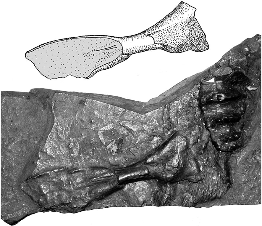
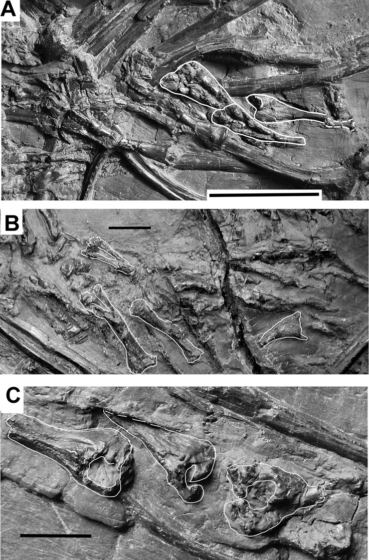
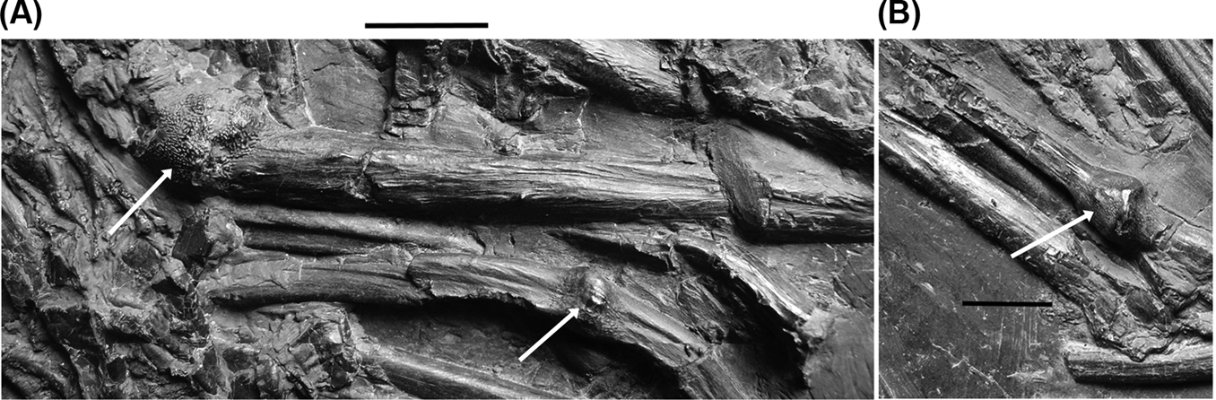
The Carboniferous of the United Kingdom has yielded four genera of fossil lungfish: Ctenodus Agassiz, Reference Agassiz1838, Sagenodus Owen, Reference Owen1867, Straitonia Thomson, Reference Thomson1965 and Uronemus Traquair, Reference Traquair1873a. Straitonia has recently been restudied (Sharp & Clack Reference Sharp and Clack2012), and Sagenodus is under review by the present authors. Ctenodus is widely distributed in time and space, being the only genus to be found in both Lower and Upper Carboniferous deposits. It ranges from the Tournaisian of Scotland to the Moscovian (Westphalian C) of Yorkshire. This paper presents a revision of the British Ctenodus species, including the description of a newly discovered specimen of an almost complete, articulated individual.
First described by Agassiz in Reference Agassiz1838, Ctenodus was the subject of much controversy in the 19th Century, particularly in its affinities with the other dipnoans, Dipterus and Ceratodus. Ctenodus has been the subject of numerous studies, notably by Hancock & Atthey (Reference Hancock and Atthey1868a, Reference Hancock and Attheyb, Reference Hancock and Attheyc, Reference Hancock and Atthey1870, Reference Hancock and Atthey1871, Reference Hancock and Atthey1872), Barkas (Reference Barkas1869a, Reference Barkasb, Reference Barkasc, Reference Barkasd, Reference Barkase, Reference Barkasf, Reference Barkas1870a, Reference Barkasb, Reference Barkasc, Reference Barkas1872, Reference Barkas1873a, Reference Barkasb, Reference Barkas1877a, Reference Barkasb, Reference Barkasc, Reference Barkasd, Reference Barkas1878a, Reference Barkasb), Traquair (Reference Traquair1873a, Reference Traquairb, Reference Traquairc, Reference Traquair1878, Reference Traquair1890a, Reference Traquairb, Reference Traquairc) and Fritsch (Reference Fritsch1889). Numerous species were erected for specimens of doubtful affinity, several of which ultimately became synonymised within Sagenodus when their generic distinctiveness was finally recognised by Woodward (Reference Woodward1891). After the publication of his catalogue, the name Ctenodus was generally assigned to those tooth plates with about 12 or more ridges. Since then, few studies have related specifically to the genus, or indeed to any British Carboniferous lungfish. The most detailed recent descriptions of Ctenodus were by Watson & Gill (Reference Watson and Gill1923) and by Thomson (Reference Thomson1965), who erected a new species on the basis of material from a Tournaisian locality in Berwickshire. Sagenodus is the most commonly used genus in comparative studies because it is better known, based on studies of the American species S. copeanus, especially that by Schultze & Chorn (Reference Schultze and Chorn1997).
The skull roof of Ctenodus was first described in detail by Hancock & Atthey (Reference Hancock and Atthey1868a, Reference Hancock and Attheyb, Reference Hancock and Attheyc). They did not figure this material, although they did draw attention to the differences between the skull roofs of C. interruptus Barkas, 1869 and C. cristatus Agassiz, Reference Agassiz1838 compared to that of C. obliquus, later (Woodward Reference Woodward1891) synonymised with Sagenodus inaequalis. Woodward (Reference Woodward1889) figured the holotype of C. interruptus and provided the first proper description of the skull roofs of the two genera. He pointed out that Ctenodus has one median occipital plate (a single B-bone), whereas Sagenodus has two (both the B- and C-bones being single).
The best description to date of the skull roof of Ctenodus is still that of Watson & Gill (Reference Watson and Gill1923). Their work concentrated on the description of Sagenodus, although they did not identify any of their described specimens to species level. They did not describe the skull roof bones of Ctenodus in detail, but used a reconstruction to represent the cranial morphology. They did not explicitly state which specimens this reconstruction was based upon, but they were probably NMS 1894.155.12 from the Lower Carboniferous, and NHMUK P 7300 from the Upper Carboniferous. Their descriptions were less detailed than those of Sagenodus, as they believed a comparison of the two genera to be sufficient. They concluded that the skull roof of Ctenodus was more primitive than that of Sagenodus and that the latter was derived from the former.
Westoll (Reference Westoll, Jepsen, Mayr and Simpson1949) described the skull roof of Ctenodus, Sagenodus and Uronemus, but these descriptions were based primarily on the work of previous authors and not on his own observations. Thomson's (Reference Thomson1965) reassessment of the taxonomic issues surrounding C. interruptus led to his illustrating what was the most complete skull roof of Ctenodus then known (NMS 1894.155.12), stating that “The skull roof of C. interruptus is of exactly the same type as found in the typical Upper Carboniferous Ctenodus” (Thomson Reference Thomson1965, p. 225). This is discussed further below.
One of the notable features of Ctenodus in comparison with Sagenodus is the relative paucity of skull roof material readily attributable to the genus. Despite the wide chronological distribution of the taxon, there is very little known from any locality. While this might not be surprising for most localities, at Newsham, whereas Sagenodus is known from a (relatively) large quantity of skull material, Ctenodus is represented by far fewer specimens.
Because of the paucity of articulated specimens, at present, only the prearticular tooth plate can be diagnostic as to species. Isolated elements, or those without associated tooth plates, can only be assigned to species by inference, on the grounds of locality, age or frequency of occurrence.
The tooth plates of Ctenodus have been a taxonomically complex and somewhat controversial collection of specimens. From the time of Woodward's (Reference Woodward1891) catalogue, the genus was diagnosed as having one medial occipital plate (B only single) and dental plates of triangular, ovate or elliptical form, which had numerous strong, outwardly directed ridges, tuberculated and crenulated. However, the original diagnoses of the various species are variable and imprecise, thus there is great variation in the morphology of tooth plates attributed to the genus.
These problems are compounded because the initial descriptions of most of these specimens were originally made without the association of skull roof material. Below, we attempt to clarify the originally erected species and give detailed descriptions of the morphology and variability in the tooth plates assigned to each species of Ctenodus. This is followed by descriptions of cranial and postcranial elements, most of which have never been figured or described previously.
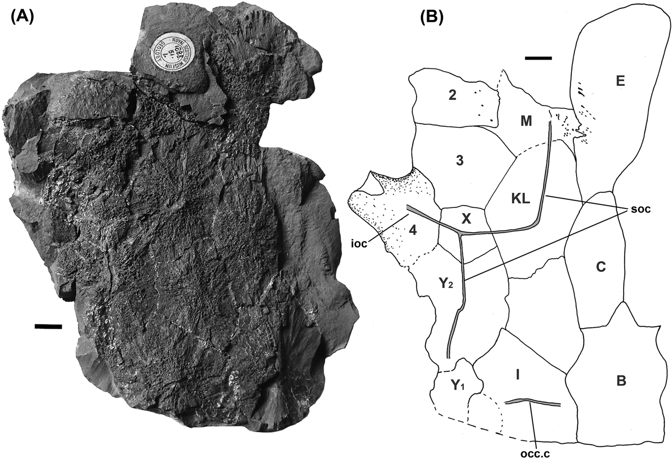
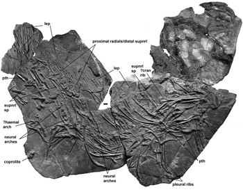
The current study is based on a re-examination of the known specimens of Ctenodus, plus a large, almost complete specimen from the early Namurian (Pendleian) of Ayrshire, Scotland, discovered in 2009, which can be identified as Ctenodus interruptus on the basis of its prearticular toothplates. Specimens previously identified as C. interruptus, such as NMS.1894.155.12 (Watson & Gill Reference Watson and Gill1923; Thomson Reference Thomson1965), on the basis of their tooth plate morphology, do not belong to this species (Barkas Reference Barkas1869c; Woodward Reference Woodward1891; Sharp Reference Sharp2007). Ctenodus interruptus was previously represented only by toothplates, which makes the new find especially informative and important. The new specimen, GLAHM 131502, can lay claim to be one of the most complete and best-preserved post-Devonian fossil lungfish ever found. Preserved elements include a complete skull with all four tooth plates in articulation, and most of the postcranial skeleton, including vertebral elements and caudal lepidotrichia.
Despite the new study, species definitions still remain based on tooth plate morphologies. Skull roof morphologies are typically only known from a sample of fewer than four good specimens, and these are all incomplete in different ways. Given the range of variation found among lungfish skull roofs, the sample is insufficient for reliable diagnoses. The best postcranial specimens usually lack tooth plates or skull roofs and are thus also undiagnostic.
One of the most interesting aspects of this genus is its distribution in the Carboniferous, with its wide temporal range through the period. The dentitions of species from the Lower and Upper Carboniferous are distinctive; although this was noted by earlier authors, it has never been investigated. The fact that Ctenodus is often placed at the base of the post-Devonian radiation of lungfishes, makes a redescription of this genus and a review of the included species both necessary and timely.
Institutional abbreviations. CAMSM, Sedgwick Museum, Cambridge; GLAHM, Hunterian Museum, Glasgow; MM, Manchester Museum, Manchester; NEWHM, Great Northern Museum (previously the Hancock Museum), Newcastle upon Tyne; NHMUK P, Natural History Museum, London; NMS, National Museum of Scotland; UMZC, University Museum of Zoology, Cambridge; YORYM=Yorkshire Museum.
1. Material and methods
Numerous described and figured, as well as previously undescribed, specimens have been studied. Two newly discovered specimens are included in the revised descriptions. The first of these, UMZC 2007.2, was collected by the late Mr S. P. Wood in 1983 from the Viséan Broxburn Shale of Murieston Water (North Bank) Mid Calder, West Lothian, Scotland. The specimen was mechanically prepared by ELS using a dental mallet and an airbrasive unit. The second, GLAHM 131502, was collected and prepared by Feona McIvor and donated to the Hunterian Museum, Glasgow in 2009. It was recovered from Powgree Burn near Longbar, Glengarnock, Ayrshire, Scotland, stratigraphically equivalent to Manse Burn Formation, Limestone Coal Group, Pendleian (Namurian E1) Serpukovian, Lower Carboniferous. It has been further prepared by JA and RNG Clack using an airbrasive unit and mounted needle.
1.1. Specimens used in the study
1.1.1. Ctenodus cristatus (all Ctenodus from Newsham regarded as C. cristatus)
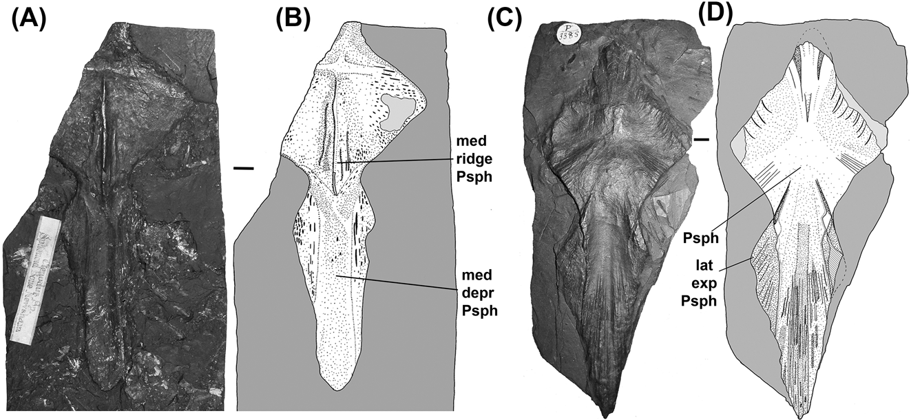
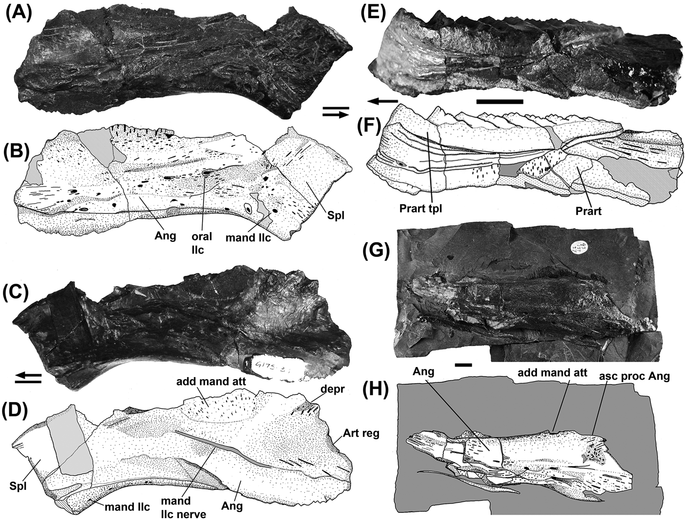
All specimens below are from the Low Main Coal Seam, Newsham, Northumberland (Upper Carboniferous, Westphalian B) unless otherwise stated: NEWHM G61.24, neotype, left pterygoid and tooth plate; NEWHM G22.66, left prearticular tooth plate; NEWHM G22.67, right prearticular tooth plate; NEWHM G24.23, right pterygoid tooth plate; NEWHM G40.97, left opercular; NEWHM G41.15, right prearticular tooth plate; NEWHM G59.25, skull roof in dorsal and ventral view, incomplete, palate in buccal view, incomplete; NEWHM G59.27, parasphenoid; NEWHM G59.28, parasphenoid; NEWHM G59.67, E and F bones; NEWHM G59.70, skull roof; NEWHM G59.79, I and Z bones; NEWHM G61.19, prearticular and pterygoid tooth plates in articulation; NEWHM G61.20, right and left prearticular toothplates with mandibular elements; NEWHM G61.21, left pterygoid tooth plate and ribs; NEWHM G61.26, right prearticular tooth plate; NEWHM G61.61, right cleithrum; NEWHN G151.13, cleithrum and ribs; NEWHM G172.33, rib; NEWHM G175.34, right angular and splenials; NEWHM G175.78, right pterygoid tooth plate; NMS 1878.45.9, cleithrum; NMS 1878.45.22, left pterygoid and tooth plate in dorsal view; NMS 1878.45.26, prearticular tooth plate and lower jaw; NMS.1888.33.2, pterygoid tooth plate and lower jaw; NMS 1888.33.3, left pterygoid tooth plate impression; NMS 1888.33.15, partial skull roof; NMS 1888.33.16, B-bone; NMS 1888.33.21, cleithrum; NMS 1893.139.27, partial lower jaw; NMS 1968.17.47, cleithrum; CAMSM E4524, (probably) Low Main Seam, Newsham, suboperculum; NHMUK P 3385, parasphenoid.
Specimens not from the Low Main Seam, Newsham: NHMUK P 7300, Knowles Ironstone, Fenton, Staffordshire (Upper Carboniferous, Westphalian C), skull roof, left and anterior portions, right side obscured by operculum, right prearticular tooth plate present; NMS 1885.57.1, Loanhead, Blackband Ironstone, prearticular tooth plate and mandibular elements.
1.1.2. Ctenodus romeri
All from Lower Calciferous Sandstone Cementstone Group, Coldstream, Berwickshire (Tournaisian, Lower Carboniferous): NMS 1896.81.41, holotype, isolated left pterygoid tooth plate; NMS 1896.81.42, paratype, isolated right pterygoid tooth plate; NMS 1896.81.43, paratype, isolated left prearticular tooth plate; NMS 1896.81.44, paratype, isolated left prearticular tooth plate; NMS 1896.81.45, small tooth plate; NMS 1896.81. 47, parasphenoid.
1.1.3. Ctenodus allodens sp. nov.
All from Straiton, near Edinburgh, Midlothian, Scotland (Dunnet Shale, Asbian, Viséan) except where otherwise stated: Holotype, NMS 1888.51.1, skull roof in dorsal view, including some left lateral circumorbitals and a left lower tooth plate; NMS 1894.155.12, eroded skull roof in part and counterpart: NMS 1891.98.1, skull roof and prearticular tooth plate and part of the lower jaw: NMS 1895.184.5 and NMS 1895.184.6, pterygoid tooth plate in part and counterpart; NMS 1896.38.4, prearticular tooth plate partial lower jaw and other bones: NMS 1897.30.1 and NMS 1897.30.2, pterygoid tooth plate in part and counterpart; NMS 1906.108, semi-articulated postcranium in ventrolateral view; UMZC 2007.2, Broxburn Shale, Murieston Water, Mid Calder, West Lothian (Lower Carboniferous, Asbian), disarticulated elements, including E-bone, tooth plates, clavicle and ribs.
1.1.4. Ctenodus interruptus
GLAHM 131502, Powgree Burn near Longbar, Glengarnock, Ayrshire, Scotland (Manse Burn Formation equivalent, Limestone Coal Group, Pendleian (E1) Serpukovian, (Namurian), Lower Carboniferous), almost complete, articulated individual with skull, palate and all tooth plates; NMS 1894.168.2, Broxburn Shale, Broxburn, Midlothian (Lower Carboniferous, Asbian), parasphenoid, pterygoid in buccal view with prearticular tooth plate; NMS 1893,139.27, South Parrot coal, Niddrie, left angular; NMS 1886.87.15, Loanhead No. 2 Ironstone, Loanhead, Midlothian (Lower Carboniferous, Namurian), eroded tooth plate; NMS 1885.57.1, Burghlee Pit Ironstone, Loanhead, Midlothian (Lower Carboniferous, Namurian), prearticular tooth plate and lower jaw; NHMUK P 11494, Loanhead No.2 Ironstone, Loanhead, Midlothian (Lower Carboniferous, Namurian), right prearticular tooth plate; MM L.8377, horizon unknown, Borough Lee, near Edinburgh, left prearticular tooth plate. Horizon and locality unknown: MM L.10412, left pterygoid tooth plate; MM L.10413, right prearticular tooth plate; MM 10414, left prearticular tooth plate.
1.1.5. Ctenodus murchisoni
NHMUK P 518, holotype, Limestone, Upper Coal Measure, Leebotwood, Shropshire (Upper Carboniferous, Westphalian C), left pterygoid tooth plate. Described by Ward (Reference Ward1890); NHMUK P 5031, Great Row Coal, Longton, Staffordshire (Upper Carboniferous, Westphalian C); skull roof, right side, split within the bone. Horizon unknown, Longton, Staffordshire: NHMUK P 7304, right pterygoid tooth plate; NHMUK P 7309, prearticular and pterygoid tooth plate in articulation; NHMUK P 7310, prearticular and pterygoid tooth plate in articulation; NHMUK P 7311, right pterygoid tooth plate; NHMUK P 7312, left pterygoid tooth plate; MM L.8343, left pterygoid tooth plate; MM L.8345, right prearticular tooth plate. Horizon unknown, Ardwick, near Manchester: MM L.10224, right pterygoid tooth plate; MM L.10254, right pterygoid tooth plate.
1.1.6. Ctenodus spp.
NMS 1893.139.27, Niddrie, shale overlying South Parrot Coal, left angular; NMS 1968.17.47, “Mussel band, Airdrie, (Upper Carboniferous)”, left cleithrum; NMS 1957.1.5738, “Mussel band, Airdrie, (Upper Carboniferous)” pterygoid; NMS 1957.1 5739, horizon unknown, Annick Lodge, Kilwinning, Irvine, Ayrshire, pterygoid; CAMSM E12969, “Airdrie, Lanarkshire”, possibly Palace Craig Ironstone (Similis–pulchra palynozone) or Airdrie Blackband Ironstone (upper modiolaris palynozone), both Westphalian B, Duckmantian (fide Panchen Reference Panchen1972), palate and operculum; MM LL.4980, “from between the Lower Florida and the Pigeon House coal seams, collected in the underground tunnel between Bickershaw and Golborne colliery workings,” right clavicle.
2. Systematic palaeontology
Osteichthyes Huxley, Reference Huxley1880
Sarcopterygii Romer, Reference Romer1955
Dipnomorpha Ahlberg, Reference Ahlberg1991
Dipnoi Müller, Reference Müller1845
Family Undesignated
Genus Ctenodus Agassiz, Reference Agassiz1838.
Diagnosis. Emended diagnosis after Woodward (Reference Woodward1891). Tooth plates with 8–23 ridges. Ridges bear individual teeth which are typically unfused, often with notable separation between teeth, apices of denticles pointed. Tooth plates, above and below, are semicircular, triangular, irregularly ovate, or elliptical in form, with numerous strong, outwardly directed ridges, tuberculated or crenulated.
Differential diagnosis. Differs from Sagenodus, with which is it most commonly found associated, by: (i) posterior and lateral skull bones ornamented with ridges, tubercles and anastomosing and ramifying grooves; (ii) plesiomorphic features: B-bone tapers anteriorly between paired C-bones (also in Scaumenacia); C-bones separated by a single D-bone; elongate E-bones that are anteriorly expanded with fimbriated or wavy anterior margins and are longer than the C-bones (also in Scaumenacia); single F-bone anteriorly flared; large KL-bone anterior to J-bone that contacts C-bone; J-bone contacts C-bone (as in Scaumenacia, Straitonia and Tranodis but not in Sagenodus); (iii) Lepidotrichia in posterior part of caudal region segmented (as in Scaumenacia and Fleurantia).
Type species. Ctenodus cristatus Agassiz, Reference Agassiz1838.
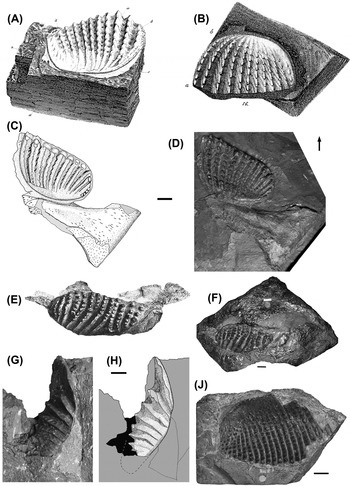
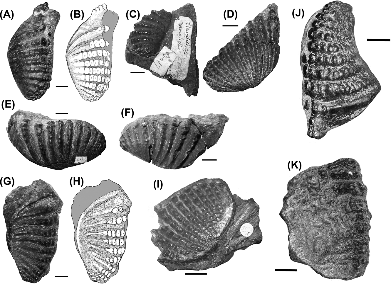
Holotype. Missing, presumed lost in World War II. Original, a pterygoid tooth plate, Leeds museum, registration number unknown (Fig. 1A, B).
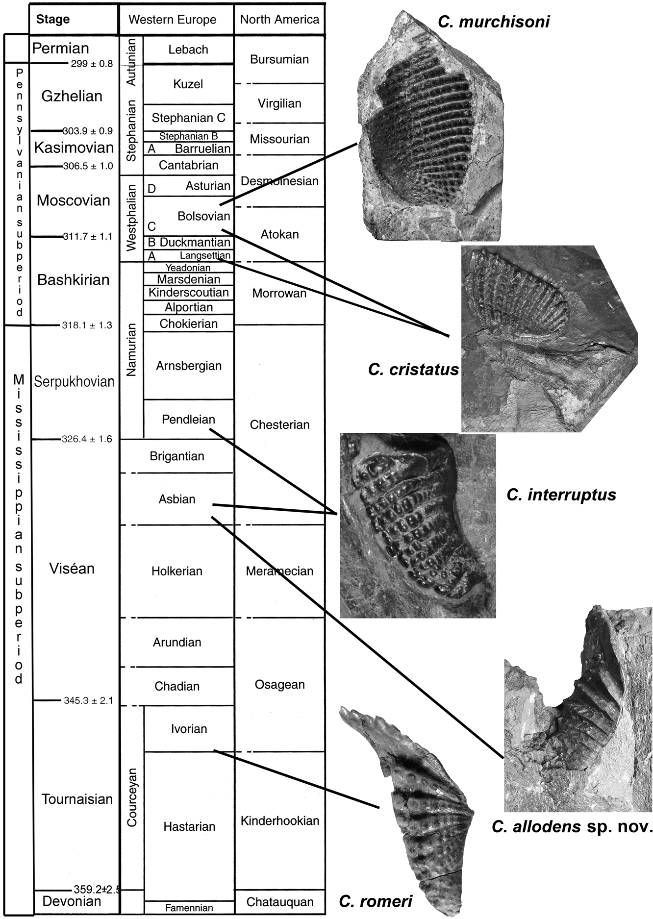
Ctenodus Agassiz Reference Agassiz1838: (A) figure of holotype of C. cristatus from Sowerby and George Reference Sowerby and George1826 (No scale is given on the figure in the description); (B) figure of holotype of C. cristatus from Agassiz Reference Agassiz1838 (No scale is given on the figure or description); (C–D) neotype of C. cristatus, NEWHM G61.24, interpretive drawing and photograph; (E) figure of holotype of C. interruptus Barkas, Reference Barkas1869c from Woodward Reference Woodward1889; (F) photograph of holotype of C interruptus, YORYM YM 445 (courtesy of the Yorkshire Museum); (G–H) holotype of C. allodens sp. nov., NMS 1894.155.12, photograph and interpretive drawing; (J) holotype of C. murchisoni Ward, Reference Ward1890, NHMUK P 518. Scale bars=10 mm.

Type locality and horizon. Tong, near Leeds, in Yorkshire. Beeston Seam, 80 yards above the Black Bed Coal or Low Moor Upper Seam. Stage unknown.
Neotype. NEWHM G61.24. Right pterygoid and tooth plate (Fig. 1C).
Neotype Locality and Horizon: Newsham, Northumberland. Low Main Seam, Duckmantian (Westphalian B, Bashkirian), Upper Carboniferous
Stratigraphic range of genus. Tournaisian–Bolsovian (=Westphalian C, Moscovian).
Remarks. The genus Ctenodus and the specific name cristatus were founded by Agassiz in Reference Agassiz1838, based on a single tooth plate from the coal measures of Tong in Yorkshire (Sowerby & George Reference Sowerby and George1826). The holotype was originally housed in the Leeds Museum, which was heavily bombed in World War II. Although some of their collections were sent to London to the British Museum (Natural History) (now the Natural History Museum UK) prior to that, it is not known whether the holotype of C. cristatus was among them. Neither they, nor the Leeds Museum has any subsequent records of the specimen (J. Nunney pers. comm. 2006).
Although the specimen is missing, there is a description, two figures and locality data, which make it possible to identify other specimens with confidence. The horizon given by Sowerby & George (Reference Sowerby and George1826) for this specimen is the Beeston Seam, which overlies the Black Bed or Low Moor upper seam by 80 yards. The latter may be the same horizon as that from which the holotype of Pholiderpeton scutigerum derived, which is dated as Westphalian A or Langsettian (Clack Reference Clack1987).
Woodward (Reference Woodward1891) diagnosed the genus as “body depressed…with large thin scales, almost rounded in shape, but having the angles rounded”. He noted the absence of a ganoine layer and a single median occipital plate (B-bone), with a pair of plates immediately adjoining in front (C-bones). He also noted the “strong, outwardly directed ridges, tuberculated or crenulated”. Watson and Gill (Reference Watson and Gill1923) described and illustrated many features of Ctenodus, but did not present a formal diagnosis.
2.1. Ctenodus cristatus Agassiz, Reference Agassiz1838, 137
Stratigraphic range. ?Langsettian (Bashkirian)–Bolsovian (=Westphalian C, Moscovian)
Diagnosis. Emended diagnosis after Woodward (Reference Woodward1891). Tooth plates with 10–14 sub-parallel ridges. Teeth on ridges are fused, but retain undulating crests where fusion is incomplete. Distal teeth on each ridge are unfused and show some lateral compression. Teeth have conical or pointed apices and circular bases. Ridges radiate anteriorly and are parallel posteriorly. Prearticular tooth plates are convex with a sub-straight lingual margin and asymmetrically curved labial margin. Ridges parallel, being curved proximally and straight distally. Pterygoid tooth plates are asymmetrically ovate with almost straight labial margins. Anteriorly expanded E-bones have an undulating margin – concave medially and convex laterally (compared with continuous rounded convex profile in Ctenodus allodens sp. nov. from Straiton and Broxburn). Bone margin is ragged.
Synomyms:
Ctenodus concavus Barkas, 1869
Ctenodus ovatus Barkas, 1869
Ctenodus tuberculatus Atthey, Reference Atthey1868
Sagenodus cristatus Sternberg, Reference Sternberg1841
Sagenodus corrugatus Woodward Reference Woodward1891
Ctenodus corrugatus Atthey, Reference Atthey1868
See Schultze (Reference Schultze1992a) for further details.
Remarks. Woodward (Reference Woodward1891) described C. cristatus as having broadly ovate or elliptical teeth with between 12 and 14 acute, prominently tuberculated ridges, only slightly radiated with laterally compressed tubercles. He noted the coronal surface to be flat or slightly concave.
Newsham in Northumberland has the largest assemblage of dipnoans of all the British Carboniferous localities. There, two genera of dipnoans are present and the volume of material far exceeds that from any other locale. As a result, it shows a high level of intraspecific variation. Newsham has classically been the location from which the most intensely studied and best preserved material of both Sagenodus and Ctenodus was collected, which in turn means that without tooth plate associations, cranial and postcranial elements may be difficult to assign to one or the other genus. However, specimens of Ctenodus are distinguished from Sagenodus by their larger size, and in differences from the jaw of Sagenodus (Schultze & Chorn Reference Schultze and Chorn1997; ELS pers. obs. S. inaequalis). All specimens from Newsham described here are thus regarded as C. cristatus.
2.1.1. Tooth plates
Tooth plates of C. cristatus from the Upper Carboniferous of Northumberland are of slightly larger average size than those from Lower Carboniferous localities, although not as large as those from the Staffordshire coal fields.
The tooth ridges are subparallel and curved posteriorly, the curvature becoming greater towards the internal half of the plate. They number between 10 and 14, with the posterior-most ridges closer together, more worn, and less highly curved than those that are anteriorly situated. For much of the tooth plate, the tooth ridges are fused into a dorsally rounded shearing crest. Unlike the ridges of Sagenodus, the ridges are not entirely straight but curved or undulating.
The most lateral or distal portions of the ridges are usually punctuated by one or two unfused, distinct teeth which are set slightly away from the buccal surface. This gives the teeth an almost conical shape except for a slight compression of the tip in line with the rest of the tooth ridge. The bases of the teeth are rounded, except in a few specimens (e.g. Fig. 4C, D) where the teeth are more pyramidal.
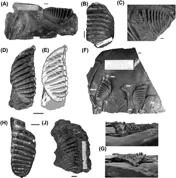
The prearticular tooth plates of specimens from Newsham (Fig. 2D–J) are comparable to those from other localities. They are narrower than their pterygoid counterparts, but less strongly convex than those from other localities. The tooth plates are a rounded triangle in shape, with the broadest portion of the tooth plate slightly posterior to the centre and the labial or lateral margin slightly S-shaped. The prearticular plates (e.g. NEWHM G22.67: Fig. 2D, E) are not symmetrical where they occluded with the pterygoid tooth plates. The ridge numbers observed are either 11 or 12, although the most posterior regions of the tooth plate are always worn, making accurate counting difficult.
Ctenodus cristatus tooth plates: (A–C) Pterygoid tooth plates: (A) NEWHM G175.78, right tooth plate; (B) NEWHM G24.23, right tooth plate; (C) NEWHM G61.21, left tooth plate; (D–J) Prearticular tooth plates: (D) NEWHM G 22.67, right tooth plate; (E) interpretive drawing of (D); (F) NEWHM G61.20, splenial and scattered postcranial elements with both tooth plates; (G) NEWHM G61.20, close up of left tooth plate in oblique view to show denticles; (H) NEWHM G22.66, left tooth plate; (J) NEWHM G61.26, right tooth plate. Scale bars=10 mm.

Similar to the pterygoid tooth plates, the most lateral extent of each tooth ridge is punctuated by one well-developed and isolated tooth, which is again conical with a rounded base. Specimen NEWHM G61.26 (Fig. 2J) shows an unusual condition in which a number of the most distal teeth remain unfused to the tooth ridge, and in NEWHM G22.66 (Fig. 2H), the teeth are not fully fused. Whereas specimens from the Lower Carboniferous of Loanhead display ‘longitudinal compression’ (sensu Thomson Reference Thomson1965, p. 226), in Newsham specimens, such as the latter, the teeth are compressed so that the long axis lies in line with the tooth ridge itself.
Some tooth plates from Newsham are extremely large (e.g. NEWHM 61.20, Fig. 2F). While most specimens are between 60 mm and 70 mm in length, some reach nearly 100 mm. However, they do not have a correspondingly higher number of tooth ridges: they have 12 visible ridges, well within the average for those found at Newsham, and fewer than those much larger tooth plates from the Staffordshire coal fields. In some specimens from Newsham (e.g. NEWHM G61.20, Fig. 2F), the tooth ridges are entirely unfused and the individual teeth are preserved. These prearticular tooth plates show the inclination of ridge 1 anteriorly, and the well-formed conical shape of the individual teeth.
A single specimen from the Knowles Ironstone at Fenton, Staffordshire, which consists of a tooth plate associated with a skull roof (see skull roof description for figure), is assigned to C. cristatus on the basis of tooth shape. This prearticular tooth plate is almost 60 mm in length and 20 mm wide, giving it an unusually high length to width ratio of 3:1. It is convex over its buccal surface and has 14 tooth ridges, with most of the teeth of the ridges conical and separated from each other. Ridge 1 is slightly elongated relative to the rest of the tooth plate, but not significantly. The ridges appear virtually parallel, with the only degree of convergence between them being at the most posterolingual region of the plate where they are considerably worn, the plate is smooth and the ridges curve posteriorly.
2.2. Ctenodus romeri Thomson, Reference Thomson1965
Holotype. NMS 1896.81.41 pterygoid tooth plate, Thomson Reference Thomson1965, p. 227 and plate III, G, H.
Type locality and horizon. Coldstream, Berwickshire. Ballagan Formation (“Lower Calciferous Sandstones” (Thomson Reference Thomson1965)).
Stratigraphic range. Tournaisian.
Other localities. Burnmouth, Berwickshire.
Referred specimens. NMS 1896.81.42, NMS 1896.81.43, NMS 1896.81.44 (Thomson Reference Thomson1965, plate III, I & J); NMS 1896.81.45, NMS 1896.81.47.
Diagnosis. Emended from Thomson (Reference Thomson1965). Tooth plates small, 4–6 cm maximum, with 11 or 12 strongly radiating tooth rows on each tooth plate. Denticles longitudinally compressed, with the exception of those on the expanded first and second rows of the lower plates which are laterally compressed. Nine to 12 denticles per row. Lower plates with a marked angle on the upper margin, upper plates lacking this feature.
Remarks. Though dated as Arundian in Thomson (Reference Thomson1965), the Ballagan Formation at Coldstream and Burnmouth has since been securely dated as Tournaisian (Waters et al. Reference Waters, Browne, Dean and Powell2007; Smithson et al. Reference Smithson, Wood, Marshall and Clack2012).
2.2.1. Tooth plates
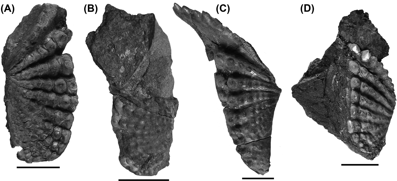
The pterygoid tooth plates (Fig. 3A, B) are oval and almost symmetrical. There is no obvious angle on the lingual margin as in other specimens of Ctenodus and the lingual margin forms a smooth curve. The labial margin is also a smooth convex curve, but is punctuated by the large teeth that reach the extremities of the plate. The tooth rows radiate from a point two-thirds anteriorly along the lingual margin of the tooth plate and on NMS 1896.81.41, 12 tooth rows carry at least 10 teeth each.
Ctenodus romeri tooth plates: (A) NMS 1896.81.41, pterygoid tooth plate, holotype; (B) NMS 1896.81.42, pterygoid tooth plate; (C) NMS 1896.81.44, left prearticular tooth plate; (D) NMS 1896.81.43, left prearticular tooth plate. Scale bars=10 mm.

The lingual teeth are compressed in a lingual–labial direction (longitudinal compression of Thomson, Reference Thomson1965, p. 226) meaning that there is a small but distinctive gap between the teeth at the extremities of the plate, giving each denticle the appearance of an ovate base whose long axis lies perpendicular to the tooth ridge. The more posterior teeth are considerably worn, and the most anterior ridge (ridge 1) is slightly longer than the other ridges of the tooth plate. The pterygoid plate is almost flat, rather than concave as in other Ctenodus species.
The prearticular tooth plates (Fig. 3C, D) are convex and triangular in shape. The lingual margin is marked by an angle of approximately 135°, situated half way along the inner tooth plate margin. The lingual margin anterior to the angle is composed of tooth ridge 1, which is angled slightly away from the rest of the tooth plate towards the symphysis. More posteriorly, the lingual margin is gently concave until it meets a lingual extension of the tooth field. At this point, the margin is crenulated slightly.
The labial margin forms a gently sigmoid curve, convex posteriorly and concave anteriorly. The external margin of the elongated ridge 1 is almost twice the length of the other ridges. One specimen (NMS 1896.81.44, Fig. 3C) has approximately 13 ridges. The number of teeth on each ridge is unclear, may be ten or fewer. The tooth ridges of the prearticular plate radiate from a large area at the lingual margin. The teeth are longitudinally compressed, with the exception of those on ridge 1 which are more laterally compressed, and with their denticles angled slightly towards the distal end of the tooth ridge.
These tooth plates are, as Thomson (Reference Thomson1965) suggested, of a more ‘primitive’ morphology than others known from Carboniferous Britain and bear a number of similarities to some Devonian tooth-plated species, such as the retention of rounded teeth on the pterygoid as opposed to cutting ridges (Ahlberg et al. Reference Ahlberg, Smith and Johanson2006). As a consequence, it seems prudent to retain these specimens as a distinct species, Ctenodus romeri. This is corroborated by other specimens subsequently collected from this locality.
2.3. Ctenodus allodens sp. nov.
Etymology. From ‘allo’, different or strange and ‘dens’, tooth.
Holotype. NMS 1894.155.12, Skull roof in dorsal view with lower jaw and prearticular tooth plate. (Figs 1G, H, 4, 8).
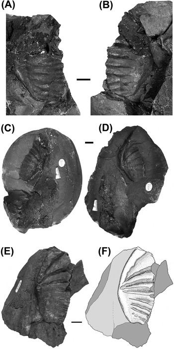
Ctenodus allodens sp. nov. tooth plates: (A–B) NMS1895.184.5 and NMS1895.184.6, right ptergyoid tooth plate in part and counterpart; (C–D) NMS.1897.30.1 and NMS.1897.30.2, left pterygoid tooth plate, part and counterpart; (E–F) UMZC.2007.2, right pterygoid tooth plate, photograph and interpretive drawing. Scale bars=10 mm.

Type horizon and locality. Straiton, near Edinburgh, Midlothian, Scotland. Dunnet Shale, Asbian, Viséan.
Stratigraphic range. Asbian.
Other localities. Broxburn Shale, Murieston Water (North Bank) Mid Calder, West Lothian.
Diagnosis. Tooth plates with between eight and twelve ridges with barely evident individual teeth. Pterygoid tooth plates narrow, oval, and with a concave occlusal surface. Elongate ridge 1. Lingual margin thickened and flattened. Prearticular tooth plate elongate oval with convex occlusal surface. Elongate ridge 1.
Referred specimens. NMS 1891.98.1, NMS 1895.184.5, NMS 1895.184.6, NMS 1897.301.1, NMS 1897.301.2, NMS 1888.51.1, NMS 1896.38.4, NMS 1906.108, UMZC 2007.2.
2.3.1. Tooth plates
The pterygoid tooth plates from the Asbian of Straiton, Midlothian (Fig. 4A–D) and Murieston Water (Fig. 4E, F) are distinguished by the sharp tooth ridges more or less lacking separate denticles and by the elongate ridge 1 on both upper and lower plates. This ridge is wider and flattened compared with the other ridges and also makes the lingual margin concave in buccal view.
On the prearticular plates, the ridges are not entirely uniform: there is some evidence of individual tooth crests, but no entire teeth are preserved. Unlike in those from Coldstream, ridge 1 seems to be turned slightly towards the tooth plate posterolabially, as do the more anterior ridges. In most specimens, the ridges are asymmetrical, with the anterior face at a shallower angle. In the proposed holotype specimen, the prearticular toothplate has been forced up through the skull roof so the posterior region is obscured. Eight ridges are visible, but there may have been as many as ten or 12, which corresponds to the pterygoid plates from this locality.
As these tooth plates are distinguishable from all other described species from the Carboniferous, we propose a new species, C. allodens sp. nov., for this newly recognised morphology. The proposed holotype, NMS 1894.155.12, was catalogued as C. interruptus, but evidently does not belong to this species.
A similar suggestion was made by Watson & Gill (Reference Watson and Gill1923, p. 196) who stated, referring to Woodward's (Reference Woodward1891) description of C. interruptus, “If [his] diagnosis of this species accurately represents Barkas's intention in founding the species, the name cannot properly be applied to the specimens in the Royal Scottish Museum which Traquair referred to it”. Further support for this suggestion is the stratigraphic separation of the Straiton and Murieston Water strata from the Loanhead locality. Murieston Water is a new locality for Ctenodus.
2.4. Ctenodus interruptus Barkas, 1869
Holotype. YORYM YM 445, an isolated prearticular tooth plate (Fig. 1E, F)
Type locality and horizon. Gilmerton, near Edinburgh, Midlothian, Scotland. “Lower Limestone Group” Brigantian, Viséan, Lower Carboniferous.
Stratigraphic range. Viséan – Pendleian (Serpukhovian).
Other localities. Broxburn; Loanhead, Burghlee Pit; Gilmerton, Midlothian, Scotland.
Synonym. Prosagenodus interruptus Romer & Smith, Reference Romer and Smith1934.
See Schultze (Reference Schultze1992a) for further details.
Diagnosis. Emended from Barkas Reference Barkas1869c and Thomson Reference Thomson1965. Pterygoid and prearticular tooth plates with 12–14 ridges. Denticles prominent, well separated in the outer parts of the ridges, each denticle longitudinally compressed, apices rounded. Tooth ridges almost parallel or slightly radiating from a point half way along the median rim. Outline of plates triangular to ovate. Vomerine tooth plates triangular, with variable number of teeth and ridges. Skull roof with conspicuous ramifying grooves as ornament on bones B, I, J, XY and upper circumorbital bones.
Referred specimens. GLAHM 131502, NMS 1894.168.2, NMS 1893.139.27, NMS 1886.87.15, NMS 1885.57.1, NHMUK P 11494, MM L.8377, MM L.10412, MM L.10413, MM 10414.
Remarks. Woodward (Reference Woodward1891) described the dental plates of C. interruptus as closely resembling those of C. cristatus, being variable in character with 12–14 ridges. The illustration in Woodward (Reference Woodward1889) was adequate to recognise the species from prearticular tooth plates, but nonetheless, several specimens have been attributed to this species with prearticular plates that do not resemble those of the holotype, or in which prearticular tooth plates are not preserved; they cannot be assigned to this species. The holotype of C. interruptus YORYM YM 445, an isolated prearticular tooth plate, has recently been relocated in the Yorkshire Museum. The associated label gives the horizon only as “Coal Measures”.
2.4.1. Tooth plates
The prearticular tooth plates of GLAHM 131502 (Fig. 5J, K) are recognisably the same species as the holotype. Furthermore, it has associated pterygoid tooth plates which allow attribution of specimens bearing only pterygoid tooth plates to this species for the first time. The many tooth plate specimens from Loanhead, Burghlee and Gilmerton can now be securely identified as C. interruptus, a proposal first made by Traquair (Reference Traquair1890a).
Ctenodus interruptus tooth plates: (A–B) NHMUK P 11494, right prearticular tooth plate, photograph and interpretive drawing; (C) MM L8377, left prearticular tooth plate; (D) MM L10414, left prearticular tooth plate; (E) MM L10413, right prearticular tooth plate; (F) MM L10414, left prearticular tooth plate; (G–H) MM L10412, photograph and interpretive drawing; (I) NHMUK P 7505, left pterygoid tooth plate; (J–K) GLAHM 131502: (J) pterygoid tooth plate; (K) prearticular tooth plate. Scale bars=10 mm.

Prearticular tooth plates are sub-triangular in their gross shape. The lingual, internal margin is a smooth, slightly asymmetrical curve, more pronounced in the pterygoid tooth plates. They are barrel shaped: the buccal surface occurs over a curve of almost 180° and has an appearance more like that of a roller than a simple flat crushing surface. The surface is highly polished in GLAHM 131502 and resembles a corn cob. The number of denticles on each ridge, and their prominence, varies with age and wear, with up to 16 in NHMUK P11484 (Fig. 5A, B).
A consistent feature of C. interruptus tooth plates is the spacing of the individual teeth on the tooth ridge. In all specimens, the most distal teeth on the ridge are distinctly separated, giving each tooth a somewhat square base. This results from the compression of the teeth, resulting in an ovate appearance such that the long axis of the tooth is parallel with the long axis of the tooth plate, and that the teeth are separated from each other in this outer portion of the tooth plate. Thomson (Reference Thomson1965, p. 226), amongst others, referred to this as ‘longitudinal’ compression (Fig. 5). Similarly, the most distal teeth are slightly removed from the main body of the tooth plate and are not incorporated into the tooth ridges completely.
The Loanhead specimens have a smaller number of ridges than the holotype, but despite their variability the compression of the denticles is consistent and diagnostic.
In many C. interruptus tooth plates, the inner regions of the buccal surface are smooth, with little of the tooth ridge remaining. This is more obvious in the posterior and lingual regions of the tooth plate (Fig. 5 A–B, J–K). The region is particularly evident in GLAHM 131502, possibly a result of wear in a large and presumably old individual.
The prearticular tooth plates have 12 identifiable tooth ridges. The anterior ridge 1 is the most prominent and is inclined away from the body of the tooth plate, a feature also seen on other specimens such as NHMUK P11494 (Fig. 5A–B); it has four very prominent teeth on ridge 1 and these are inclined towards the symphysis to an even greater degree than the rest of the ridge.
2.5. Ctenodus murchisoni Ward, Reference Ward1890
Holotype. NHMUK P 518 left pterygoid tooth plate (Fig. 1J).
Type locality and horizon. Leebotwood, Shropshire: “Spirorbis Limestone” (Woodward Reference Woodward1891 p. 255), Upper Coal Measures, Bolsovian (=Westphalian C, Moscovian).
Stratigraphic range. Upper Coal Measures.
Other localities. Longton, Staffordshire; Ardwick, Manchester.
Synonym. Ctenodus alatus Agassiz, Reference Agassiz1843.
See Schultze (Reference Schultze1992a) for further details.
Diagnosis. Emended from Woodward (Reference Woodward1891). Very large tooth plates with between 17 and 23 tooth ridges. Ridges usually unfused, denticles on tooth ridges conical with little compression. Ridges sub-parallel. Pterygoid tooth plates ovate with concave occlusal surface and tapered anterior margin. Lingual extension of tooth plate devoid of ridges. Prearticular tooth plates an elongate ellipse with tapered posterior and anterior margins. Straight symphysial margin. Convex occlusal surface with teeth inclined towards labial margin.
Referred specimens: NHMUK P 5031, NHMUK P 7304, NHMUK P 7309, NHMUK P 7310, NHMUK P7 311, NHMUK P 7312, MM L.8343, MM L.8345, MM L.10224, MM L.10254.
Remarks. Although Agassiz used the name, it was Ward (Reference Ward1890, p.156) who first diagnosed the species as distinct from C. cristatus, stating that he had only seen specimens from the shale overlying the Bassey Mine Ironstone, Longton. Despite this, the holotype specimen is from a different locality and Ward never actually referred to NHMUK P 518. Woodward (Reference Woodward1891 p. 255), in his catalogue of fossil fishes, nevertheless referred to this specimen as the holotype of the species. He identified the handwriting on the specimen label as being Agassiz's. We assume that similar evidence led Ward (Reference Ward1890) to attribute this specimen to Agassiz's species.
2.5.1. Tooth plates
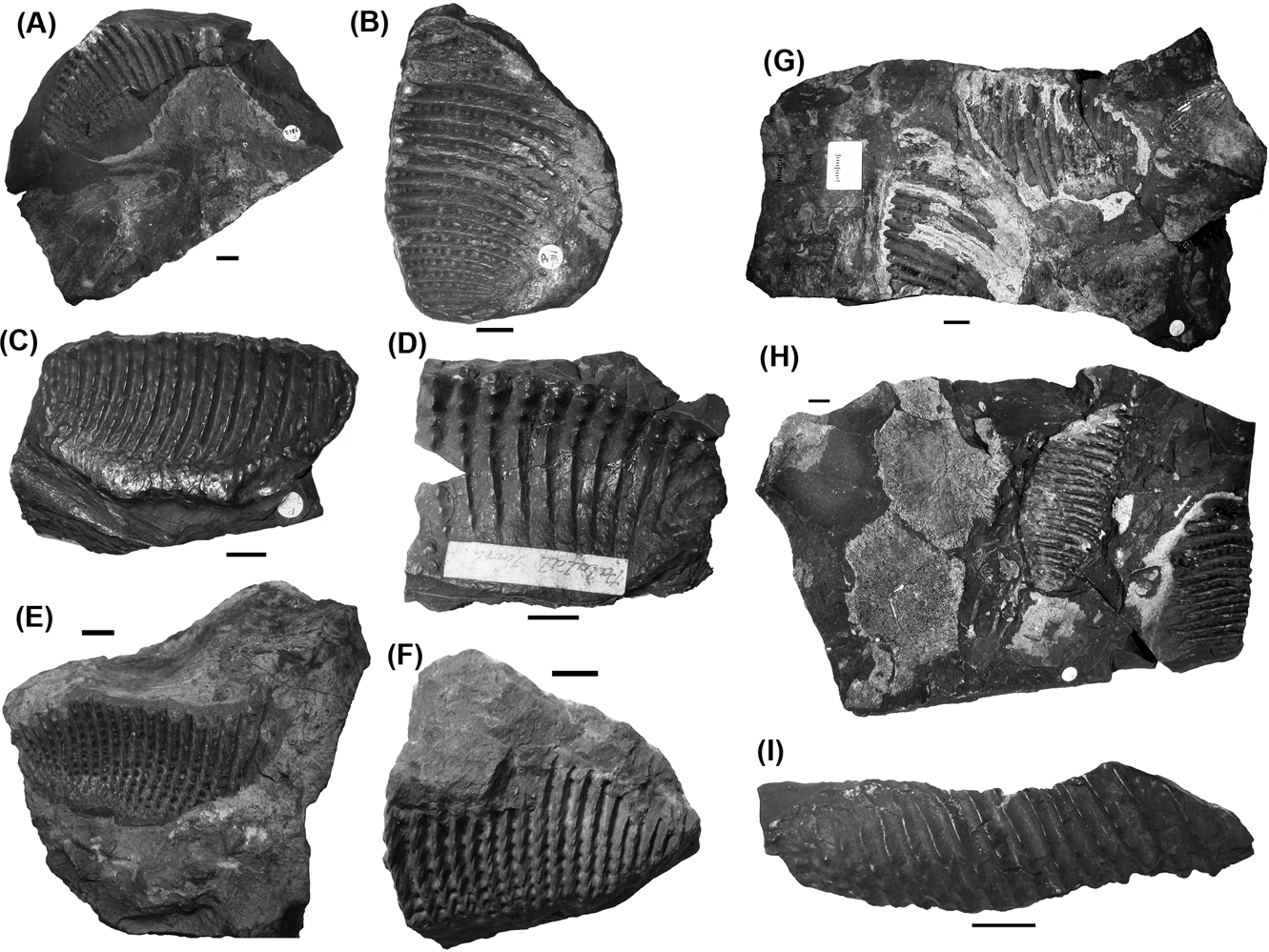
The holotype of C. murchisoni (Fig. 1J) has between 20 and 22 gently curved, almost entirely parallel ridges. The ridges are spaced closely and composed of fine teeth, fused in some places, but otherwise conical and slightly separated from each other. A large portion of the lingual region of the tooth plate is very worn, with the tooth ridges only faintly visible. The posterior margin of the tooth plate is somewhat truncated but it would probably have been an almost symmetrical oval.
The majority of tooth plates from Staffordshire are from the Bassey Mine Ironstone at Longton. They are extremely large, with higher ridge numbers than those from other localities (Fig. 6). The plates are between 8 cm and 10 cm in length. The pterygoid tooth plates are unusually wide, between 63 mm and 67 mm. Their length to width ratio is between 1.35:1 and 1.45:1, which is very low, even for the pterygoid tooth plates, which are usually much broader than the corresponding prearticular plates.
Ctenodus. murchisoni tooth plates: (A) NHMUK P 7312, left pterygoid tooth plate, impression of buccal surface; (B) NHMUK P 7311, right pterygoid tooth plate, impression of buccal surface; (C) NHMUK P 7304, right pterygoid tooth plate, oblique view; (D) MM L8343, incomplete left pterygoid tooth plate; (E) MM L10254, right pterygoid tooth plate; (F) MM L10224, right pterygoid tooth plate; (G) NHMUK P 7310, pterygoid tooth plates in articulation, buccal surface; (H) NHMUK P 7309, prearticular (left on specimen) and pterygoid (right on specimen) tooth plates; (I) MM L8345, incomplete right prearticular tooth plate. Scale bars=10 mm.

The pterygoid tooth plates have a distinct expansion of the lingual margin (e.g. Fig. 6B) in the posterior two-thirds of the plate. There are no ridges more prominent then others, or they may be very worn in this region. This gives the tooth plate an unusual shape. While the anterior regions are more tapered than the posterior, as is common to the tooth plates of many other taxa, the posterior region is relatively much expanded. Specimens NHMUK P 7312 (Fig. 6A) and, NHMUK P 7310 (Fig. 6G) show the unusual orientation of the bones on the palate.
The number of ridges on the pterygoid tooth plates varies between 17 and 23, with those in the most posterior region more worn and indistinct. The ridges are all curved, their distal point being anterior to the proximal origins of the ridge. The ridges are also more highly curved in the lingual portions of the tooth. There is variation in the preservation of teeth on the buccal surface. Many specimens (e.g. Fig. 6C) show ridges where the teeth are mostly fused into a crest, others show individual teeth (e.g. Fig. 6D).
The buccal surface of the pterygoid tooth plates is concave and symmetrical. No examples of complete pterygoid and prearticular tooth plates from the same individual are known. The only known example of a prearticular tooth plate from Longton is MM L.8345 (Fig. 6I). This specimen is long, with 20 tooth ridges visible and a slightly convex buccal surface. It is 8.6 cm long and 3.8 cm wide, with a length to width ratio of approximately 2.3:1. Its overall shape is a slightly elongated and distorted oval. The labial margin is almost straight (excluding denticles visible on this margin), turning inwards posteriorly. The lingual margin is symmetrical relative to the labial for two-thirds of its length, but the anterior third of the inner margin turns outwards at a much greater angle, one of almost 45°, and meets the labial margin at a much more prominent point than the posterior margin.
On the prearticular tooth plate, the teeth and ridges are slightly flattened towards the labial margin, most clearly seen in those regions where the teeth remain unfused. As a consequence, the labial tooth margin is highly scalloped. Lingually, the ridges are worn and much less prominent than along the rest of the plate. The shape of the upper and lower tooth plates from Longton suggests the pterygoid plate would have fitted, and almost surrounded, the prearticular plate, with the internal margins of both the upper and lower tooth plates being more closely apposed than elsewhere.
The specimens examined from Staffordshire have all been identified as C. murchisoni Agassiz (Reference Agassiz1839), but were not described until 1890 by Ward. However, the holotype of C. murchisoni is not from Fenton or Longton, but from Leebotwood in Shropshire, making it more difficult to attribute material from Staffordshire accurately to the species. Furthermore, Ward (Reference Ward1890) claimed that the only specimens of C. murchisoni he had observed were from the Bassey Mine Ironstone. He also described specimens of C. cristatus from both the Bassey Mine Ironstone and the Knowles Ironstone. Ward noted that the major difference between these two species is that the pterygoid tooth plates of specimens of C. cristatus had between 12 and 14 tooth ridges, whereas those of C. murchisoni had from 18 to 20. Of those specimens examined, where it is possible to determine, all of those from Longton have 17 or more tooth ridges. This includes MM L.8345 (Fig. 6I), which was attributed to the Knowles Ironstone.
The Upper Carboniferous locality of Ardwick, near Manchester has also yielded material attributed to Ctenodus, including two very large tooth plates identified as C. murchisoni (Fig. 6E, F). They are large, with a high ridge number: MM L.10224 (Fig. 6F) is incomplete and has 17 ridges, while MM L.10254 (Fig. 6E) has 23, the greatest number observed in any Ctenodus specimen.
2.6. Other named species
Ctenodus robertsoni Agassiz, Reference Agassiz1840
Ctenodus tardus Fritsch, Reference Fritsch1889
C. robertsoni and C. tardus are here considered to be nomina dubia. C. robertsoni is poorly described and lacks type material (Schultze Reference Schultze1981, Reference Schultze1992a; Sharp Reference Sharp2007). C. tardus is probably dipnoan, but is not attributable to Ctenodus (Watson & Gill Reference Watson and Gill1923; E. L.Sharp, pers. obs.).
3. Description
3.1. Dermal skull roof
The skull roof of Ctenodus is broad and fairly long, being less square than that of Sagenodus. The skull has a gently convex profile, the bones are relatively firmly associated and they are quite heavily ornamented on their dorsal surface. There are few examples of the ventral surface of any of the bones, but there is some evidence of the ventral lappets common to most dipnoans. As with many other dipnoans, the form of the skull roof is variable between individuals. The following description is compiled from information from the most complete specimens, noting that the sample is insufficient to distinguish specific differences that may exist.
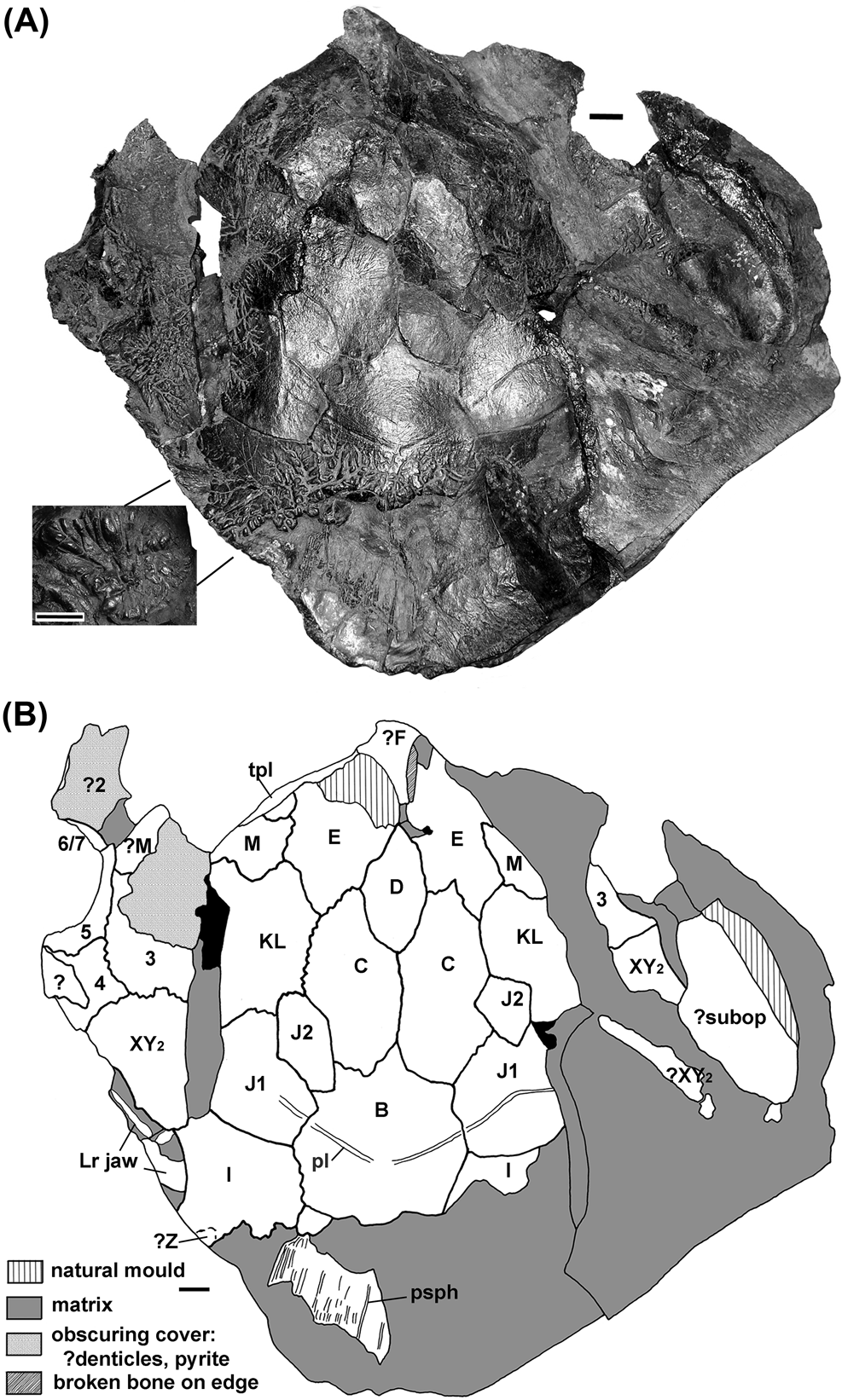
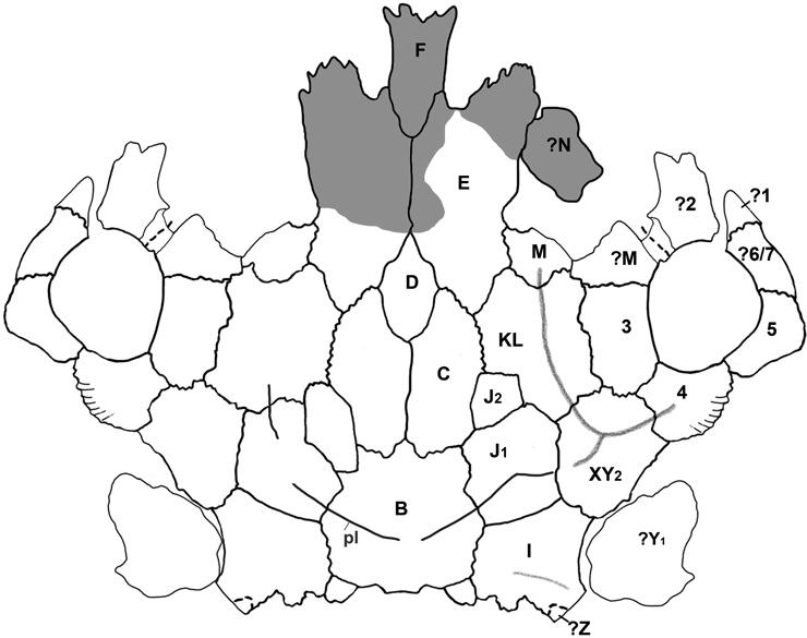
Specimen GLAHM 131502, C. interruptus, shows the tubercles and ramifying grooves on the posterior part of the B-bone that are typical for Ctenodus and were illustrated by Watson & Gill (Reference Watson and Gill1923). It also shows them extending not only onto the I-bone posteriorly, but onto the lateral portions of the J-, KL-, XY- and M-bones, and further onto the circumorbital 2-, 3- and 4-bones. More centrally on the C-, D- and E-bones, medially on the J- and KL-bones and anteriorly on the I-bone, the surface is much smoother, marked by fine radiating striations. As far as can be judged, specimens from the Lower Carboniferous that preserve good bone surface, GLAHM 131502 (C. interruptus) and NMS 1894.155.12 (holotype of C. allodens sp. nov., Fig. 8), have a notably greater degree of dermal ornament of the dorsal surface than those from the Upper Carboniferous.
In GLAHM 131502 (C. interruptus, Fig. 7), pit line grooves are traceable on the B-bone, passing from the radiation centre of the fine striations, positioned towards the posterior half of the bone, and running obliquely anteriorly to enter J1-bones. Short stretches are traceable on the lateral side of the J1-bone, splitting into two branches. One passes across the J1– KL–bone boundary and the other may pass onto XY2-bone. The presence of extensive pit lines is unusual in Carboniferous lungfishes. Lateral lines are not generally visible on specimens of Ctenodus, but the I-bone carries the occipital commissure from the Z-bone to the B-bone, seen in broken specimens and NMS 1888.51.1 (C. allodens sp. nov., Fig. 9).
Ctenodus interruptus, GLAHM 131502, skull roof: (A) photograph; (B) interpretive drawing. (Inset) possible displaced Y1 from region between the skull and postcranial blocks, leaders indicate position as preserved. Scale bars=10 mm. Capital letters and numbers refer to bone identifications. Abbreviations: Lr=lower; pl=pit line; psph=parasphenoid; subop=suboperculum; tpl=tooth plate.

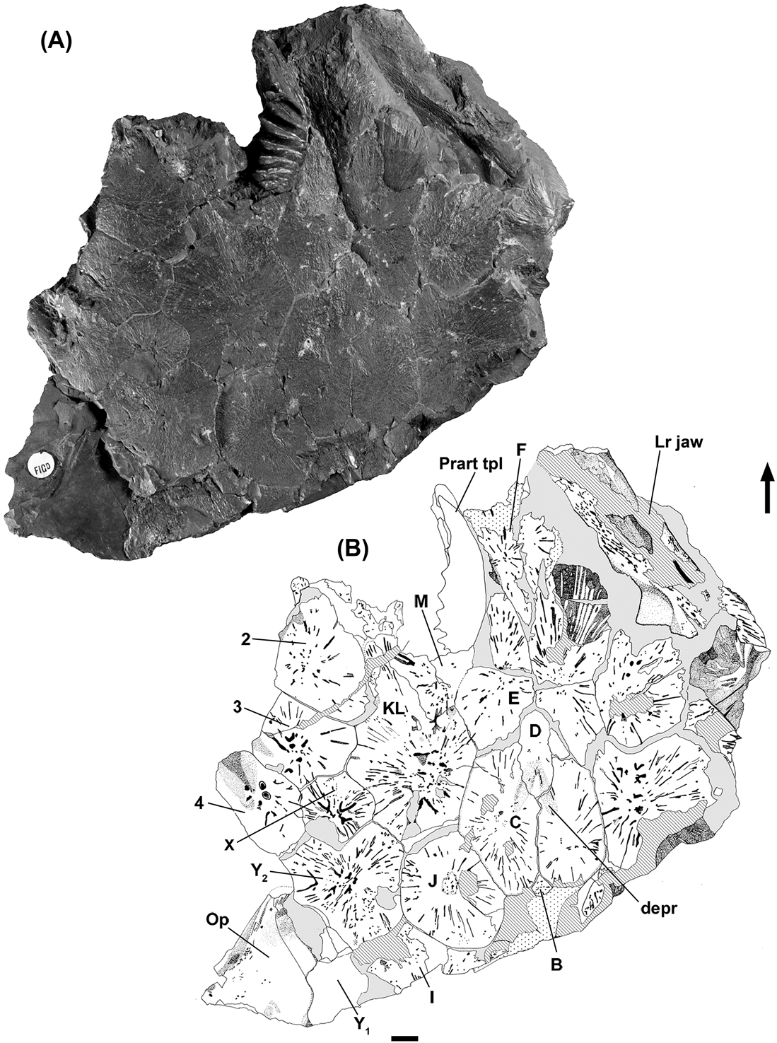
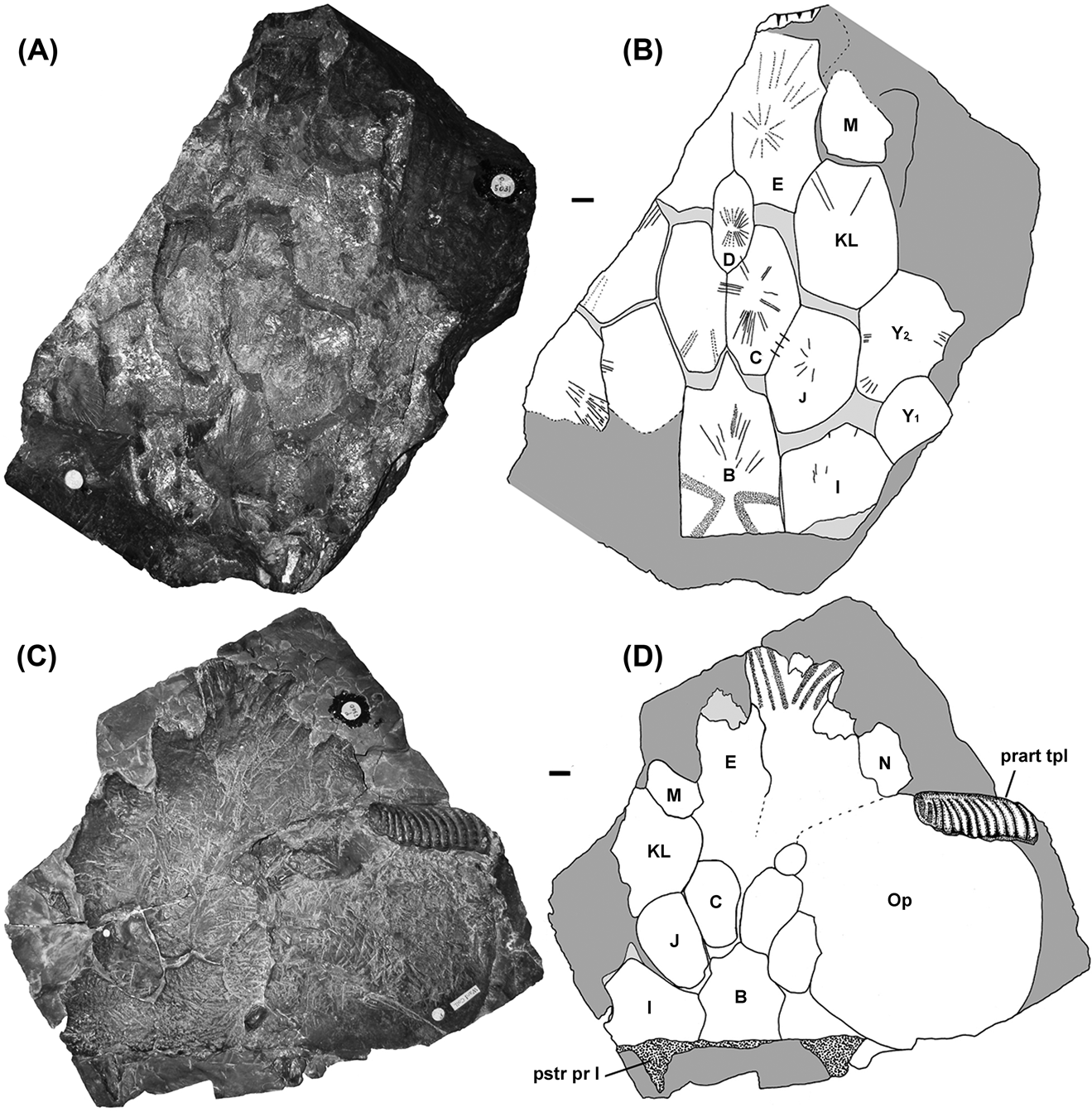
The B-bone of Ctenodus is similar to that of the Late Devonian dipnoans and is the largest element of the posterior region of the skull roof. The anterior margin of the B-bone forms points that pass anteriorly between the adjacent bones to a variable degree. In NMS 1894.155.12 (C. allodens sp. nov., Fig. 8), very little of the B-bone separates the paired C-bones. In contrast, in NHMUK P 5031 (C. murchisoni, Fig. 10A, B), NEWHM G59.70 (C. cristatus, Fig. 11) and GLAHM 131502 (C. interruptus, Fig. 7), the C-bones are parted by the posterior element to a much greater degree. Variation in this bone does not seem to be related to different localities.
Ctenodus allodens. sp. nov., NMS 1894.155.12, skull roof: (A,) photograph; (B) interpretive drawing. Scale bar=10 mm. Capital letters and numbers refer to bone identifications. Abbreviations: depr=depression; Lr=lower; Op=operculum; Prart=prearticular; tpl=tooth plate.

Ctenodus allodens. sp. nov., NMS 1888.51.1, skull roof: (A) photograph; (B) interpretive drawing. Scale bars=10 mm. Capital letters and numbers refer to bone identifications. Abbreviations: ioc=infraorbital canal; occ.c=occipital canal; soc=supraoccipital canal.

Ctenodus skull roofs: (A–B) NHMUK P 5031, Ctenodus murchisoni, photograph and interpretive drawing; (C–D) NHMUK P 7300, Ctenodus cristatus, photograph and interpretive drawing. Scale bars=10 mm. Capital letters and numbers refer to bone identifications. Abbreviations: Op=operculum; prart=prearticular; pstr pr I=posterior process of the I-bone; tpl=tooth plate.

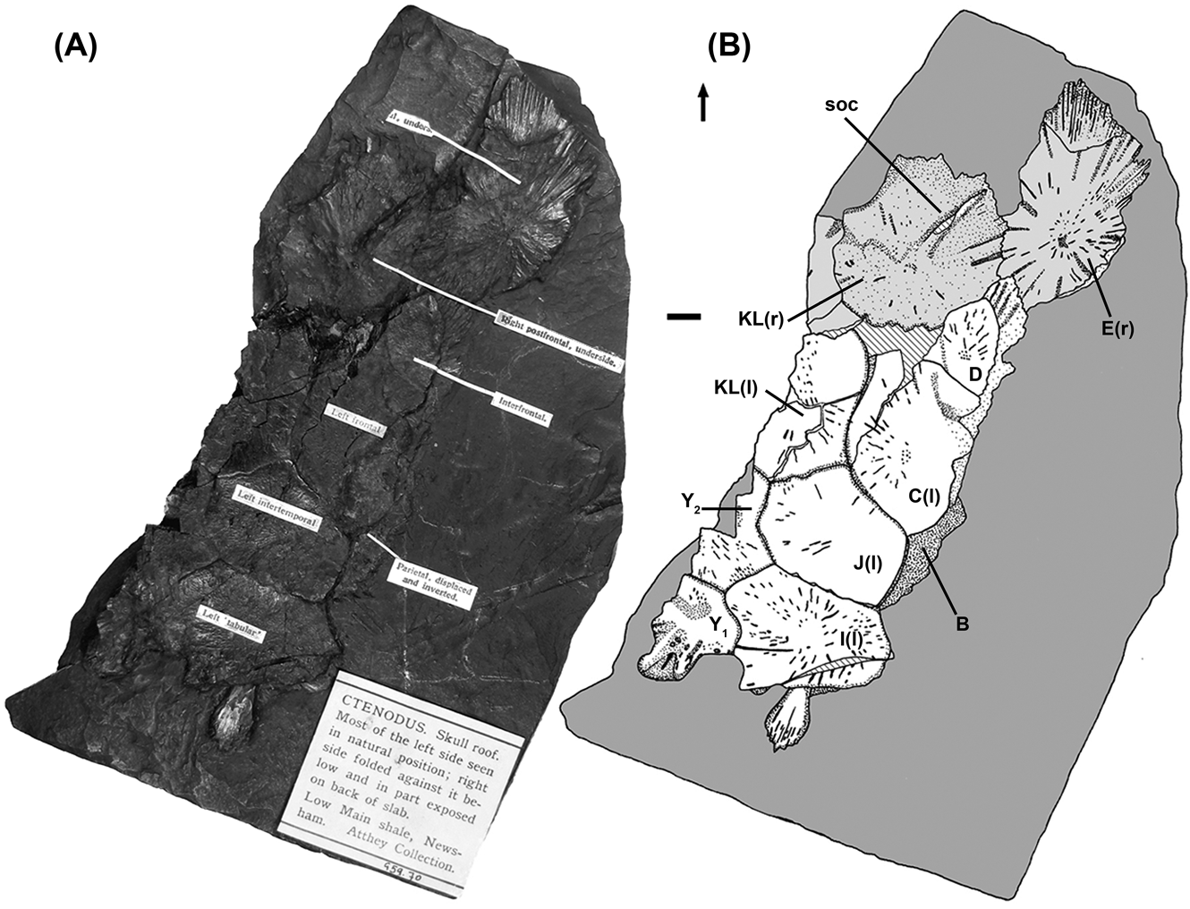
Ctenodus cristatus, NEWHM G59.70, skull roof: (A) photograph; (B) interpretive drawing. Arrow indicates anterior and direction of midline. Scale bar=10 mm. Capital letters and numbers refer to bone identifications. Abbreviation: l=left; r=right; soc=supraoccipital canal.

In NHMUK P 7300 (C. cristatus, Fig. 10C, D) and NEWHM G59.25 (C. cristatus, Fig. 12), there are occipital flanges projecting from the ventral surface of the bone, although these cannot be seen in GLAHM 131502 (C. interruptus). According to Schultze & Chorn (Reference Schultze and Chorn1997, referring to Sagenodus) these flanges would have served for the insertion of the epaxial muscles. The posterior region of the B-bone in C. interruptus carries the occipital sensory line canal and is often broken in this region showing the route of the canal.
Ctenodus cristatus, NEWHM G59.25, skull roof and partial palate: (A–B) dorsal surface, photograph and interpretive drawing; (C–D) palatal surface, photograph and interpretive drawing. Arrow indicates longitudinal axis of body. Scale bars=10 mm. Capital letters and numbers refer to bone identifications. Abbreviations: med ridge=median ridge; Psph=parasphenoid; pstr pr I=posterior process of the I-bone; Pter= pterygoid.

Specimen NHMUK P 5031 (C. murchisoni, Fig. 10) shows a partial ventral impression of the B-bone, with a number of prominent striations visible on the anterior half of the bone. These radiate from a position slightly anterior to that of the striations on the dorsal surface of the bone. Two large, depressed areas forming a ><; shape may correspond to two ridges on the ventral surface of the B-bone that contacted the cristae by which the braincase would have been suspended beneath the dermal skull roof. Alternatively, they could be the courses of the lateral line or pit line system exposed by erosion. There is more evidence for these features in NMS 1888.51.1 (C. allodens. sp. nov., Fig. 9A), a part and counterpart specimen that has been split through the bone.
As in most other fossil Palaeozoic lungfish, the B-bone is surrounded by six other bones, in contrast to Straitonia, in which there are five, and Sagenodus, in which there are seven. There are also seven in one specimen of Tranodis illustrated by Schultze & Bolt (Reference Schultze and Bolt1996, fig. 2D).
The C-bones in Ctenodus are paired, the plesiomorphic condition for the dipnoans. Where known, the dorsal surface of the C-bone is marked by fine radiating striations that often pass across sutures onto adjacent bones. In NMS 1894.155.12 (C. allodens sp. nov., Fig. 8), the surface of the C-bone is more highly ornamented. The degree of interdigitation of the sutures between the C-bones and those adjacent varies somewhat from simple in NHMUK P5031 (C. murchisoni) to apparently interdigitated in GLAHM 131502 (C. interruptus). The overall form of the C-bones does not differ in any significant way between specimens from the Lower and Upper Carboniferous.
The D-bone is a single midline element and is an elongate oval with sub-straight edges (Figs 7, 8, 10).
The paired midline E-bones are the most characteristic bones of Ctenodus, although varying slightly between C. cristatus and specimens from the Lower Carboniferous. They are the longest elements of the skull roof of Ctenodus and the anterior portions are incompletely preserved in GLAHM 131502 (Fig. 7).
There is some variation in the nature of the posteromedial margin between E- and D- bones between specimens (compare C. allodens sp. nov. NMS 1894.155.12, Fig. 8 with C. interruptus GLAHM 131502, Fig. 7 and C. murchisoni NHMUK P 5031, Fig. 10).
Specimens NHMUK P 5031 (C. murchisoni), P7300 (C. cristatus) (Fig. 10) and NEWHM G59.67 (C. cristatus, Fig. 13) show the anterior expansion of the E-bones, flaring laterally and anteriorly into a fan-shape. In GLAHM 131502 (C. interruptus), although the E-bones are incomplete anteriorly, there is a natural mould of the left element that appears to terminate in an undulating margin without an extensive anterior expansion. In some specimens, the accessory branches of the lateral line penetrate the E-bones from the KL- and M-bones which surround them.
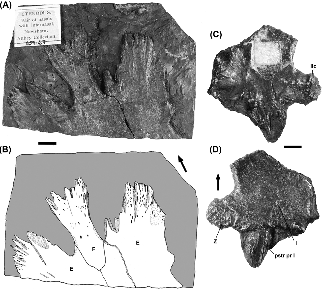
Ctenodus cristatus individual bones: (A– B) NEWHM G59.67 E- and F-bones, photograph and interpretive drawing; (C–D) NEWHM G59.79 I- and Z-bones, internal and external (dorsal) views. Arrows indicates longitudinal axis of body. Scale bars=10 mm. Capital letters refer to bone identifications. Abbreviations: llc=lateral line canal; pstr pr I=posterior process of the I-bone.

In the Upper Carboniferous NEWHM G59.70 (C. cristatus, Fig. 11), the posterior region of the left half of the skull roof is preserved on the block in dorsal view, but the B-bone is missing. The E-bones of this specimen appear to be expanded into a fan-shaped lobe.
The anterior portion of the skull of Ctenodus is relatively poorly known, but as seen in NEWHM G59.67 (C. cristatus, Fig. 13), seems to be composed of a single median F-bone which parts the E-bones anteriorly. An anterior prenasal ‘fan’, which surrounds the F-bone and the anterior margin of the E-bones is seen in C. cristatus (NHMUK P 7300, Fig. 10C, D and NEWHM G59.30, Fig. 13A, B) as illustrated by Watson & Gill (Reference Watson and Gill1923). This fan is of thick bone whose surface is punctuated by a series of grooves and troughs, although there is no good evidence for the supraoccipital canal, usually assumed to have been present in the rostral regions of the skull.
The posteriorly narrow F-bone flares anteriorly, possibly characteristic of specimens from the Upper Carboniferous (Fig. 13A, B). In contrast, the F-bone of NMS 1895.155.12 (C. allodens sp. nov., Fig. 8) from the Lower Carboniferous seems larger and more triangular between the two correspondingly broader E-bones. In GLAHM 131502 (C. interruptus), there may be the remains of an F-bone, but it is incomplete anteriorly.
The I-bones are relatively large but not as large as in Sagenodus. Ventrally, the I-bone of Ctenodus has characteristic posterior prongs (amongst Upper Carboniferous genera at least), referred to by Watson & Gill (Reference Watson and Gill1923) as ‘tabular horns’ (e.g. C. cristatus, Figs 11, 12). An isolated I-bone is exposed in both dorsal and visceral view (C. cristatus, Fig. 13C, D). Visceral views of skull bones, when available, are particularly valuable for identifying isolated bones from other taxa, as they often carry distinctive processes and facets for attachment of internal structures or for housing the lateral line canal.
The anterior margin of the I-bone where it joins the J1-bone is typically a smooth curve (GLAHM 131502 C. interruptus, Fig. 7). However, in some, such as NEWHM G59.25 (C. cristatus, Fig. 12), it is more closely interdigitated than that of the medial margins. In GLAHM 131502, contact with the B-bone is more interdigitated. The union of the I-bone with the Y2-bone is usually short, but is not represented in GLAHM 131502. In that specimen, there is a combined Y2- and X-bone. The margin between the I-bone and Z-bone is tightly interdigitated, so that the two are often preserved together, as in GLAHM 131502 and NEWHM G59.79 (Figs 7, 13).
The posterior margin of the I-bone is usually concave and bears a ventral flange in addition to the ventrally projecting prongs (Fig. 13C, D). In visceral view, the posterior prong of the I-bone is concave suggesting the passage of some structure, possibly a blood vessel or ligament. These cannot be seen in GLAHM 131502, (C. interruptus) but may lie beneath the matrix. However there is a very irregular posterior margin on the left, with an ornamented lappet extending a little way beyond the posteromedial margin. No other Lower Carboniferous specimens preserve the posterior process of the I-bones and they may have been absent.
The shape of the J-bones varies somewhat among the specimens, and even within individuals. In GLAHM 131502 (Fig. 7), there are two on each side, a larger J1-bone and a smaller J2-bone. Whereas on the left, the J2-bone contacts the B-bone, on the right it does not. The J1-bone contacts the C-bone on the right, but not on the left. Its posteromedial border contacts the anterior half of the B-bone and the anterormedial border contacts the C-bone in most other specimens. This is the plesiomorphic condition for dipnoans as seen in Straitonia (Sharp & Clack Reference Sharp and Clack2012) and Scaumenacia (Cloutier Reference Cloutier, Schultze and Cloutier1996), and it contrasts with the arrangement of skull roof bones in Sagenodus, in which the J-bones do not contact the single C-bone. In GLAHM 131502 (C. interruptus), the J bones bear conspicuous pit lines, continuations of those on the B-bone.
The anterior extent of the J-bone in Ctenodus generally exceeds that of the B-bone, extending to the mid point of the C-bone (in GLAHM 131502 (C. interruptus), only if both J-bones are included); again in contrast to the situation seen in Sagenodus. From here, the anterolateral margin contacts the posterior margin of the KL-bone and it is in this region that the J-bone is most variable in its morphology. In NMS 1894.155.12 (C. allodens sp. nov., Fig. 8) from the Lower Carboniferous and NHMUK P 5031 (C. murchisoni, Fig. 10) from the Upper Carboniferous, the margin from this point is laterally curved, with no distinct angles on the margin. In contrast, in C. cristatus, NEWHM G59.70 (Fig. 11) and NEWHM G59.25 (Fig. 12), both Upper Carboniferous, have a more angled margin in this region.
The KL-bone is the largest of the skull roof after the E-bone, with a highly variable morphology. The most conspicuous difference is the degree to which the M-bone penetrates the KL-bone, and also the degree of interdigitation present at the margins.
Specimen NEWHM G59.70 (C. cristatus, Fig. 11) preserves what is considered to be the visceral surface of the right KL-bone, in articulation with the right E-bone. A single large, prominent ridge that houses the lateral line arises at the centre of the bone and continues towards the margin just lateral to the E-bone.
The M- and N-bones are rarely completely preserved: the N-bone may be present on NHMUK P 7300 (C. cristatus, Fig 10C, D).
The Z-bone is a very small bone that, unlike those in Sagenodus, is exclusively restricted to the posterior margin of the skull roof. It is thicker than its near neighbours and is round, or sub-rounded, with a pocked dorsal surface and a small, crenulated posterior margin that is confluent with that of the I-bone. This bone may be represented on GLAHM 131502 (C. interruptus), although it is incomplete and tightly sutured to the I-bone.
The lateral line usually enters the skull through the Z-bone ventrally, bifurcating into a medial branch as the occipital commissure, which travels in parallel with the posterior margin of the skull roof, and an anterior one as the cephalic division of the main lateral line canal. The latter travels through the Y1- and Y2-bones, and into the X-bone, where it bifurcates again. The isolated example of an I-bone plus a Z-bone (NEWHM G59.79 C. cristatus, Fig. 13C, D) shows the pitting that indicates the presence of a sensory canal below the surface, while the visceral surface of this bone shows the three-pronged thickening which housed it.
The Y-bone normally forms the posterolateral corner of the skull roof, and is usually smaller than the Y2-bone. Independent Y2-bones are absent in GLAHM 131502 (C. interruptus), but are combined with the X-bone into the XY2-bone. What is probably a displaced Y1-bone lies posterior to and somewhat below the main skull roof (Fig. 7 inset). It is approximately oval and bears the typical ridge and groove ornament of the other posterior skull bones, but its fit with the other bones is hard to judge.
In C. cristatus (NEWHM G59.70, Fig. 11), the Y1-bone bears deep pits indicative of the cephalic division of the lateral line. The visceral surface of the Y1-bone bears a large and prominent ridge that runs from the posterior margin of the skull roof close to the Z-bone and into the posterior margin of the Y2-bone. This ridge is much larger than the thickening on the Z-bone, but is interpreted as the canal that carries the lateral line forward into the anterior regions of the skull roof.
The Y2-bone lies lateral to the J-bone and anterior to the Y1-bone, except in GLAHM 131502 (C. interruptus), in which it is conjoined with the X-bone (termed here the XY2-bone) (Fig. 7). The anterior margin of the Y2-bone contacts two sides of the small, approximately pentagonal X-bone, seen in C. allodens sp. nov. (NMS 1894.155.12, Fig. 8 and NMS 1888.81.1, Fig. 9), both from Straiton in the Lower Carboniferous. It is not possible to assess whether the form of this union is typical for Ctenodus, or restricted to the Lower Carboniferous species. It is, however, consistent with the fusion of the X-bone and Y2-bone in GLAHM 131502 (C. interruptus).
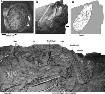
The posterolateral margin of the Y2-bone generally bears a smooth embayment, or notch, confluent with the lateral margin of the Y1-bone. This is probably where the tabulate process of the operculum articulated. In GLAHM 131502, the XY2-bone contacts the 3- and 4-bones in a gentle convex curve, but is interdigitated.
The visceral surface of the Y2-bone, seen in NMS 1888.51.1 (C. allodens sp. nov.) and NEWHM G59.25 (C. cristatus) (Figs 9, 12), bears the continuation of the ridge from the Y1-bone carrying the main branch of the lateral line canal. The dorsal surface of the bone, where known (and XY2-bone in GLAHM 131502), is deeply ornamented and pitted, as is typical for a bone that carries the main branch of the lateral line canal.
The X-bone is preserved as a complete and separate bone only in two specimens, both C. allodens sp. nov.: NMS 1894.155.12 and NMS 1888.51.1A (Figs 8, 9). Watson & Gill (Reference Watson and Gill1923, p. 191) did not recognise this bone as belonging to the skull roof proper; instead they believed it to be an element of the orbit, unique to C. interruptus.
The X-bone is usually the point at which the main cephalic branch of the lateral line system bifurcates. In NMS 1888.51.1A (C. allodens sp. nov., Fig. 9), the supraorbital canal passes from the X-bone into the KL-bone and through the lateral bones of the skull roof. Medially and ventrally, the X-bone carries the infraorbital lateral line canal into the 4-bone, where it again divides. This bone is usually the most cratered and punctured by foramina, and carries a number of accessory canals into its neighbouring bones. There are no specimens showing its visceral surface, although there is evidence from Sagenodus that the lateral line is more deeply buried than in the more posterior Y1- and Y2-bones.
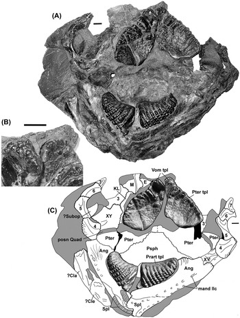
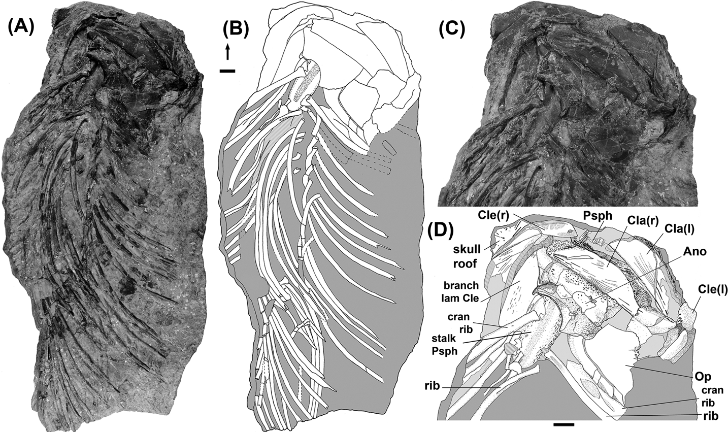
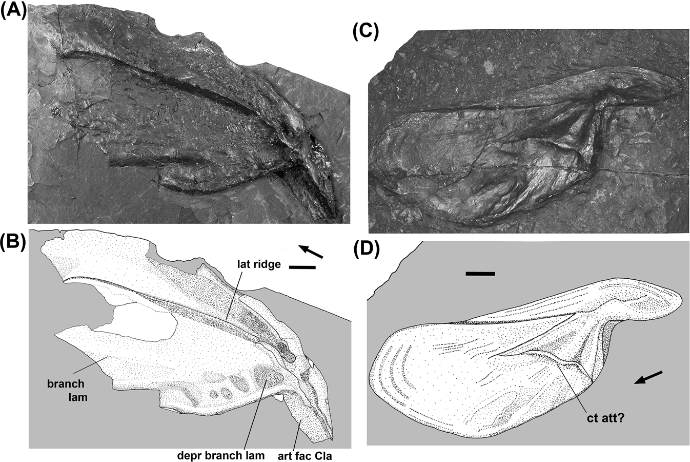
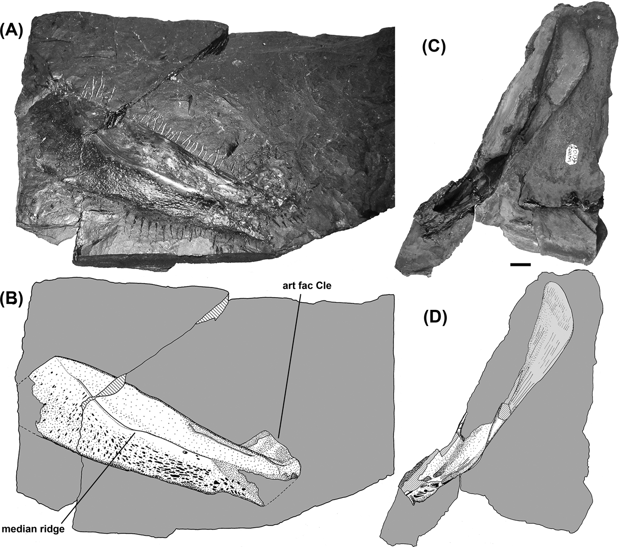
The orbital region of Ctenodus, hitherto very poorly known, is represented in GLAHM 131502 (C. interruptus), in which most or all bones are in articulation. Many of them are visible in both external and internal views (Figs 7, 14). On its left side, the 5-, 6- and 7-bones have been displaced dorsally to lie slightly beneath the 4-, 3- and 2-bones, and are twisted so that they appear in internal view on both sides of the palatal surface of the specimen.
Ctenodus interruptus, GLAHM 131502, palate, lower jaws and associated tooth plates: (A) photograph; (B) closeup of vomerine tooth plates; (C) interpretive drawing. Scale bars=10 mm. Capital letters and numbers refer to bone identifications. Abbreviations: Ang=angular; Cla=clavicle; Cle=cleithrum; llc=lateral line canal; mand=mandibular; posn=position; Prart=prearticular; Psph=parasphenoid; Pter=pterygoid; Quad=quadrate; Spl=splenial; Subop=suboperculum; tpl=tooth plate; Vom=vomer.

The most posterior element of the orbit is the 4-bone (sometimes identified as a postorbital). It is visible in both external and visceral views on the left side of GLAHM 131502 (C. interruptus, Fig. 7), and in external view on the left side of C. allodens sp. nov. (NMS 1894.155.12, Fig. 8 and NMS 1888.51.1, Fig. 9), split into part and counterpart. The external surface is ornamented, and the bone is irregular in shape. In NMS 1894.155.12, it is roughly an elongate hexagon. In GLAHM 131502, it is overlain by another, unidentified, bone. The visceral surface is seen on both sides of that specimen, where it appears to be broadly crescentic. It has a shorter concave margin roughly parallel to the orbit margin, and a longer convex margin posterolaterally, which has a fimbriated edge suggesting interdigitations. A short, straighter margin joins to the 5-bone ventrally. A prominent ridge curves from its medial-most point onto the adjacent circumorbital 5-bone, seen also in the counterpart of NMS 1888.51.1 in visceral view. This probably supported the infraorbital lateral line.
The 4-bone contacts the XY2-bone or the separate X- and Y-bones posterodorsally. Its posterior margin is straight and smooth in C. allodens sp. nov. (NMS 1894.155.12), but interdigitated in C. interruptus (GLAHM 131502, Fig. 14), and is confluent with the lateral margin of the Y2-or XY2-bones, together with which it would have braced against the operculum.
In C. allodens sp. nov. (NMS 1894.155.12 Fig. 8), the dorsal portions of the external face are smooth, with a gently convex surface; in contrast to this there is a large depression in the middle portions of the ventral region of the bone, leaving the margins of the bone considerably raised relative to the centres of the bone. While it could be considered as an artefact of preservation, this condition is also observed in C. interruptus (GLAHM 131502 and C. cristatus (NEWHM G59.25) (Figs 7, 12).
The infraorbital lateral line canal enters the orbit via the 4-bone in C. allodens sp. nov. (NMS 1888.51.1 and NMS 1894.155.12). There are large numbers of deep sensory pores on the surface of the 4-bone in NMS 1894.155.12 (Fig. 8).
The dorsalmost and one of the largest bones of the orbital series is the 3-bone. In GLAHM 131502 (C. interruptus), the contacts with adjoining circumorbitals and with the XY2 and ?M-bones can be seen in both internal and external views, where they differ from each other (Figs 7, 14). Most are interdigitated, except for the 3–XY2-bone junction on the right external surface, which appears straight. They are straighter in the other specimens that show these bones. In NMS 1894.155.12 (C. allodens sp. nov.), the 3-–2-bone junction is completely straight, similar to that between the 3a and 3b bones of Sagenodus.
Bone 3 in NMS 1894.155.12 (C. allodens sp. nov.), like bone 4, has numerous sensory pores on its surface, as it receives a short branch of the infraorbital canal from the latter bone (Fig. 8). These pores are offset towards the orbit and the 4-bone, although accessory branches ramify into the surrounding bone and its neighbours from here. Similarly, the 2-bone has some sensory pits offset towards the orbital margin and a pattern of ornament which radiates from this region.
The 2-bone is larger than the 3-bone in NMS 1894.155.12 (Fig. 8), but its contribution to the orbit does not seem to be greater. Only a small area of the surface of 2-bone can be seen in GLAHM 131502 (C. interruptus), because it is covered with a shagreen of a denticle-like field which may be a pyritic deposit or remains of a denticulated dermal covering (Fig. 7). Part of the orbital margin is preserved on the left side.
In GLAHM 131502 (C. interruptus), the 5-bone forms the posteroventral margin of the orbit and is crescentic in shape, but narrower than the 4-bone (Fig. 14). It carries the continuation of the ridge seen internally on the 4-bone. Its narrowest point lies at the most ventral part of the orbit margin. Posterior and anterior to this point, it broadens to form an external lamina; the anterior section is excavated internally into a depression. The lamina is interdigitated for contact with the 4-bone and 6-bone. The 6-bone is essentially similar. The external surfaces of these bones cannot be adequately seen, but there is no evidence of lateral line pores, even though they might be expected. The 7-bones on each side are short and triangular, continuing the supporting internal ridge. Although neither is quite complete, there appears to be little missing such that their short length is not an artefact (Fig. 14).
The collection of the Great Northern Museum contains a large number of isolated bones which are certainly attributable to the orbit of either Ctenodus or Sagenodus, but as they are from Newsham, it would be impossible to identify the genus to which they belong. The new information may help to identify these bones.
3.2. Palate
Vomerine and pterygoid tooth plates of Ctenodus are seen for the first time in C. interruptus (GLAHM 131502), and the associated pterygoids and parasphenoid can be confidently attributed to this species, again for the first time (Fig. 14).
Both vomerine tooth plates are present, more or less fused onto the corresponding pterygoid (Fig. 14, inset). The better-preserved right element bears two or three rows of small tubercular teeth; on the outermost margin there are at least two, although the margin is broken. The next row bears seven teeth, reducing in size medially along the row, and the third row, three. There are one or two additional teeth on the vomerine plate that do not appear to be in rows. The bone is triangular, narrowing to a point posteriorly.
The pterygoid tooth plates of GLAHM 131502 appear very worn, and bear what may be pathologies: numerous pits over the surface of the plate, largest towards the medial margins and smaller towards the posterolateral margins. Such pitting has not been seen on any other tooth plates. The plates are concave, with the lateral, toothbearing margin a shallow curve and longer than the medial, posteromedial and posterolateral margins. On the right, these are also shallowly curved, but on the left they are somewhat sigmoid. There are ten to 12 rows of teeth (the two sides vary slightly). Of these, the most anterior two are longest, bearing about three or four cusps before each row is fused into a ridge. They appear closely apposed and the rows possibly fuse to each other medially. The third row also bears three or four cusps before again fusing to a ridge, with the cusps becoming indistinct. This row is set a little apart form the first two rows on each plate. On the right plate, rows four and five are conjoined, with only two cusps readily distinguishable, and the subsequent seven or eight rows each bear only one or two teeth at the anteriormost point of its ridge. The ridges soon fade out into the body of the plate. The left plate has more strongly developed rows four and five, bearing three cusps, but otherwise the two plates are similar.
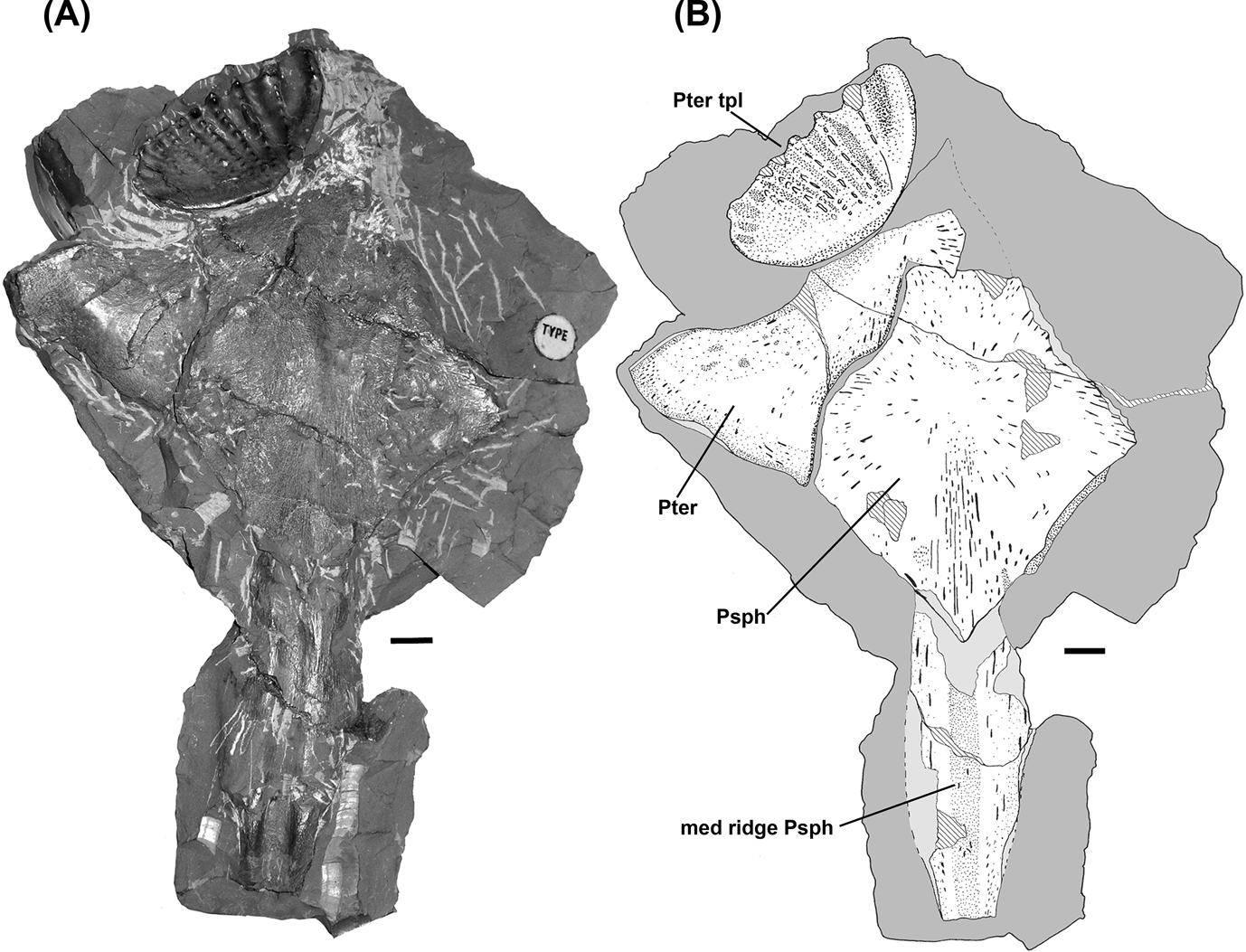
These pterygoid tooth plates are quite unlike those known from other Ctenodus species or also those of Sagenodus. Unless they have extensive pathology or wear, they should be easily distinguishable if found isolated, or in association with other material, for example as in NMS 1894.168.2 (C. interruptus, Fig. 8). The latter specimen shows a pterygoid tooth plate associated with a pterygoid and parasphenoid (Fig. 15). The tooth plate closely resembles that of GLAHM 131502, though less worn. It is from the Asbian locality of Broxburn, contrasting with the later Pendelian localities of Loanhead and Gilmerton.
Ctenodus interruptus, NMS 1894.168.2, parasphenoid, pterygoids and associated pterygoid tooth plate in visceral view: (A) photograph; (B) interpretive drawing. Scale bars=10 mm. Abbreviations: med ridge= median ridge; Psph=parasphenoid; Pter=pterygoid; tpl=tooth plate.

The paired pterygoids are the main toothbearing bones of the palate and these elements are intimately associated, fusing at an early stage of development and, therefore, often being found preserved together, as in NMS 1984.168.2 (C. interruptus, Fig. 8) and NEWHM G61.24 (C. cristatus, Fig. 1C, D).
Given the close apposition between the pterygoid tooth plates in GLAHM 131502 (C. interruptus), the anteromedial margins of the bones would have articulated in the mid-line of the palate. In NMS 1897.30.2 (C. allodens sp. nov., Fig. 4C, D), the anteromedial margin is sub-parallel with the margin of the more straight and elongate tooth ridge 1.
At the midpoint of the pterygoid, the medial margin turns laterally at a sharp angle. This angle varies between specimens and localities and, again, NMS 1897.30.2 (C. allodens sp. nov.) shows a somewhat different condition. In this specimen, the angle between the two margins is shallow whereas in other specimens, the angle is approximately 130°. In those specimens where the posteromedial margin is preserved, there is a distinct curve of the margin posteriorly, and the buccal surface is unfinished and roughened where the parasphenoid would have contacted the pterygoid.
The posterior portion of the pterygoid is very characteristic in lungfishes, consisting of a delta-shaped quadrate ramus positioned laterally to the tooth plate. In life, this ramus would be angled ventrally to meet the quadrate. Although this feature is usually preserved flattened in a single plane, in NMS 1897.30.2 (C. allodens sp. nov., Fig. 4C, D) the ramus is strongly curved and resembles that in the extant Protopterus. The lateral portion curved ventrally away from the plane of the palate, as it does to some extent in GLAHM 131502 (C. interruptus). In that specimen, the position of the quadrate is marked by a rugose margin.
The visceral, or dorsal, surface of the pterygoid is known from a single specimen, NMS 1878.45.22 (C. cristatus), and appears to be entirely confluent with the visceral surface of the tooth plate. No junction is visible between the two, indicating the complete fusion between these two elements.
The parasphenoid of Ctenodus is a large bone, even compared with that of other post-Devonian dipnoans. Isolated parasphenoids of Ctenodus from Newsham can be distinguished from those of Sagenodus by their broader and more flared stems, which also have a more densely ridged and grooved visceral surface. The parasphenoid of Ctenodus is illustrated in buccal view in NMS 1894.168.2 (C. interruptus, Fig. 15), NEWHM G59.28, and in dorsal or visceral view in NHMUK P3385 (C. cristatus, Fig. 16). Specimen GLAHM 131502 (C. interruptus) preserves part of the body in buccal view and part of the stalk in visceral view. The junction with the pterygoids medially is a straight transverse suture, as Watson & Gill (Reference Watson and Gill1923) noted in their description of ‘C. interruptus’. They may have examined NMS 1894.168.2 from the Lower Carboniferous of Broxburn, which is one of the few specimens to show this feature.
Ctenodus cristatus parasphenoids: (A–B) NEWHM G59.28 in buccal (ventral) view, photograph and interpretive drawing; (C–D) NHMUK P 3385 in dorsal view, photograph and interpretive drawing. Scale bars=10 mm. Abbreviations: lat exp=lateral expansion; med depr=median depression; med ridge=median ridge; Psph=parasphenoid.

The corpus is a slightly elongated diamond shape, with the anteroposterior length slightly greater than the width. The anterior extent of the parasphenoid is not known, but is probably composed of a truncated and slightly rounded tabulate process. In NHMUK P3385 (C. cristatus), the anterior point is more elongate and rounded when the parasphenoid is viewed in visceral view (Fig. 16). The lateral corners of the corpus are also rounded, making an angle of slightly more than 90° in most cases, while the posterior corner is slightly less than 90°, which makes the corpus elongated anteroposteriorly. The exception to this is CAMSM E12969 (Ctenodus sp.), in which the parasphenoid appears wider than it is long. In general, the lateral margins are unfinished, and here the pterygoids would have overlapped the parasphenoid.
In GLAHM 131502 (C. interruptus), the buccal surface is marked by very fine ridges and wrinkles radiating in a cruciform pattern from a midpoint level with the widest part of the bone. In other specimens, it is punctuated with delicate pits and grooves, with no extensive ornamentation of the bone as is seen in some specimens of Sagenodus. Instead, the central region has small pits which laterally become fine grooves oriented towards the corners of the corpus.
The corpus of Upper Carboniferous parasphenoids can be characterised by a large ridge running anteroposteriorly along the buccal surface, confluent with the median ridge found anteriorly on the stalk (e.g. C. cristatus NEWHM G59.25, Fig. 12C, D; NEWHM G59.28, Fig. 16A, B; and NEWHM G59.27).
The median ridge at the junction of the corpus and stem may be the point of divergence for the dorsal aorta into two lateral dorsal aortae. Watson & Gill (Reference Watson and Gill1923, p.191, described as “club shaped”) noted that in the largest specimens the ridge at the junction of the corpus and stalk was much expanded compared with smaller specimens.
In specimens of Ctenodus in which almost all the parasphenoid is preserved, the corpus is slightly longer than the stalk, for example in C. cristatus (NEWHM G59.28, Fig. 16A, B), in buccal view. The most conspicuous feature of the stem in buccal view is the median depression that may indicate the path of the dorsal aorta. The surface of the bone in this region is relatively smooth, with fine pits and grooves parallel to the direction of the stem.
The visceral surface of the parasphenoid stalk is more prominently ridged than the buccal surface, although there variations shown in C. cristatus (NHMUK P3385, Fig. 16 and NEWHM G59.27). The former specimen shows a concave stem with a distinct trough running the entire length from the posterior point onto the corpus, with which it is confluent. Specimen NEWHM G59.27 (C. cristatus) shows a much less troughed parasphenoid stem, but one which is more sculptured with prominent ridges and grooves, many of which penetrate onto the body. A dense pattern of fine pores is present on the surface of this specimen and NHMUK P3385 (C. cristatus, Fig. 16C, D). In GLAHM 131502 (C. interruptus), the central trough bears a denticle field.
The most prominent features of the visceral surface of the corpus are three sets of radiating ridges emanating from the centre of the corpus anteriorly and posterolaterally. Anterolaterally, the body is slightly depressed relative to the centre of the bone and the more posterior regions.
The relationship between the parasphenoid and the pterygoid is variable. In NMS 1894.168.2 (C. interruptus, Fig. 15), the parasphenoid is positioned dorsal to the pterygoids, confirmed by CAMSM E12969 (Ctenodus sp.). An alternative configuration is seen in NEWHM G59.25 (C cristatus, Fig. 12), in which the left wing of the parasphenoid is ventral to the left pterygoid. In this case, it must be presumed that anteriorly the pterygoid comes to underlie the parasphenoid, allowing the tooth plates to project ventrally into the mouth. This second condition is what has been described for Sagenodus, although no specimens with the parasphenoid in articulation with the pterygoids have been noted for this genus. The paucity of articulated material makes it impossible to comment with certainty on whether this is a true morphological difference between this and the other specimens, or an artefact of preservation.
On account of their shape and large size, parasphenoids of Ctenodus from some Upper Carboniferous localities have in the past been misidentified as the interclavicle of tetrapods (Clack et al. Reference Clack, Sharp, Long, Jørgensen and Joss2011). Watson & Gill (Reference Watson and Gill1923) noted that the larger specimens could be up to a foot (c.300 mm) in length.
It has long been noted that there is no evidence of ossification of the braincase in Ctenodus or Sagenodus, making both genera directly comparable in this respect with Neoceratodus. Watson & Gill (Reference Watson and Gill1923) stated that where specimens were found with the skull partially intact, the palate and the skull roof lie in direct apposition with “hardly a trace even of matrix between them” (Watson & Gill, Reference Watson and Gill1923, p.171). Consequently, the visceral faces of the palatal bones are rarely preserved and have only rarely been observed.
3.3. Lower jaw
It is rare for the bones of the lower jaw to be found in articulation in Carboniferous lungfishes, but is shown in GLAHM 131502 (C. interruptus). The identification of many isolated elements as Ctenodus is by inference. For example, NMS 1893.139.27 (C. cristatus, Fig. 17G, H) is from the Upper Carboniferous South Parrot Coal Shale, from Niddrie, Midlothian. Sagenodus is unknown at this locality and, along with the large size of the element in question, allows it to be identified as Ctenodus with some confidence.
Ctenodus mandibles: (A–D) Ctenodus cristatus, NEWHM G175.34, right angular and splenials: (A–B) lateral view, photograph and interpretive drawing; (C–D) medial view, photograph and interpretive drawing; (E–F) Ctenodus cristatus, NEWHM G22.67, right prearticular and tooth plate in medial view, photograph and interpretive drawing; (G–H) Ctenodus sp., NMS 1893.139.27, left angular in lateral view, photograph and intepretive drawing. Arrows point anteriorly. Scale bars=10 mm. Abbreviations: add mand att=adductor mandibulae attachment; Ang=angular; asc proc Ang=ascending process of angular; Art reg=articular region; depr= depression; llc=lateral line canal; mand= mandibular; Prart=prearticular; Spl=splenial; tpl=tooth plate.

The largest element of the lower jaw is the angular, which forms most of the lateral portion of the mandible. It is an outwardly convex bone with a generally triangular shape, its anterior margin tapering to an irregular point. Its dorsal margin is also slightly concave, although not to the same degree as in Sagenodus, giving the dorsal and ventral margins a sub-parallel appearance. In other genera, the posterodorsal margin of the angular seems to be formed from two distinctly inclined margins. The posterior region of the bone is incomplete in all Ctenodus specimens examined, with the articular facet of the bone not preserved. Dorsal to this, however, the margin is intact in NMS 1893.139.27 (C. cristatus, Fig. 17G, H) and seems to be formed by a margin inclined at approximately 50° to the horizontal. This is steeper than is seen in other genera, and seems to extend for more than half the depth of the articular bone. Dorsally, this margin leads to the ascending process of the angular (Fig. 17H). This area is not particularly pronounced in GLAHM 131502 (C. interruptus, Fig. 14), without the distinct crest that is seen in other genera, and leads smoothly into the more anterior dorsal margin of the angular. The ascending process is irregular in its margin, with a number of small notches in the more dorsal regions. It is possible that one of these more anteriorly placed depressions marks the region for the attachment of the adductor mandibulae.
The most prominent of the lateral lines on the lower jaw is the oral canal, of which six well-developed pores are present in GLAHM 131502 on the left element. This canal traverses the bone anteriorly just below the midline of the bone. The mandibular canal is also carried on the angular, where three pores can be seen (Fig. 14).
The lateral face of the angular shows a distinct thickening in the region of the lateral line, suggesting that the canal was robustly embedded in bone, and some of the pores open via slightly raised tubercles. The bone is convex in section, more prominently so than in Sagenodus, and in GLAHM 131502 (C. interruptus, Fig. 14) more obviously on the right side. The medial face of the angular is seen in specimen NEWHM G175.34, an isolated lower jaw (C. cristatus Fig. 17A–D) showing its strong concave curvature. A furrow or canal in the bone (in-filled with matrix), also seen in other specimens, is similar to one that in S. copeanus was described (Schultze & Chorn Reference Schultze and Chorn1997) as carrying the mandibular lateral line nerve forward in the lower jaw. Anterior to this, at the dorsal margin of the bone, a distinctly depressed area would probably have received the adductor mandibulae (Fig. 17D).
The junction between the angular and the splenial is highly intimate, and is usually difficult to see. In GLAHM 131502 (C. interruptus, Fig. 14), the anterior portion can be distinguished. It is straight for the most part, with the splenial overlapping the angular on the lateral margin to a moderate extent, judging by the unfinished nature of the anterior region of NMS 1893.139.27 (C. cristatus Fig. 17G, H). In GLAHM 131502, five pores of the mandibular lateral line lie on the anterior part of the splenial. The splenial is a triangular bone, tapering strongly posteriorly, but forming most of the lower jaw symphysis. The mandibular canal continues parallel to the ventral margin of the splenial, until meeting its counterpart at the symphysis. The most anterior three pores on the splenial are borne on a slightly raised area in GLAHM 131502.
The prearticular is the inner bone of the lower jaw and bears the lower tooth plate, which is fused, or ankylosed, to it to a greater or lesser extent. There are no complete specimens of the prearticular known for Ctenodus. The medial surface of the lower jaw is not visible in GLAHM 131502. The prearticular is a long “strap-shaped” bone which forms the whole inner face of the lower jaw and sandwiches the Meckel's cartilage between it and the articular. Specimen NEWHM G22.67 (C. cristatus, Fig. 17E, F) shows a medial view with the tooth plate still attached.
The union between the tooth plates and prearticular is so intimate that tooth plates are rarely found without some of the bone still associated. It is more common for the prearticular itself to be broken than for the two elements to be dissociated entirely. These two elements appear to fuse during development and, as a consequence, the join between the two can only be identified in section, as a result of the different materials that compose the bone and teeth.
3.4. Branchial region
The branchial region of Ctenodus is not well known. The operculum is known from C. cristatus in NHMUK P 7300 and NEWHM G40.97 (Fig. 18A). Most of the branchial region would have been cartilaginous and is not often preserved. A ceratoyhyal is recorded for Sagenodus (Schultze & Chorn Reference Schultze and Chorn1997). It is assumed that the branchial region of Ctenodus conforms broadly with that of Neoceratodus or, at the very least, is more similar to that of Neoceratodus than that of the Devonian lungfishes. For a good account of the comparative morphology of the branchial region Devonian and recent dipnoans, see Miles (Reference Miles1977).
Ctenodus opercular, subopercular and other associated elements: (A) Ctenodus cristatus, NEWHM G40.97, left opercular; (B–C) Ctenodus cristatus, CAMSM E4524, suboperculum, photograph and interpretive drawing; (D) Ctenodus interruptus, GLAHM 131502, portion of postcranial block with cranial, branchial and postcranial elements: ceratohyal, possible opercular, possible cleithrum, parasphenoid stalk and cervical neural arches. Scale bars=10 mm. Abbreviations: Cle=cleithrum; cran=cranial; Op=operculum; Psph=parasphenoid; tab pr=tabular process; Y1=Y1 bone.

The operculum is comparable to that in most dipnoans and is a large, approximately circular bone which is vertically oriented and situated at the back of the skull against the lateral wall of the gill chamber. It has a tabulate process anteriorly, which would have articulated with the embayment in the lateral margin of the Y1 and Y2 bones. The bone is thick, particularly in its anterior regions, and is laterally convex. The external surface of the bone is punctuated with small pores and ornament (NEWHM G.40.97, Fig.18A). This isolated example is 154 mm in height and is attributed to C. cristatus on the grounds of size.
Dorsal to the tabulate process, the bone is almost perfectly circular. At the ventral margin the curve tightens, leading to the region where the suboperculum would have overlapped the bone ventrally. The anteroventral margin of the operculum is somewhat triangular and forms a very shallow obtuse angle with the ventral portion of the tabulate process, and then an angle of approximately 125° with the anterior margin.
Apart from the operculum, the elements of the operculogular series are very poorly known in the majority of dipnoans, but particularly so in the case of the post-Devonian dipnoans. Modern lungfishes and Permian dipnoans (e.g. Conchopoma; Schultze Reference Schultze1975) have lost all but the opercular and subopercular bones and reduced the submandibular bones (Miles Reference Miles1977), and the same may be so in Carboniferous forms.
Schultze & Chorn (Reference Schultze and Chorn1997) described the suboperculum for S. copeanus, and Watson & Gill (Reference Watson and Gill1923) identified a number of small elements as the same. The suboperculum of Ctenodus has not previously been described. What may be parts of opercula or subopercula are present in GLAHM 131502 (Figs 14, 18D). The right element is represented in part by a natural mould, but is incomplete, so its full outline is unknown and its identity uncertain. The left element is broken and its shape cannot be determined, although in the position expected of an opercular bone.
A possible suboperculum is preserved in CAMSM E4525 (probably C. cristatus, Fig. 18B, C). The bone is 100 mm in length, too large to pertain to Sagenodus, and is an elongate teardrop shape, with the narrow end missing. The broader (and presumed anterior) end is also incomplete, but an impression suggests that the bone would have formed a curve punctuated by a notch. The tapering end of the bone appears to be asymmetrical, with the dorsal margin descending at a shallower angle than the ventral one. The bone is punctuated by a series of well-defined canals or channels, as well as a number of pores on the surface. The bone surface is undulating, and in the posteroventral region is depressed. This bone also resembles that described by Watson & Gill (Reference Watson and Gill1923) as a gular. Schultze & Chorn (Reference Schultze and Chorn1997) believed these authors to be mistaken and that the element is a suboperculum.
The ceratohyal has so far been poorly known for post-Devonian dipnoans, with the exception of S. copeanus where it is preserved in three dimensions. GLAHM 131502 (C. interruptus) preserves a good example (Fig. 18D), which resembles S. copeanus (Schultze & Chorn Reference Schultze and Chorn1997, fig. 30). The bone is cylindrical, with a flared posterior and anterior end, and a constricted middle portion. The posterior end of the bone is flared to a much greater degree than anteriorly, and asymmetrically so; the dorsal portion is more enlarged than ventrally. The outer/lateral face of the bone is strengthened by a longitudinal ridge, whereas the posterodorsal face of the inner/medial face is somewhat depressed and unfinished.
3.5. Pectoral girdle
Little material can be attributed to the pectoral girdle of Ctenodus. The disarticulated and scattered remains are much less common than those of Sagenodus. As a consequence, pectoral elements of Ctenodus are identified mainly by comparison to Sagenodus. This is well characterised from North American material of S. copeanus. Therefore dipnoan pectoral material from the Carboniferous of the United Kingdom, where Sagenodus is absent, is more likely to be attributed to Ctenodus than to the rarer Uronemus or Straitonia. Where both genera are found, such as Newsham, Northumberland, they can usually be distinguished by the larger size of Ctenodus.
The dipnoan pectoral girdle is composed of three dermal elements: the anocleithrum, cleithrum and clavicle. A limited amount of information is available from the two new specimens of Ctenodus from the Lower Carboniferous, UMZC 2007.2 (C. allodens) and GLAHM 131502 (C. interruptus), and from an articulated specimen of Ctenodus from Straiton that is referred to C. allodens (NMS.1906.108, Fig. 19). In general, the pectoral girdle elements of Ctenodus are conspicuously robust.
Ctenodus allodens sp. nov. NMS 1906.108, cranial and postcranial articulated elements: (A) whole specimen photograph; (B) whole specimen interpretive drawing; (C) anterior portion detail photograph; (D) anterior portion detail interpretive drawing. Scale bars=10 mm. Abbreviations: Ano=anocleithrum; branch lam=branchial lamina; Cla=clavicle; Cle=cleithrum; cran rib=cranial rib; l=left; Op=operculum; Psph=parasphenoid; r=right.

The anocleithrum (post-temporal of Watson & Gill Reference Watson and Gill1923) is the element of the pectoral girdle that articulates with the posterior process of the I-bone. That of Ctenodus is poorly represented. A partially exposed example is preserved on NMS 1906.108 (Fig. 19).
The cleithrum is a large triangular bone with a large blade or branchial lamina, which in life would have braced the posterior portion of the gill chamber. Identification of the cleithrum of Ctenodus is somewhat problematical – there are no certain examples of cleithra known from the Lower Carboniferous, where Ctenodus is the most abundant dipnoan. Possible exceptions are partial examples of such elements in GLAHM 131502 (C. interruptus).
Preserved in anterior/external view, the cleithrum has a highly developed and broad branchial lamina shaped like an asymmetrical fan. The branchial lamina of Ctenodus is considerably larger than that in Sagenodus, and its dorsal margin broader and a little more truncated. In NMS 1968.17.47 (C. cristatus, Fig. 20A, B) from the Upper Carboniferous Mussell Band of Airdrie, Lanarkshire, a locality from which Ctenodus is the only described dipnoan, the dorsal margin is incomplete, but NEWHM G61.61 (C. cristatus, Fig. 20C, D) from New-sham, also identified as the external face of a cleithrum of Ctenodus, shows the rounded dorsal portion of the cleithrum well.
Ctenodus cleithra: (A–B) NMS 1968.17.47, Ctenodus cristatus, left cleithrum in external view, photograph and interpretive drawing; (C–D) NEWHM G61.61, Ctenoous cristatus, right cleithrum in internal view, photograph and interpretive drawing. Arrow indicates longitudinal axis of body. Scale bar=10 mm. Abbreviations: art fac=articular facet; branch lam=branchial lamina; Cla=clavicle; ct att?=connective tissue attachment point; depr=depression; lat=lateral.

The external surface of the branchial lamina bears a large lateral ridge which passes from the articular overlap to (presumably) the dorsomedial margin. This ridge is consistently seen on those cleithra attributed to Ctenodus, whereas it is not seen on specimens of Sagenodus (ELS pers. obs. S. inaequalis). The medial margin of the external/anterior face is also punctuated by a notch and a thickened ridge, in parallel to the ventral margin of the branchial lamina.
This anterior margin is thickened, dorsal to which is a series of depressions (Fig. 20). Ventrally, the articular overlap for the clavicle is a thin and depressed area which turns slightly laterally. Specimen NEWHM G61.61 (C. cristatus, Fig. 20C, D) shows the full margin of the branchial lamina. On the internal face, the site for the attachment of some connective tissue or muscle can be seen.
The clavicle is less well known than the cleithrum and only a few specimens have been described. An articular region provides attachment for the cleithrum dorsally, and a long shaft or blade curves ventrally and contacts the operculum. A right clavicle in specimen MM LL4980 (Ctenodus sp., Fig. 21A, B) is exposed in posteroventral view; NMS 1906.108 (C. allodens sp. nov., Fig. 19) preserves both clavicles in ventral view; UMZC.2007.2, C. allodens sp. nov., preserves an isolated left clavicle (Fig. 21C, D), and GLAHM 131502 preserves parts of both elements (Figs 14, 18D).
Ctenodus clavicles: (A–B), MM LL4980, Ctenodus sp., right clavicle in posteroventral view, photograph and interpretive drawing, length of clavicle about 90 mm; (C–D) UMZC 2007.2, Ctenodus allodens sp. nov., left clavicle, Photograph and interpretive drawing. Scale bar=10 mm. Abbreviation: art fac Cle=articular facet of cleithrum.

The blade broadens gradually along its length, ending in a truncated margin at its ventral end. Externally, a ridge separates a region of ornamentation on the clavicle from a smoother area bearing only some fine ornament. The ornamentation suggests that the bone was close to the skin surface, as in the condition seen in temnospondyls (Witzmann et al. Reference Witzmann, Scholz, Müller and Kardjilov2010). Posteriorly, the bone rotates obliquely such that the articular surface of the clavicle contacts the facet on the cleithrum. On GLAHM 131502 (C. interruptus), part of the right clavicle is lodged beside the right lower jaw, possibly overlain by part of the corresponding cleithum. The clavicle is stout and its distal tip ends in a depressed area and a concave margin. What is probably the left cleithrum is represented by a finely sculptured bone, although it has a small round embayment in its margin that would be unusual for such an element (Fig. 18D).
3.6. Axial skeleton
The postcranium of Ctenodus has, until now, been virtually unknown. This study has identified new specimens that allow postcranial elements to be described, including large numbers of ribs and elements of the axial skeleton.
All extant lungfishes have one or two pairs of modified ribs orientated horizontally in life and which articulate with the occipital region of the braincase (Goodrich Reference Goodrich1930) rather than with the vertebrae (Miles Reference Miles1977; Bemis Reference Bemis1986; Long Reference Long1993). These are the cranial ribs and, as well as being present in the living lungfishes, have been identified in a number of dipnoan genera from the Middle Devonian onwards, including Dipterus (Ahlberg & Trewin Reference Ahlberg and Trewin1995), Barwickia and Howidipterus (Long Reference Long1993), Scaumenacia (Goodrich Reference Goodrich and Lankester1909; Cloutier Reference Cloutier, Schultze and Cloutier1996) and Sagenodus (Schultze & Chorn Reference Schultze and Chorn1997).
According to Long (Reference Long1993, p. 201), the morphology of the cranial ribs is distinctive with “a slightly expanded flat head, narrow neck and flat shaft” which can be distinguished from the body ribs by their “broader and flatter shape” and their location as the first pair of ribs behind the head. Given that the braincase of the Carboniferous dipnoans is not preserved, cranial ribs can only be identified by this distinctive morphology. For example, Schultze & Chorn (Reference Schultze and Chorn1997) described a cranial rib of S. copeanus which matches the description given by Long (Reference Long1993).
Specimen NEWHM G172.33 (C. cristatus, Fig. 22) shows a specimen that may be a cranial rib of Ctenodus from Newsham. The specimen is isolated, but it is present on the same block as a partial tooth plate, probably a prearticular tooth plate of C. cristatus. The bone is between 60 mm and 70 mm long. It has an asymmetrical hourglass shape, with two flared ends connected by a constricted neck. One end, the dorsal head, is about half the length of the ventral shaft and a little more flared. The connecting region between them is approximately equal in length to the head of the rib. The majority of the bone is crushed and flattened. The bone resembles the cranial rib previously identified in Sagenodus (Schultze & Chorn Reference Schultze and Chorn1997. fig. 37). Compared with that of S. copeanus, its anterior region appears to be twisted slightly relative to the posterior part of the shaft, giving the bone a semi-ovate shaped cross section. In life, the bone would have shown considerable torsion, with the two ends oriented at approximately 45° to each other, though this specimen is crushed. Specimen NMS 1906.108 (C. allodens sp. nov.) also shows a probable cranial rib (Fig. 19), and GLAHM 131502 (C. interruptus Fig. 18D) preserves a robust element with an expanded end, which may be another example.
Ctenodus cristatus cranial rib: Photograph and interpretive drawing of NEWHM G172.33, rib approximately 60 mm long.

Homologies of the axial elements are difficult to determine in the caudal (post-anal) region, but we attempt the following, based mainly on GLAHM 131502 (C. interruptus), and using Arratia et al. (Reference Arratia, Schultze and Casciotta2001) for a modern comparison. Combined neural arches and spines, supraneural spines and proximal radials (or distal supraneurals) are preserved, but no centra. A group of elements from the anterior part of the column are probable cervical neural arches (Fig. 18D), comparable to those figured for Conchopoma (Schultze Reference Schultze1975, fig. 3). Neural arches with short spines are preserved roughly in line in the central part of the body, with some showing an articulatory facet for a radial. Supraneural spines from the anterior region are long, straight and robust, but the neural arches and spines are rather delicate (Figs 23, 24). Neural arches from the more posterior region seem to be more robust, with little in the way of a spine preserved. No certain haemal arches are identifiable, although elements towards the posterior portion of the specimen could be haemal arches (Fig. 23).
Ctenodus interruptus GLAHM 131502, specimen completely assembled. Scale bar=10 mm. Abbreviations: cran rib=cranial rib; lep=lepidotrichia; pth=pathology; supnrl sp=supraneural spine.

Ctenodus interruptus, GLAHM 131502, neural arches, elements outlined in white: (A) anterior elements; (B) mid-body elements; (C) posterior elements. Scale bars=10 mm.

The large body (pleural) ribs of dipnoans are highly characteristic and are found in all lungfishes for which whole body features are known, serving principally to protect the lungs and viscera (Long Reference Long1993). In extant dipnoans, they are long and extend for most of the trunk, ending where the pelvic fins begin. The degree of curvature varies throughout the length of the body (Long Reference Long1993). The ribs are relatively invariant in their morphology between taxa and, consequently, it is difficult to assign isolated ribs to a particular genus. Nevertheless, there are a number of examples of ribs associated with easily identified material of Ctenodus. Specimens GLAHM 131502 (Fig. 23) and NMS 1906.108 (C. allodens sp. nov., Fig. 19) preserve articulated sequences of pleural ribs.
The ribs are composed of three general regions with subtly differing morphologies. The most proximal region is the head of the rib, which flares where it contacts the vertebrae. The head is often offset dorsally from the main shaft so that the proximal end has a sigmoid appearance (e.g. Fig. 23). The main part of the rib then arches dorsally. In cross-section, the proximal half of the rib is not cylindrical, but is slightly compressed anteroposteriorly, with the central sections deeply grooved. This gives the bone a figure-of-eight shape in cross-section and, presumably, served as the site for muscle attachment close to the body. As the ribs arch ventrally to surround the body cavity, this median depression becomes broader and less prominent, until the marginal ridges disappear and the rib becomes slightly flattened. At about half of its length, it expands again to become cylindrical in shape, which is retained for the rest of the length. It is this more distal portion of the rib that is preserved in the specimens. Finally, the ribs taper slightly towards the ends. Several pleural ribs preserved on GLAHM 131502 have been straightened during preservation.
Several ribs and supraneural spines show evidence of breakage followed by rehealing, associated with a callus (Fig. 25).
Ctenodus interruptus, GLAHM 131502, elements showing healed breaks indicated by white arrows: (A) supraneural spine and rib from mid body region; (B) supraneural spine from posterior region. (See Fig. 24.) Scale bars=10 mm.

Paired fin skeletons are unknown in Ctenodus and until now there have been no elements of the median fins known for this genus. Specimen GLAHM 131502 preserves much of an articulated dorsal fin, complete with lepidotrichia. The proximal radials are of an elongate hour-glass shape as in Howidipterus (Long & Clement Reference Long and Clement2009), not sigmoid as in Scaumenacia (Cloutier Reference Cloutier, Schultze and Cloutier1996) (Fig. 23). This specimen shows that, as with later lungfishes, and as in Straitonia (Sharp & Clack Reference Sharp and Clack2012), Sagenodus and all later taxa, there was a combined first and second dorsal fin. However, Ctenodus retains the primitive condition of having distally segmented lepidotrichia that usually bifurcate at the point where segmentation begins (Figs 23, 26). The lepidotrichia increase in width and robusticity in more posterior elements. They taper distally, with the more anterior examples more slender and flexible. All are grooved to some degree, although preparation has not always exposed this feature. In the most anterior part of the fin, no segmentation is evident, but begins at the point marked by the arrow in Figure 26. It is then sporadic in distribution, until becoming more evident in the most posterior region preserved.
Ctenodus interruptus, GLAHM 131502: (A) anterior part of tail showing continuation of lepidotrichia across the break in the specimen, indicating a combined first and second dorsal fin. Arrow indicates segmented lepidotrichium; (B) photograph of most posterior section of tail; (C) interpretive drawing to same scale as (B); (D) enlargement of segmented and bifurcated lepidotrichia. Scale bars=10 mm.

The condition of the caudal fin remains unknown, but ventrally it does not appear to have extended as far anteriorly as does the second dorsal fin, to judge by the distribution of radials and lack of haemal spines. In this respect, it differs from that of Straitonia, in which the ventral caudal fin extends almost as far anteriorly as the combined caudal and dorsal fins.
4. Discussion
We found no significant differences from the reconstruction of the skull roof of Ctenodus by Watson & Gill (Reference Watson and Gill1923), although we can now add details from the circumorbital bones. A revised reconstruction, based on C. interruptus, but with additional information from C. cristatus, is given in Figure 27. Specimen GLAHM 131502 measures approximately 730 mm in length, of which the skull roof accounts for approximately 150 mm from B to E-bones. This specimen is not the largest example of Ctenodus, which is represented by NHMUK P5031 (C. murchisoni) and NHMUK P 7300 (C. cristatus), with an equivalent part of the skull roof measuring about 210 mm. The extent of the caudal fin in Ctenodus remains unknown, but the proportions indicated by these specimens suggest that the body length could reach well over one metre.
Reconstruction of skull bones of Ctenodus based mainly on GLAHM 131502, with additional information on the N-, E-, and F-bones from NEWHM G59.67 and NHMUK P 7300 (shaded). Capital letters and numbers refer to bone identifications. Abbreviation: pl=pit line. Lateral lines, shown on the right side, are taken from Ctenodus allodens sp. nov., NMS 1888.51.1. The orientation of the probable Y1-bone is uncertain.

Ctenodus is the only genus of lungfish that crosses the boundary between Lower and Upper Carboniferous deposits, being found from the Tournaisian to the Bolsovian. The species C. interruptus ranges from the Asbian to Pendleian; the latter is Late Mississipian in age.
Of the five valid species, three (C. romeri, C. allodens sp. nov. and C. murchisoni) apparently have a very narrow stratigraphic range, being confined to a single substage, whereas two (C. interruptus and C. cristatus) have a slightly broader distribution (Fig. 28). Ctenodus interruptus and C. allodens sp. nov. overlap in the Asbian. The locality of Straiton has yielded the single specimen of Straitonia waterstoni, but this apparently mature individual is much smaller than examples of C. allodens sp. nov. Although the single specimen of Straitonia does not show tooth plates, it can be recognised on the basis of skull roof characters, including a single C-bone and absence of a D-bone, that differ from Ctenodus (Sharp & Clack Reference Sharp and Clack2012). Large specimens from Straiton that lack preserved tooth plates but preserve skull roofs are thus referred to C. allodens sp. nov.
Stratigraphic table showing the distribution of Ctenodus species through the Carboniferous. Table from Gradstein et al. (Reference Gradstein, Ogg and Smith2004).

Ctenodus romeri occurs in the Tournaisian Ballagan Formation, a series of altenating cycles of mudstones, sandstones and dolostones, that represent a marginal marine system deposited in an arid climate setting. The environmental interpretation seems to be as a low-relief muddy floodplain, including small fluvial channels and ephemeral lakes that pass into coastal mudflats (Stephenson et al. Reference Stephenson, Williams, Monaghan, Arkley and Smith2002). It was probably subjected to occasional marine incursions, as shown by the presence of anhydrites in the sequences (D. Millward, pers. comm. 2012). The exact horizon from which the specimens derived is unrecorded, so that whether C. romeri inhabited marine, marginal marine or freshwater conditions is unknown.
Similar uncertainty pertains to the specimens from most of the Scottish deep mine localities, in which the precise horizon was never recorded. The Asbian-age localities of Broxburn, Burdiehouse, Straiton and Gilmerton form part of the West Lothian Oil Shale Formation, interpreted as fluviodeltaic. The oil shales themselves were probably formed in large freshwater lagoons rich in algae and other organic matter (Browne et al. Reference Browne, Dean, Hall, McAdam, Monro and Chisholm1999). Lungfish from these localities could, like C. romeri, have lived in marine marginal marine or freshwater conditions. Specimens from the Pendleian locality of Loanhead are variously recorded as from the Burghlee Ironstone and the Loanhead Ironstones Nos 1, 2 or 3, part of the old ‘Limestone Coal Formation’. This is characterised by delta-top facies in which there are well-developed coal seams (Whyte Reference Whyte, Cossey, Adams, Purnell, Whiteley, Whyte and Wright2004). Only two main marine incursions are found within this group, and the lungfish may have been found in the deltaic or marginal marine environments of the time.
In the Late Devonian, lungfishes are found in both fully marine localities such as Gogo, and freshwater ones such as Mount Howitt (Long Reference Long1992). It may be that during the Early Carboniferous, their tolerance of fully marine conditions was reduced. There is a break in the record of fossil lungfishes between the Early Mississippian Pendleian (Bashkirian) stage and the Middle Pennsylvanian Langsettian stage; this corresponds to a similar faunal break in the record of tetrapods (Smithson Reference Smithson1985). Deposits from the latter stage appear to lack marine influence (O'Mara & Turner Reference O'Mara and Turner1999). Lungfishes occurring later than this are mainly found in freshwater deposits, although Sagenodus and Gnathorhiza are found in the North American localities of Hamilton and Robinson in Kansas, which are marginal marine sequences (Maples & Schultze Reference Maples, Schultze, Mapes and Mapes1989).
The Langsettian stage locality of Newsham, exemplified by the Low Main Seam deposit, was a deep isolated lake, probably an old ox-bow meander cut off from the main river channel. Its fine-grained coal shale has yielded many lungfish specimens, but also many tetrapod taxa (Boyd Reference Boyd1984). It is the only known locality in which Ctenodus and Sagenodus occur together. Sagenodus seems to have been a genus more tolerant of marine conditions than Ctenodus became in the Late Carboniferous.
The localities of the Staffordshire coalfield, such as Fenton and Longton, are all Late Carboniferous, Bolsovian stage, and part of the “Upper Coal Measures” of that area. They were not significantly influenced by marine incursions (Rees & Wilson Reference Rees and Wilson1998). Mudstones from this age and area have yielded a diverse fish fauna, including rhizodonts, chondrichthyans, acanthodians and actinopterygians (Ward Reference Ward1890; Traquair Reference Traquair1903). The largest lungfish tooth plates known, those of C. murchisoni, come from this region.
The lungfish dentition is highly derived and unique amongst osteichthyans, but its functional significance in terms of feeding, especially considering the range of variation among Palaeozoic forms, is not clear. Modern lungfish have sectorial cutting ridges, apparently used for shearing and crushing; reports of the feeding habits of the extant Neoceratodus show them to be relatively unspecialised in their diets (Kemp Reference Kemp1986). A number of Palaeozoic taxa (e.g. Griphognathus minutidens Schultze, Reference Schultze1969) are well characterised as being durophagus, having denticulate palates, basibranchial ‘rasps’ (Campbell & Barwick Reference Campbell and Barwick1987; Friedman Reference Friedman2007) and a large cranial vault to house an increased volume of adductor musculature. Schultze (Reference Schultze1992b) suggested that short-snouted, tooth plated dipnoans use the mechanical advantage of a shorter jaw to produce a more powerful bite, which might explain the reduction in the size of their cranial vault and, presumably, volume of adductor musculature. However, if the majority of short-snouted, tooth-plated lungfish were in possession of a powerful crushing bite, it is hard to explain the differing dentitions among Ctenodus species. Nothing like the variety and types of morphologies of these tooth plates are found in post-Carboniferous lungfish taxa. Even Sagenodus has what appears to be a more generalised form of lungfish dentition. Its fused, sectorial, ‘radiating’ ridges are highly reminiscent of those of Neoceratodus, as is the short, slightly flattened form of the skull with what seems to be a relatively truncated snout. If this is the case, then its diet might be hypothesised to be fairly unspecialised, similar to Neoceratodus. Species of Ctenodus, however, vary greatly. The earliest known species, C. romeri, has a dentition that bears the greatest resemblance to some of the Late Devonian forms. The limited quantity of known material of this species shows ridges with closely packed teeth, and those ridges are notably divergent from each other. The tooth plates have buccal surfaces that are near flat or very slightly convex. The prearticular tooth plates exhibit a highly elongate ridge 1 and a notable angle on the inner margin.
The other species of Ctenodus display pterygoid tooth plates that are concave on their buccal surface and prearticular tooth plates that are convex. Ctenodus cristatus has distinctly pointed teeth along its ridges, whereas C. allodens sp. nov. is characterised by reduction of the individual teeth. The species with the most distinctive tooth plates are C. interruptus and C. murchisoni. The corn-cob- and barrel-shaped prearticular tooth plates of C. interruptus could be compared with the crushing dentition seen in some batoids. The inner regions of the tooth plates are very smooth and the form of denticle compression (as defined by Thomson (Reference Thomson1965)) unusual for dipnoans. This dentition is likely to be predominantly specialised for crushing hard-bodied and shelled animals.
An even more extreme example of a crushing dentition is exhibited by C. murchisoni (Fig. 6), in which the tooth plates are highly enlarged and characterised by a large flattened and worn median heel with numerous ridges and many teeth along each. In this species, the pterygoid tooth plates are also concave, with a correspondingly convex prearticular tooth plate, although of lower profile than in C. interruptus. There seems to be a much closer correspondence between the shape of the opposing plates in this species, suggesting a fairly accurate occlusion. It seems improbable that a dentition such as this, and that seen in C. interruptus, would have been involved in catching fish to a great extent. More plausible is that the dentition of C. murchisoni was specialised for crushing hard-bodied invertebrates, probably molluscs. The need for a specialised dentition for this is demonstrated by Kemp (Reference Kemp1986), who observed that Neoceratodus had difficulty in retaining and masticating snails, which she reported as often being expelled through the opercular flap. It is also possible that the anterior elongation of the E-bones in Ctenodus is related to this, providing a stronger rostral region in concert with different forces being exerted on the palate.
The function of the cranial ribs is still poorly understood, at least in the extinct lungfishes. In the extant dipnoans breathing and feeding occur via suction – the mouth opens, followed by the rapid depression of the hyoid apparatus, which increases the volume of the buccal cavity and leads to the sucking into the mouth of prey, or the inspiration of air (Bemis 1987). After the depression of the hyoid apparatus, the pectoral girdle moves ventrally and posteriorly to draw air into the posterior part of the buccal cavity (Bishop & Foxon Reference Bishop and Foxon1968; Bemis 1987). The cranial rib serves for the origin of muscles which depress the hyoid apparatus, and also for muscles that insert on the pectoral girdle; they are believed to brace and stabilise the pectoral girdle and assist in the depression of the hyoid apparatus during the gulping cycle (Bemis 1987; Long Reference Long1993). The presence of cranial ribs in fossil dipnoans has therefore been associated with the inference of air breathing (Long Reference Long1993). However, the same actions and musculature are involved in the lungfishes' specialised suction feeding mechanism. The relative importance of one of these functions over the other in the evolution of the lungfish skull, hyoid and pectoral apparatus cannot be differentiated on present evidence (Clack et al. Reference Clack, Sharp, Long, Jørgensen and Joss2011).
Ctenodus retains a number of primitive characters, such as the retention of paired C-bones, separated anteriorly by a D-bone. This is consistent among those skull roof specimens in our study that show the region adequately. However, in Tranodis (Schultze & Bolt Reference Schultze and Bolt1996, figs 2D, 3A) a Mississippian genus roughly contemporary with C. interruptus, two of the six illustrated skull roofs retain a separate D-bone, whereas the other four do not. In one of the Tranodis specimens (Schultze & Bolt Reference Schultze and Bolt1996, fig 2D), the bone labelled D is in a position different from that in other taxa, being situated between the B-bone and the paired C-bones, instead of anteriorly between the paired C- and E-bones. In both characters, the contemporary genera Straitonia and Sagenodus are derived in lacking the D-bone in the known specimens. The fate of the D-bone is discussed in Sharp & Clack (Reference Sharp and Clack2012).
Ctenodus also retains another conspicuously primitive character, lepidotrichia, in which the distal ends of some are segmented and bifurcate at the point at which segmentation begins. Segmented and bifurcating or branched lepidotrichia were previously known only in Devonian forms. In the Middle Devonian Howidipterus (Long & Clement Reference Long and Clement2009), the lepidotrichia consist of unsegmented and segmented regions of comparable length, with the latter bifurcating. In the Late Devonian Scaumenacia (Cloutier Reference Cloutier, Schultze and Cloutier1996), the lepidotrichia are unsegmented proximally, with the distal half segmented, but only the most distal parts are branched. In the Late Carboniferous Sagenodus, lepidotrichia are unbranched and unsegmented. Ctenodus shows an intermediate condition between Scaumenacia and Sagenodus. It also shows that the appearance of segmented lepidotrichia was sporadic in the more anterior part of the fin, and that only the most distal parts of any lepidotrichia were segmented or bifurcated, thus suggesting a gradual pattern of loss of these features, occurring from cranial to caudal and proximal to distal. Segmented lepidotrichia imply greater flexibility of the fin tip than unsegmented ones, as seen in fossil coelacanths, for example (Wendruff & Wilson Reference Wendruff and Wilson2012). Functionally therefore, it appears that fossil lungfish increased the stiffness of their dorsal fins, probably alongside the change in distribution of the fins into a single, smooth, symmetrical shape.
By contrast, as shown in GLAHM131502, Ctenodus has acquired the more derived condition of a combined first and second dorsal fin, as found in Straitonia, Sagenodus and all later lungfishes. Regrettably, the condition of the caudal fin is unknown: the possibility remains that the caudal fin could have been separate from the combined dorsal fin and from the anal fin.
This unique combination of primitive and derived characters is consistent with the occurrence of Ctenodus in the Early Carboniferous, and represents an intermediate condition between Late Devonian taxa and Sagenodus and later forms. Future work will include a phylogenetic analysis of Carboniferous and other lungfish genera, in a review of the species Sagenodus inaequalis.
5. Acknowledgements
We thank Dr D. Pemberton, Mr M. Lowe, R. Long and M. Dorling (Sedgwick Museum, Cambridge), Drs M. Taylor and R. Paton (National Museum of Scotland, Edinburgh), Drs P. E. Ahlberg, V. T. Young, M. Richert and Z. Johanson (Natural History Museum, London), Dr J. Liston (Hunterian Museum, Glasgow), Dr S. McLean and Mrs S. Humphries (Great Northern Museum, Tyne and Wear) and Dr R. Smith (Manchester Museum) for permission to examine, borrow and prepare specimens in their care. Sarah Finney, of the Sedgwick Museum Specimen Conservation Unit, greatly assisted us both with preparation and access to their facilities. We are grateful to Isla Gladstone (Yorkshire Museum) for rediscovering the holotype of Ctenodus interruptus and photographing it for us. The late Mr S. P. Wood discovered the specimen of C. allodens sp. nov. that was purchased by the UMCZ, and we thank him for his insight into lungfish localities and his help with locality data. Ms F. McIvor and her partner discovered and prepared the new specimen of C. interruptus from Ayrshire and generously donated it to the Hunterian Museum; we owe her especial thanks for that. Dr T. R. Smithson contributed to our discussions and loaned us lungfish tooth plate material from Coldstream that he had collected, and ELS thanks Dr M. Friedman for discussions about lungfish during her PhD work. Drs J. Chorn and D. Miao (Natural History Museum, Lawrence, Kansas) kindly provided access for ELS to examine material of Sagenodus copeanus. We also thank our reviewers, Z. Johanson and H-P. Schultze for their very helpful comments on the manuscript. ELS was supported by BBSRC grant number 02/A1/G/08088.