Implications
Experimental models show that severe stress during gestation can impair some maternal and offspring characteristics important for neonatal survival. However, whether improving maternal welfare in pig herds can have beneficial effects on maternal aptitudes, neonatal health and piglet survival remains to be investigated. In this study, contrasted survival rates were recorded between piglets born from sows reared in poor and welfare-friendly gestation environments. Physiological variables of both sows (part I) and piglets (part II) were related to these differences and reveal complex mechanisms whereby environmental conditions influence neonate robustness. These findings suggest that improving piglet survival partly relies on environmental enrichment of sows during gestation.
Introduction
In most European farms, one out of seven piglets dies between birth and weaning, and about two-thirds of these deaths occur within the first 72 h (KilBride et al., Reference KilBride, Mendl, Statham, Held, Harris, Cooper and Green2012). It is recognized that housing and management practices used during gestation in intensive pig farms are detrimental for sow welfare. Feed restriction, leg disorders, fear from human and social stress that may be due to competition for access to the feeder activate the maternal neuroendocrine stress system, and favour the development of abnormal behaviours (Kranendonk et al., Reference Kranendonk, Mulder, Parvizi and Taverne2008; Rutherford et al., Reference Rutherford, Donald, Arnottt, Rooke, Dixon, Mehers, Turnbull and Lawrence2012). Exposure of females of different species, including ungulates, to severe experimental stressors during gestation can alter maternal parameters important for neonatal survival such as colostrum production and immunoglobulin (Ig) content, as well as maternal care to the newborns (Merlot et al., Reference Merlot, Quesnel and Prunier2013). However, even if some clues in the literature suggest that poor sow welfare could increase piglet mortality, a majority of studies have failed to prove it.
We have previously shown that, compared with a system on slatted floor with low space allowed per sow, an enriched system with deep straw bedding and enlarged space per sow was associated with reduced stress in gestating sows and lower piglet preweaning mortality (Merlot et al., Reference Merlot, Calvar and Prunier2017). The latter difference likely had its origins during gestation since all sows were housed in comparable lactation pens after farrowing and litters were managed in the same way. We hypothesized that the gestational environment could have affected piglet survival via changes in sows’ physiology with consequences on the lacteal secretions, and (or) in foetal development with deleterious changes in piglet physiology at birth. The present study aimed at testing the first hypothesis by comparing the performance and physiological characteristics of sows (part I). The effect on neonate physiology is presented in a companion paper (part II; Quesnel et al., Reference Quesnel, Père, Louveau, Lefaucheur, Perruchot, Prunier, Pastorelli, Meunier-Salaün, Gardan-Salomon, Gondret and Merlot2018).
Material and methods
Animals and experimental design
The experiment was carried out in the experimental farm of the Chambre Régionale d’Agriculture de Bretagne (CRAB, Saint-Nicolas du Pélem, France). The farm was divided into two adjacent independent farrow-to-finish units. Each unit had a group-housing gestating room for 24 sows equipped with an automatic electronic feeding station. The first unit was on concrete slatted floor and offered 58 m2 of space. It was denominated ‘conventional system’ (C) because 95% of French farms use comparable housing for gestating sows. The enriched unit (E) was on deep straw and offered 84 m2 of space. The C and E lactation units had identical housing design (slatted floor, 10 individual farrowing crates per room) and animal management practices from late pregnancy to weaning.
In each of the three successive batches, 24 C and 24 E Landrace × Large White sows of comparable parity ranks were inseminated. Sister sows and semen from 4 to 5 Piétrain boars were equally distributed among C and E groups, and boars were different in each batch. After 4 weeks, ultrasound examination were carried out and only pregnant sows were kept in the groups (supplementary Table S1; 13, 17 and 19 C sows in batches 1, 2 and 3 and 19 E sows per batch), resulting in a surface per sow of 4.4 m2 in the E group and of 3.0 to 4.4 m2 in the C group.
Animal management
Sows were introduced into the gestating rooms a few days before the day of first insemination (DG0) and transferred to farrowing rooms at ∼105 days of gestation (DG105). On DG114, parturition was induced by an intramuscular injection of cloprostenol (2 ml of Planate®; MSD Santé Animale, Beaucouzé, France). Sows were injected with Sergotonine® (5 ml, i.m.; Mérial SAS, Villeurbanne, France) and then Dynolytic® (2 ml, i.m.; Zoetis, Paris, France) 36 to 48 h after parturition to stimulate uterus emptying. Litter size was standardized 24 h after farrowing to a maximum of 11 to 13 piglets by cross-fostering within treatments. Iron injection, tail and tooth clipping, and preventive treatment against diarrhoea were performed within 24 h after birth, except a subset of litters (C: n = 18, E: n = 19, i.e., ∼6 sows per treatment and per replicate) for which no interventions were performed within the first 24 h to allow physiological measurements. Males were castrated after analgesic treatment (méloxicam; Metacam®, Boehringer Ingelheim, Pacé, France) 5 days after birth. Piglets were weaned at 28 days.
Sows were fed a conventional gestation diet (9.29 MJ of net energy (NE)/kg, 12.0% crude protein (CP), 0.6% lysine, 6.2% crude fibre) up to the 4th day of lactation (DL4, DL0 being the day of farrowing) and then a lactation diet (9.8 MJ of NE/kg, 14.0% CP, 0.9% lysine, 4.8% crude fibre). During gestation, the average feed allowance was 2.6 kg/day to C and 2.7 kg/day to E sows to compensate for the lower backfat thickness observed in E sows at previous weaning. After farrowing, feed allowance was adapted to the number of piglets. Sows and piglets had free access to water. Piglets had also access to creep feed from DL10 onwards.
Measurements on animals
Sows were weighed and their backfat thickness was measured using an IMAGO apparatus (ECM, Angoulême, France) on DG105 and DL28. Their rectal temperature was recorded between the birth of the 2nd and 3rd piglet and 24 h later. All piglets were counted and individually weighed within 12 h after birth. The number and weight of weaned piglets were recorded, as well as the date and causes of piglet death (weakness, crushing, splayleg, anaemia, bitten by the sow, other causes) during lactation.
Sampling
Saliva samples were collected on DG35, and before (DG105) and after (DG107) the transfer to farrowing rooms at 09:00 am using cotton buds (Salivette®; Sarstedt 51588 Nümbrecht, Germany). Blood samples were collected from the jugular vein into heparin and ethylenediaminetetraacetic acid (EDTA) vacutainers after the transfer on DG105. Additional samples were collected from the subset of sows monitored after birth (C: n = 18, E: n = 19) on DL1 for blood and on DL4 for saliva. Blood was collected from one male and one female piglet per litter on DL4 (C: n = 36, E: n = 37). From the subset of 37 sows, colostrum was collected ∼1 h after the onset of parturition, and milk was collected at DL4, after an intramuscular injection of 20 IU of oxytocin (Ocytovem; Céva Santé Animale, Libourne, France). Samples (50 ml) were manually obtained from several functional teats located in the anterior, middle and posterior parts of the udder.
Salivary, blood and plasma analyses
Salivary cortisol was assessed using a luminescence immunoassay kit (LIA, IBL, Hamburg, Germany). The intra- and inter-assay CV were 6% and 8%, respectively, at 2.1 ng/ml. The assay sensitivity was 0.15 ng/ml. The total numbers of lymphocytes and granulocytes were measured in EDTA-blood with a haematology automatic cell counter (MS-9®; Melet Schloesing Laboratories, Osny, France). Haptoglobin was assayed in EDTA plasma (Tridelta Ltd, Maynooth, Ireland). Plasma concentrations of IgG and IgA were assessed using porcine kits (Bethyl Laboratories, Montgomery, TX, USA). The intra- and inter-assay CVs were 2% and 9% for IgG, 9% and 15% for IgA and 7% and 24% for haptoglobin. The limits of sensitivity were of 1.4 ng/ml for IgG, 7.8 ng/mL for IgA and 0.033 mg/ml for haptoglobin. Hydroperoxides (dROM-test) and antioxidant capacity of plasma (Biological Antioxidant Power test, BAP) were quantified on heparin-plasma samples by commercial kits (H&D srl, Parma, Italy). The intra- and inter-assay CV were 4% and 6% for dROM and 6% and 8% for BAP. An oxidative stress index (OSI) was calculated as the ratio between hydroperoxides and BAP.
Colostrum and milk biochemical and cellular composition
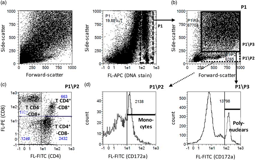
Ash, dry matter, gross energy, CP, lipids and lactose were assayed as previously described by Loisel et al. (Reference Loisel, Farmer, Ramaekers and Quesnel2013), and Na+ and K+ concentrations using an atomic absorption spectrometer (SpectrAA 220FS, Varian, Grenoble, France). IgA and IgG were assayed in triplicate with the same kits as for plasma. To determine cellular composition, pellets obtained from colostrum or milk samples (10 ml) were washed in Hank’s Balanced Salt Solution (20 min at 600×g), and re-suspended in 1 ml for cell numeration using an automatic cell counter (Vi-Cell XR; Beckman Coulter, Paris, France). No gradient centrifugation was performed, which allowed to estimate total epithelial, lymphoid and myeloid cell numbers in the suspension. Flow cytometry analysis using the DNA-selective Vybrant® DyeCycle™ Ruby stain (Thermo Fisher Scientific, Waltham, MA, USA) revealed that fat globules represented 70% to 99% of the counted events (Figure 1a). Preliminary developments showed that anti-CD3 and anti-CD21 antibodies did not bind to cells of the suspension. Moreover, in the usual mononuclear cell gate about 98% of the cells expressing CD4 or CD8 also co-expressed CD2 (porcine MAC83 clone; Bio-Rad, Oxford, UK) and could be then ascribed to the T-cell lineage. To identify T lymphocytes, a double CD4/CD8 staining was thus performed in this study (anti-porcine CD4 74-12-4 and CD8 76-2-11 clones; Clinisciences, Nanterre, France; Figures 1b and 1c). Another staining was realized for monocytes and macrophages/granulocytes using an anti-porcine CD172a antibody (74-22-15 clone; Santa Cruz Biotechnology, Dallas, TX, USA; Figure 1d). Incubations with antibodies and their corresponding isotypic controls (Clinisciences, Nanterre, France) were performed in a final 100 µl volume, after a pre-incubation with Fc-R blocking reagent (Miltenyi Biotec, Bergisch Gladbach, Germany). Cells were washed and suspended in 0.5 ml phosphate buffered saline with 1 µl of Dye Cycle Ruby Stain. After 20 min incubation, total events were acquired using the MACSQuant flow cytometer (Miltenyi Biotec) from 100 µl per cell suspension, allowing calculating backward the absolute number of cells contained per millilitre of colostrum or milk.
Strategy of cell phenotyping from lacteal secretion by flow cytometry. (a) Nucleated (DNA-positive) events were first sorted out according to vibrant dye ruby stain (APC fluorescence, P1 gate). (b) Low (P1\P2) and high (P1\P3) side-scatter gates were designed among nucleated events. (c) A double CD4 (fluorescein isothiocyanate labelled, FL-FITC) and CD8 (phycoerythrin labelled, FL-PE) staining gated on P1\P2 allowed discrimination between naive CD4+CD8− and memory CD4+CD8+ T helper lymphocytes, CD4−CD8+ cytotoxic lymphocytes (including T CD8 and T gamma–delta cells), and other CD4−CD8− mononuclear cells. (d) A simple staining for CD172a (FITC-labelled) allowed monocyte and polynuclear cell identification as CD172a+ cells in P1\P2 and P1\P3 gates, respectively.

Statistical analyses
Data, except litter size and mortality rate, were analysed by ANOVA using the MIXED procedure of SAS (SAS Institute Inc., Cary, NC, USA), with the sow as experimental unit. All models included at least the effect of the system (C or E) and the replicate (1, 2 or 3). For traits measured several times, analyses were done separately for each time-point. Cortisol concentrations and total cell numbers in mammary secretions were transformed by square root and Natural logarithm, respectively, before analysis. For body condition and reproductive performance, the model also included the effect of parity (primiparous or multiparous) and the system × parity interaction. The GENMOD procedure was used with a Poisson distribution and a Log link function for numbers of piglets and with a binomial distribution and a Logit link function for mortality rates, in models that included the effects of system, parity, replicate and the interactions. For piglet IgG concentrations, the experimental unit was the piglet and the model included the random effect of sow nested within system × replicate. Data are expressed as least-square means and SEM when analysed using the MIXED procedure, and as raw data when analysed using the GENMOD procedure. Differences between systems were considered significant if P < 0.05.
Results
Body condition and reproductive performance
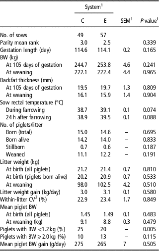
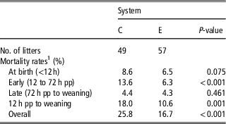
Average parity of sows and the proportions of primiparous and multiparous sows (26% and 73%, respectively) were similar in the two gestation systems (Table 1). Primiparous sows were lighter (219 v. 280 kg at DG105, P < 0.001), had less stillborn (0.4 v. 0.7, P = 0.04) and lighter litter weight (20 v. 23 kg at birth for total born piglets, P = 0.005, and 96 v. 105 kg at weaning, P < 0.001), but the system × parity interaction was never significant (P > 0.10). Sows from the two systems had similar BW, backfat thickness on DG105 and at weaning (Table 1). Gestation length averaged 114.3 ± 0.2 days. The numbers of born alive, stillborn and total born piglets were also similar in both systems (P > 0.10). However, total pre-weaning mortality was lower in E than in C litters (P < 0.001, Table 2), which was due to a trend for a decreased mortality at birth (P = 0.07) and a substantially lower mortality between 12 and 72 h postpartum (P = 0.001). By contrast, mortality from 72 h up to weaning and the distribution of dead piglets among the four classes of postnatal mortality causes were not influenced by the system (Supplementary Figure S1, P > 0.10). Litter and piglet weights at birth and daily weight gains during lactation did not differ between systems (Table 1). However, the frequency of piglets with a birth weight under 1.2 kg was greater in C compared to E system (P < 0.01).
Body condition and reproductive performance of sows that were housed during gestation in the conventional (C) v. the enriched (E) system

1 Data are expressed as least-squares means and the greatest SEM, except for numbers of piglets per litter that are expressed as raw data.
2 CV = coefficient of variation of piglet birth weight.
Rates of piglet mortality from birth to weaning when gestating sows were housed in the conventional (C) v. the enriched (E) system

1 Mortality ‘at birth’ included stillborn piglets and piglets that died within 12 h, relatively to total born piglets. Early, late and 12 h pp to weaning mortality included piglets that died during the respective periods relatively to born alive piglets. Overall mortality included stillborn piglets and piglets born alive that died before weaning, relatively to total born piglets.
Sow physiology
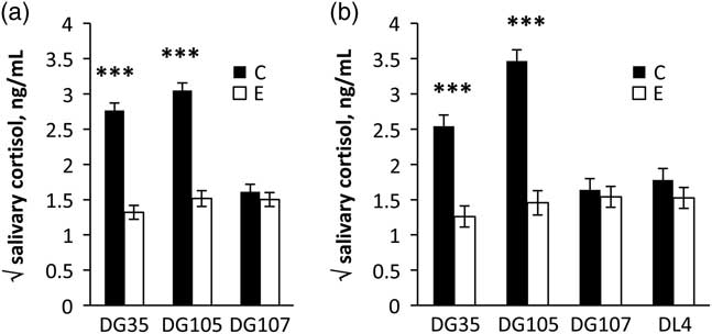
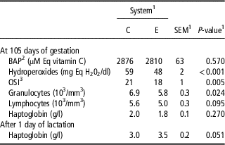
On DG35 and DG105, E sows had lower salivary cortisol concentrations than C sows (P < 0.001, Figure 2). After the transfer into farrowing rooms (DG107) and during early lactation (DL4), cortisol concentrations of C sows dropped to concentrations comparable to E sows. On DG105, IgG (45 ± 7 mg/ml) and IgA (0.89 ± 0.09 mg/ml) in plasma as well as IgA in saliva (0.18 ± 0.02 mg/ml) had similar concentrations in sows of the two systems (P > 0.10). Total white blood cells on DG105 were 11% less in E than C sows (P < 0.01), due to a decrease in granulocyte number (P < 0.001, Table 3). Plasma concentration of haptoglobin was similar in all sows on DG105 (P > 0.10) and DL1, although it was numerically greater in E compared with C sows on DL1 (P = 0.051). On DG105, plasma hydroperoxides and OSI were lower in E sows than in C sows (P < 0.01, Table 3). However, there was no difference in BAP antioxidant capacity (P > 0.10).
Salivary concentrations of cortisol in sows reared during gestation in the conventional (C) and enriched (E) systems. Saliva was collected at 0900 h on days of gestation (DG) 35, 105 and 107 from 106 sows (a). For the subset of 37 sows sampled for colostrum and milk after farrowing, additional saliva was also collected on day 4 of lactation (DL4; b). Data are expressed as least-squares means ± SEM of the square-root transformed values.

Blood markers of inflammation, oxidative stress and immune activation in sows that were reared during gestation in the conventional (C) v. the enriched (E) system

1 Data are expressed as least-squares means and the greatest SEM. The number of animals was C: n = 49 and E: n = 56 to 57, excepted for haptoglobin were n = 18 and 19, respectively.
2 BAP = Biological Antioxidant Power.
3 OSI = Oxidative stress index as the ratio between hydroperoxides (mg Eq H2O2/dl) and BAP (µM Eq vitamin C) × 1000.
Colostrum and milk composition
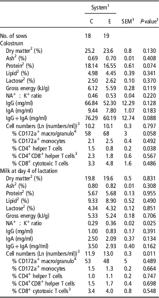
Parity and litter characteristics of the subset of sows used for colostrum and milk analyses were comparable to those of the whole set of animals (see part II, Quesnel et al., Reference Quesnel, Père, Louveau, Lefaucheur, Perruchot, Prunier, Pastorelli, Meunier-Salaün, Gardan-Salomon, Gondret and Merlot2018). The gestation system did not influence IgG (59 ± 5 mg/ml) and IgA concentrations in colostrum (8.6 ± 0.6 mg/ml), IgA concentration in milk (2.3 ± 0.2 mg/ml), and IgG concentrations in plasma of piglets on DL4 (19 ± 1 mg/ml, P > 0.10). The system did not alter the nutritional composition of colostrum and milk. Nevertheless, protein content was numerically lower in colostrum from E sows (P = 0.07, Table 4). Moreover, the Na+ : K+ ratio was greater in milk from E sows on DL4 (P = 0.025).
Colostrum and milk composition in sows that were reared during gestation in the conventional (C) v. the enriched (E) system

1 Data are expressed as least-squares means and the greatest SEM.
2 Grams per 100 g of whole colostrum or milk.
3 The statistical analysis was performed on the Natural logarithm of total cell numbers/ml blood.
4 CD172a+ macrophages and granulocytes.
The absolute cell numbers estimated with the counter were 580 ± 80 and 3400 ± 500 thousand/ml of colostrum and milk, respectively. Estimations were much lower when calculated using flow cytometry, after excluding events that did not bind the DNA-selective stain (Table 4). Using this latter analysis, an increase in cell numbers was observed between farrowing and DL4 in both groups of sows (P < 0.001). The numbers were comparable between groups in colostrum, whereas milk of E sows contained about three-fold more cells than that of C sows (452 v. 148 thousand cells/ml of milk, respectively, P <0.05; Table 4). Per cent of CD172+ monocytes, CD8+CD4+ memory and CD8+CD4− cytotoxic T lymphocytes did not differ significantly between the two groups (P > 0.05). The per cent of CD172+ macrophages and granulocytes was numerically but not statistically greater in colostrum of E sows (P = 0.06), and the percentage of CD8−CD4+ helper lymphocytes was lower (P < 0.05) than in C group.
Discussion
In the present experiment, the two housing systems for gestating sows led to contrasted piglet pre-weaning survival rates, with a lower mortality rate observed in the E system which included deep straw bedding, than in the C system which was based on a concrete floor. This finding was in agreement with previous results reported on the same systems (Merlot et al., Reference Merlot, Calvar and Prunier2017). The total mortality rates (25.8% in the C system v. 16.7% in the E system) were thus greater and smaller, respectively, than the average mortality rates in French herds (20.0 ± 4.2%, IFIP, 2015), which gives an interesting model to better understand the influence of the maternal gestation environment on the neonatal survival. The lower mortality in E litters was mainly due to a reduced mortality during the first 3 days after birth. Importantly, there were no differences in the main causes of piglet mortality between systems, and the herd was free from any specific swine disease.
Effects of the environment during gestation on sow cortisol
Present data showed that, at DG35 and DG105, sows from the C system displayed a greater salivary cortisol concentration than E sows, as previously observed (Merlot et al., Reference Merlot, Calvar and Prunier2017). The higher cortisol level in the C environment could be explained by higher social stress and frustration due to an inability to perform rooting behaviour and to satisfy hunger. Indeed, less skin injuries have been observed in E than in C sows in the same herd (Paboeuf et al., Reference Paboeuf, Gautier, Cariolet, Meunier-Salaün and Dourmad2010), suggesting less frequent and/or less severe aggressive interactions in relation with more space allowance and the presence of straw (Remience et al., Reference Remience, Wavreille, Canart, Meunier-Salaun, Prunier, Bartiaux-Thill, Nicks and Vandenheede2008). Moreover, straw is a good foraging material and can also be ingested to alleviate hunger (Spoolder et al., Reference Spoolder, Burbidge, Edwards, Simmins and Lawrence1996).
After transfer into the farrowing crate, salivary cortisol decreased in C sows to a level similar to that of E sows. This suggests that a strong stressor, likely social stress, was removed in C sows, whereas the level of stress did not change in E sows. The stability in salivary cortisol concentrations between DG105 and DG107 in E sows, despite their restraining in isolated farrowing crates with no straw, may be surprising. It may be related to the fact that in natural conditions sows isolate themselves from the group a few days before the onset of parturition to seek for a suitable nest site (Algers and Uvnäs-Moberg, Reference Algers and Uvnäs-Moberg2007).
Although the mechanisms are still poorly understood, it seems that maternal stress during gestation can increase neonatal mortality (Tuchscherer et al., Reference Tuchscherer, Kanitz, Otten and Tuchscherer2002), even if this is not always observed (Merlot et al., Reference Merlot, Quesnel and Prunier2013). Prenatal stress could directly influence foetal piglet development through maternal glucocorticoids (Kranendonk et al., Reference Kranendonk, Hopster, Fillerup, Ekkel, Mulder, Wiegant and Taverne2006). Since the morphology of the placenta has been shown to be altered in stressed ewes (Baxter et al., Reference Baxter, Mulligan, Hall, Donbavand, Palme, Aldujaili, Zanella and Dwyer2016), it is also possible that prenatal stress affects placental trophic function. In rodents and humans, stress also enhances inflammatory markers at placental level (Bronson and Bale, Reference Bronson and Bale2014; Miller et al., Reference Miller, Borders, Crockett, Ross, Qadir, Keenan-Devlin, Leigh, Ham, Ma, Arevalo, Ernst and Cole2017), which are associated with adverse foetal outcomes.
Effects of the environment during gestation on sow plasma immune markers
Sows in the C system presented an increased OSI when compared with E sows at DG105. During pregnancy, it is generally admitted that oxidative stress is elevated because of placental production of radical-oxygen species (Berchieri-Ronchi et al., Reference Berchieri-Ronchi, Kim, Zhao, Correa, Yeum and Ferreira2011). However, an excessive oxidative stress could affect placental remodelling and function (Marseglia et al., Reference Marseglia, D’Angelo, Manti, Arrigo, Barberi, Reiter and Gitto2014). A factor that might have increased the release of oxidative stress products in the C sows is immune activation (Lykkesfeldt and Svendsen, Reference Lykkesfeldt and Svendsen2007). Indeed, deteriorated housing conditions, amplifying health disorders and stress, can increase oxidative stress as recently showed in piglets at weaning (Buchet et al., Reference Buchet, Belloc, Leblanc-Maridor and Merlot2017). In the present study, no signs of diseases were reported and the serum concentrations of haptoglobin, an indicator of inflammation, and of IgG and IgA, indicators of antigenic immune stimulation, were not different between C and E sows. However, C sows exhibited a greater number of circulating granulocytes, as previously observed (Merlot et al., Reference Merlot, Calvar and Prunier2017). This can be indicative of microbial infections (Baarsch et al., Reference Baarsch, Foss and Murtaugh2000; Segales et al., Reference Segales, Alonso, Rosell, Pastor, Chianini, Campos, Lopez-Fuertes, Quintana, Rodriguez-Arrioja, Calsamiglia, Pujols, Dominguez and Domingo2001), but is also observed in pigs kept in a dirty environment without specific pathogens (Chatelet et al., Reference Chatelet, Gondret, Merlot, Gilbert, Friggens and Le Floc’h2018). Lastly, stress-induced glucocorticoid and catecholamine are also well known to modulate blood leukocyte distribution. The greater number of circulating granulocytes could then also result from a higher stress, as reported in growing pigs housed in a poor slatted-floor environment compared to animals housed in an enriched environment (Merlot et al., Reference Merlot, Vincent, Thomas, Meunier-Salaun, Damon, Robert, Dourmad, Lebret and Prunier2012) and in other situations of long-lasting exposure to stressful situations in pigs (Hjarvard et al., Reference Hjarvard, Larsen, Juul-Madsen, Jorgensen and Jensen2009; Salak-Johnson et al., Reference Salak-Johnson, DeDecker, Horsman and Rodriguez-Zas2012).
Greater oxidative stress and granulocyte count could be therefore interpreted as signals of increased immune activation, due to stress or other environmental factors. Low grade immune activation could affect the developing foetuses and their postnatal survival. Indeed, maternal systemic inflammation can generate placental and sterile intra-amniotic inflammation (Nadeau-Vallee et al., Reference Nadeau-Vallee, Chin, Belarbi, Brien, Pundir, Berryer, Beaudry-Richard, Madaan, Sharkey, Lupien-Meilleur, Hou, Quiniou, Beaulac, Boufaied, Boudreault, Carbonaro, Doan, Joyal, Lubell, Olson, Robertson, Girard and Chemtob2017). In humans and rodents, intra-amniotic inflammation was associated with preterm birth and, in full-term gestations, with morphological abnormalities, indicators of brain, heart and immune immaturity, and increased neonatal mortality (Romero et al., Reference Romero, Miranda, Chaiworapongsa, Chaemsaithong, Gotsch, Dong, Ahmed, Yoon, Hassan, Kim, Korzeniewski, Yeo and Kim2015; Miller et al., Reference Miller, Borders, Crockett, Ross, Qadir, Keenan-Devlin, Leigh, Ham, Ma, Arevalo, Ernst and Cole2017; Nadeau-Vallee et al., Reference Nadeau-Vallee, Chin, Belarbi, Brien, Pundir, Berryer, Beaudry-Richard, Madaan, Sharkey, Lupien-Meilleur, Hou, Quiniou, Beaulac, Boufaied, Boudreault, Carbonaro, Doan, Joyal, Lubell, Olson, Robertson, Girard and Chemtob2017).
Effects of the environment during gestation on sow lacteal secretions
As mortality rates were especially affected within the first 3 days after birth, the biochemical and immunological composition of colostrum and milk were of major interest to better understand the mechanisms whereby gestation housing system had influenced piglet survival. The nutritional composition of colostrum and milk was not affected, except a higher protein content of colostrum in C than in E sows, which was opposite to what could be expected to explain the greater mortality rate in C piglets.
Regarding the transfer of immune protection, there was no effect on IgG concentration in colostrum and, consequently, on IgG concentration in piglet blood. Milk also contains maternal Ig which can neutralize feed antigens and microbes in the lumen of the digestive tract of the piglet (Salmon et al., Reference Salmon, Berri, Gerdts and Meurens2009). Our data did not show significant effects of the housing environment on these components. Lacteal secretions also contain macrophages, granulocytes, lymphocytes and mammary epithelial cells (Schollenberger et al., Reference Schollenberger, Degorski and Frymus1986). As reported by others, we found that the total number of immune cells was greater in milk than in colostrum (Schollenberger et al., Reference Schollenberger, Degorski and Frymus1986; Hlavova et al., Reference Hlavova, Stepanova and Faldyna2014). Macrophages and neutrophils cells represented the majority of leukocytes in lacteal secretions, and among T lymphocytes, T CD8+ were the most abundant, followed by memory T CD4+CD8+ helper lymphocytes and finally naive T CD4+. Colostral cells ingested by the neonate can cross the gut barrier, recirculate in piglet lymphoid organs (Salmon et al., Reference Salmon, Berri, Gerdts and Meurens2009, for review) and thus protect the neonate against specific pathogens (Oh et al., Reference Oh, Seo, Han, Park and Chae2012). In our study, maternal environment during gestation had little effect on the cellular composition of colostrum. This indicates that the passive immunity transferred via colostrum was almost comparable in both groups of sows and the subtle changes in the proportions of T helper lymphocytes and granulocytes is unlikely to be involved in the observed difference in piglet mortality between C and E sows.
The role of immune cells in the milk has been poorly explored. There is some debate about the ability of milk cells to cross the neonatal intestinal epithelium but these cells might contribute to the clearance of milk and gut lumen from microbes. In vitro, milk cells prevent infection of intestinal epithelial cells with Salmonella tiphimurium (Scharek-Tedin et al., Reference Scharek-Tedin, Kreuzer, Twardziok, Siepert, Klopfleisch, Tedin, Zentek and Pieper2015). The total number of cells was increased in milk of E sows compared with C sows. This cannot be attributed to differences in dilution of milk between E and C sows, since the nutrient concentrations were similar in both groups. The relative proportions of immune sub-populations were however similar in E and C sows. This indicates that all cells, including the different immune lineages and epithelial cells, were more numerous in milk of E sows. Moreover, the greater Na+ : K+ ratio in milk of E sows suggests a greater opening of tight junction of the mammary epithelium, which could favour epithelial cell exfoliation and permeability to blood components, including immune cells (Herve et al., Reference Herve, Quesnel, Lollivier and Boutinaud2016). In ruminants, numerous factors were found to be able to modulate mammary epithelium integrity, including endocrine status, immune stimuli and management practices (Herve et al., Reference Herve, Quesnel, Lollivier and Boutinaud2016).
It cannot be excluded that the greater number of cells in E sows was an indicator of subclinical mastitis (Persson, Reference Persson1997), which would be in agreement with the tendency for a greater haptoglobin concentration at DL1 and rectal temperatures at farrowing and DL1 in these sows. However, in any case, subclinical mastitis in the mothers could certainly not explain the lower piglet mortality in this group. Moreover, the observed variations in milk cell numbers are unlikely involved in the differences in neonatal mortality between groups since they were observed at L4, whereas differences in neonatal mortality were settled during the 48 1st h postpartum.
In conclusion, the enriched environment during sow gestation reduced maternal cortisol secretion and immune stimulation during gestation, did not affect colostrum and milk nutritional characteristics but influenced cellular content in milk. Whether the better survival of E piglets was causally related to the concomitant changes in maternal physiology during gestation remains to be established.
Acknowledgements
The authors thank C. Calvar and B. Le Bris (CRAB, France), F. Robert, A. Hamard and T. Pinard (Deltavit CCPA group) for their involvement, the staff of Crécom (B. Le Guilcher, P. Lirzin, D. Lesaicherre, M. Gautier) for animal care and data collection, and the staff of UMR1348 PEGASE (N. Bonhomme, R. Comte, S. Daré, S. Philau, C. Guérin, S. Jaguelin, M. Lefèbvre, F. Mayeur, S. Tacher, F. Thomas, and C. Tréfeu) for sample collection and biological analyses. Research has received funding from the EU FP7 Prohealth project (no. 613574).
Declaration of interest
The authors declare that they have no competing interests.
Ethics statement
Animals were cared for according to the French regulations for the human care and use of animals in research. The experimental protocol (no. 02805-02) was approved by the local Ethics Committee in Animal Experiment of Rennes, France and by the French Ministry of Higher Education and Research.
Software and data repository resources
None of the data were deposited in an official repository.
Supplementary material
To view supplementary material for this article, please visit https://doi.org/10.1017/S1751731118002987