Introduction
Passive smoking is the inhalation of smoke, termed second-hand smoke or environmental tobacco smoke, produced from tobacco products used by others. A complex mixture of chemicals is generated from the burning and smoking of tobacco. The passive smoker breathes ‘side-stream’ smoke from the burning tip of the cigarette, together with ‘mainstream’ smoke that has been inhaled then exhaled by the smoker.
Second-hand smoke causes the same negative health effects as active tobacco smoking, particularly heart disease and cancer. Non-smokers who are exposed to second-hand smoke at home or work increase their heart disease risk by 25–30 per cent and their lung cancer risk by 20–30 per cent. Second-hand smoke has been estimated to cause 38 000 deaths per year, of which 3400 are deaths from lung cancer in non-smokers.Reference Pell, Haw and Cobbe1
Most studies concerning the negative effects of smoking on the respiratory tract have addressed its carcinogenic effect on the lungs and bronchi. Little information is available concerning the effect of smoked tobacco on the histopathology of the nasal mucosa, although the nose is one of the most exposed organs.Reference Dessi, Sambuc, Movlin and Iedoray2 Publications on passive smoking are even scarcer.Reference Gedikondele, Longo-Mbenza, Nzanza, Luila, Reddy and Buso3, Reference Stanković, Nikolić and Arandelović4 Vinke et al. Reference Vinke, Denjan, Severijnen and Fottens5 reported that passive smoking might cause allergic cellular infiltrates within the nasal mucosa. Nageris et al. Reference Nageris, Hadar and Hansen6 reported the deleterious effect of smoking on olfaction in children. A thorough search of the literature did not disclose any other studies addressing histopathological changes in the nasal mucosa of subjects exposed to passive smoke.
The aim of the present study was therefore to investigate the histopathological changes in the nasal mucosa of an adult population exposed to passive smoking.
Material and methods
The study included 20 women aged between 35 and 51 years, who were scheduled for non-rhinological surgical procedures and who had at least 10 years' history of prolonged passive exposure to household cigarette smoke. The duration of passive smoke exposure was recorded. Exposure to passive smoking was determined using a questionnaire on household smoking. The study was performed at Alexandria Medical School between April 2009 and February 2012.
Participants' urinary cotinine/creatinine ratio was measured, constituting an objective, validated measure of smoke exposure.
Another 10 women with a negative household smoking history were included as a control group.
All participants were non-atopic and had no family history of allergy. None had infective rhinosinusitis.
The subjects and controls were consecutive new clinic attendees who were willing to participate in the research. Informed consent was obtained from all participants for inclusion in the research. The relevant ethics committee approved the study. The study was performed in accordance with the rules of the Helsinki Declaration.
At the time of surgery, two 1-mm3 biopsies were taken from the lower border of the inferior turbinate, 1.5 cm from its anterior end, with punch forceps. Specimens were prepared for both light and transmission electron microscopy. Two pathologists, who were blinded to the smoking status of the subjects, examined the specimens.
Specimen preparation for light microscopy
The specimens were fixed in formalin and embedded in paraffin. Sections were stained with conventional haematoxylin and eosin stain and Alcian blue stain (designed to show the mucopolysaccharides or glycosaminoglycans of mucous acini and goblet cells).
Histopathological changes were considered to be present if they existed in more than 50 per cent of the examined sections. An observation was considered mild if present in 50–60 per cent of examined sections, moderate if present in 60–80 per cent of sections, and severe if present in more than 80 per cent of sections. Changes were graded as 0 if absent, 1 if mild, 2 if moderate and 3 if severe.
Specimen preparation for transmission electron microscopy
The specimens were immediately fixed in Karnovesky's fixative at 4°C for 4 hours. They were then post-fixed in 1 per cent osmium tetroxide and dehydrated in graded alcohol solutions. Specimens were then impregnated with araldite (CY212) using propylene oxide as the intermediate solvent. The specimens were finally polymerised at 60°C in embedding capsules. Ultrathin sections were cut with an ultramicrotome, stained with 4 per cent uranyl acetate and Reynold's lead acetate, and examined under a Hitachi H-600 high-resolution transmission electron microscope (Hitachi, Tokyo, Japan).
Statistical analysis
Statistical analysis was performed using SPSS for Windows version 15 software (SPSS Inc, Chicago, Illinois, USA). Mean inter-group histological scores were compared using Student's t-test. The relationship between duration of passive smoke exposure and scores for histopathological changes was analysed using Pearson's correlation coefficient.
Results
The present study included 20 women, aged between 35 and 51 years, who had a positive history of exposure to passive household cigarette smoke for at least 10 years.
The urinary cotinine/creatinine ratio of these women ranged from 35 to 71 ng/mg (mean, 45 ng/mg); in the control group, the range was 7–16 ng/mg (mean, 9 ng/mg).
The nasal mucosa of subjects exposed to passive smoking showed several histopathological changes. The mean histopathological score was 1.35 (standard deviation (SD), 1.09) in subjects and 0.40 (SD, 0.50) in controls. The difference between the two groups was statistically significant (t = 3.5411, p = 0.0011).
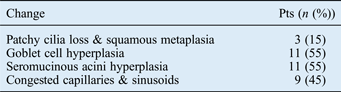
The frequency of histopathological changes is shown in Table I. These histopathological changes included hyperplasia of goblet cells (55 per cent), hyperplasia of seromucinous glands (55 per cent) and vascular congestion (45 per cent). Patchy loss of cilia and areas of squamous metaplasia were seen in three cases (15 per cent).
Histopathological changes in passive smoking patients

Pts=patients
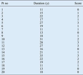
The histopathological changes tended to be more severe with increased duration of exposure to cigarette smoke (Table II). A positive correlation existed between subjects' histopathological scores and their duration of passive smoking exposure (r = 0.828596).
Patients' passive smoking duration and histopathological scores
Pt no = patient number; y = years
A detailed description of the histopathological changes observed is given below, together with figures showing representative examples of each type of change.
The nasal mucosa showed hyperplasia of the goblet cells and seromucinous acini (Figure 1a). Both the goblet cells and the seromucinous acini cells appeared distended with secretory granules (Figure 1b and 1c), indicating increased secretory activity of the cells. Patchy areas showing loss of cilia and squamous metaplasia were seen in three cases (Figure 2).
(a) Photomicrograph showing hyperplasia of nasal mucosa goblet cells and glandular acini (stained blue) (Alcian blue stain; ×200). (b) Transmission electron micrograph showing hyperplastic goblet cells distended with secretory granules (G) (×2500). (c) Transmission electron micrograph showing glandular acini distended with mucigen granules (M) (×1800).
Photomicrographs showing squamous metaplasia of the nasal mucosa surface epithelium. The basement membrane is intact. The lamina propria shows loose connective tissue with no subepithelial oedema. (H&E; ×200)
The lamina propria showed fine, irregularly arranged collagenous fibres, but there were no areas of sub-epithelial oedema. No eosinophils were seen in the lamina propria. A few intact plasma cells and mast cells were occasionally seen; these showed no evidence of involvement in any antigen–antibody reactions.
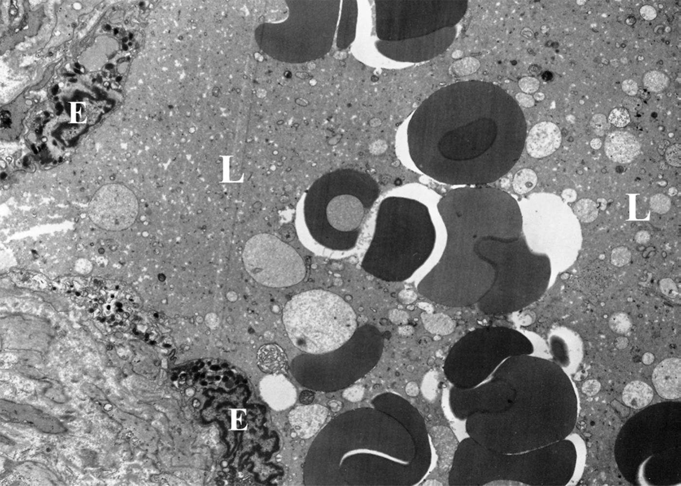
The capillaries and venous sinusoids appeared congested and dilated (Figure 3). There was no pericapillary oedema, and the inter-endothelial junctions were intact and not widely opened. The endothelial cells showed a few small pinocytotic vesicles.
Transmission electron micrograph showing venous sinusoid lined with endothelial cells (E). The lumen of the sinusoid (L) appears distended with blood. (×4500)
Discussion
Cigarette smoke is a mixture of pharmacologically active, toxic, mutagenic and carcinogenic substances.Reference Usenius, Karja and Collan7 Second-hand smoke includes the smoke from the burning end of a cigarette, pipe or cigar as well as the smoke exhaled from the lungs of smokers. It lingers in the air hours after the cigarette, pipe or cigar has been extinguished, and is involuntarily inhaled. Second-hand smoke may cause many of the diseases induced by direct smoking.Reference Samet8
As the nose lies at the entrance of the respiratory tract, it would be expected that pathological changes in the nasal mucosa may occur secondary to exposure to active or passive cigarette smoke. However, a comprehensive literature review identified only a few studies addressing this issue, despite its obvious importance.Reference Atef, Abo Zeid, Qotb and Gad El Rab9 The present study may be the first histopathological report assessing the effect of passive smoking on the respiratory nasal mucosa of adults.
In the present study, several changes were observed in the respiratory nasal mucosa of adults regularly exposed to passive cigarette smoke. Congestion of the mucosa and hyperplasia of the mucus-producing elements in the nasal mucosa were the commonest observations in our series. We used a simple, four-point grading system modified from that proposed by Torjussen et al.,Reference Torjussen, Solberg and Hogeveit10 in order to quantify the observed epithelial changes and to correlate these changes with the duration of passive smoking. The grading scheme was also necessary to detect inter-observer variation; cases for which there was disagreement between the two examining pathologists were excluded from the study.
Subjects' respiratory nasal mucosa changes appeared to be directly proportionate to their duration of passive smoke exposure. As the duration of exposure increased, the ciliary loss tended to become more widespread, and areas of squamous metaplasia and total cilia loss started to appear. This observed deleterious effect of passive smoking on nasal cilia confirms the findings of Agius et al.,Reference Agius, Smallman and Pahor11 who reported that smoking decreased ciliary beat frequency. Our observations also confirm those of Atef et al.,Reference Atef, Abo Zeid, Qotb and Gad El Rab9 who found that passive smoking had a negative impact on cilia regeneration following functional endoscopic sinus surgery.
Hyperplasia of goblet cells and seromucinous acini was a notable feature in 45 per cent of cases. Rogers and JefferyReference Rogers and Jeffery12 reported similar secretory cell hyperplasia in the bronchial mucosa. This hyperplasia of mucin-producing cells is obviously a protective mechanism aiming to wash out noxious particles and protect the delicate nasal mucosa. However, although tobacco smoke may increase mucus production of the respiratory mucosa, it may also change its physical properties, as reported by Jeffrey,Reference Jeffery13 who found that smoking altered the glycoprotein content of bronchial mucus and degraded its viscoelastic properties.
Vinke et al. Reference Vinke, Denjan, Severijnen and Fottens5 proposed that passive smoking may cause an allergic cellular infiltrate in the nasal mucosa of non-atopic children. Our findings, however, did not support this theory, for several reasons. First, we observed only a few plasma cells, eosinophils and mast cells within the affected mucosa. These are the cells typically involved in allergic reactions, and their scarcity indicates an absence of immunological stimulation. Second, the mast cells we observed were intact and fully granulated, unlike the degranulated, histamine-releasing mast cells seen in allergic cases.Reference Toppozada and Talaat14 Third, the gaps between the capillary endothelial cells were intact and narrow, with few pinocytotic vesicles. These findings are contrary to those reported in patients with allergic rhinitis,Reference Toppozada and Talaat15 and also explain the absence of the pericapillary and sub-epithelial oedema commonly described in allergic cases.Reference Elwany and Bumsted16
• Passive smoking may damage nasal respiratory mucosa
• Changes include patchy cilia loss, goblet cell and glandular acini hyperplasia, vascular congestion, and squamous metaplasia
• Structural change severity increases with passive smoking duration
• Other changes are also possible
The present study findings indicate that passive cigarette smoking may disturb the nasal defence system by impairing mucociliary clearance and altering mucosal secretory components. Similar changes have been reported in the lung and bronchi.Reference Dye and Adler17 As a result of these effects, activeReference Benninger18 and passive smokers may suffer from a higher frequency of sinonasal infections. On the other hand, the present study did not find any evidence of ongoing immunoglobulin E mediated reactions, indicating that the observed changes are likely to be due to the direct, damaging effect of injurious components of passive cigarette smoke.
The present study found no evidence of any neoplastic transformation.
Conclusion
Adults exposed to passive cigarette smoke may develop several histopathological changes in their respiratory nasal mucosa, with subsequent negative effects on its ciliary activity and mucociliary function. As a result of these effects, defence mechanisms of the nose may be disturbed or lost, with subsequent development of persistent sinonasal infections.
It is expected that exposure to passive smoking for longer periods of time may also induce other significant changes that were not detected in the present study.