Introduction
Migrated ingested foreign bodies in the aerodigestive tract can lodge within vital organs and vessels, causing potentially devastating complications.Reference Remsen, Biller, Lawson and Som1 It is often difficult to localise these foreign bodies, with extrication resulting in the requirement for open approaches which may cause significant morbidity.Reference Al-Sebeih, Abu-Shara and Sobeih2
We present the case of an ingested migrated stingray bone lodged adjacent to the vertebral artery in the upper cervical spine. This was managed via an endoscopic transoral approach, with the assistance of an image-guidance system.
Case report
Our patient was a 56-year-old man, who presented to the emergency department with odynophagia. He described having eaten grilled stingray (Dasyatidae sp.) during a meal around 3 hours prior to presentation and noted odynophagia with some haemoptysis afterwards. No neck pain, shortness of breath or fever was reported.
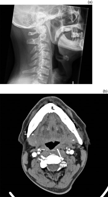
The physical examination findings were otherwise unremarkable, with no foreign body noted on nasoendoscopy. A lateral neck X-ray was conducted; this showed a linear opacity anterior to the C2 vertebra, which was suspicious for a migrated foreign body (Figure 1a). Computed tomography (CT) of the neck was performed; the scan showed a 2.3 cm linear foreign body which had migrated through the oropharyngeal mucosa and penetrated through the left C2 transverse foramina, embedding medial and adjacent to the left vertebral artery (Figure 1b). Of note, the foreign body had already migrated 1 cm deep to the overlying pharyngeal mucosa. No contrast extravasation or collection was otherwise observed on CT.
(a) Lateral neck X-ray of the migrated foreign body anterior to the C2 vertebra. (b) Axial computed tomography scan of the migrated foreign body medial to the left vertebral artery at C2.

Intravenous antibiotics were started, and the decision was made to bring the patient urgently to the operating theatre for examination under general anaesthesia and removal of the migrated foreign body. As part of surgical planning and as an adjunct to help localise the foreign body, a CT scan of the paranasal sinuses was performed, but with a specific request to extend the imaging down to the level of the C3 vertebra, just beyond where the foreign body was located. This scan was then formatted according to the image-guidance system protocol for use in endoscopic sinus surgery.
Intra-operatively, transoral inspection of the pharynx anterior to C2 was performed using a 4 mm, 0 degree rigid endoscope. However, no obvious foreign body or ulcer was visualised or palpable. We used an electromagnetic image-guidance system (Medtronic Fusion™ ENT Navigation System) for intra-operative localisation. Registration was performed using the traditional registration points for endoscopic sinus surgery around the face, and then registration accuracy down to the oral cavity was confirmed using bony landmarks.
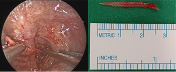
We used the straight image-guidance system probe to help localise the origin of foreign body penetration through the pharynx. Dissection through the same origin with monopolar diathermy was performed, with the subsequent encounter of a 2.3 cm serrated bone which had penetrated through the prevertebral space and into the spine. The foreign body was further exposed and removed using a pair of artery forceps, with no subsequent significant bleeding or haematoma observed (Figure 2). As there was some purulence encountered around the foreign body during dissection, the decision was made to leave the wound open, to heal by secondary intention, instead of primary closure.
• Migrated ingested foreign bodies in the aerodigestive tract can cause potentially devastating complications including death
• Very few studies have described migrated ingested foreign bodies in the high cervical spine
• This study is the first to describe a migrated ingested foreign body as high as the C2 vertebra
• Image-guidance systems can be effectively used to localise migrated foreign bodies at inaccessible areas such as the high cervical spine
• The transoral endoscopic approach is a minimally invasive, safe alternative to open approaches for removing high cervical foreign bodies adjacent to the vertebral artery
• Serrated foreign bodies should be urgently removed because of rapid migration secondary to unidirectional propulsion
Endoscopic extraction of the serrated foreign body from the left C2 transverse foramina.

The patient had an uneventful post-operative recovery. He was kept nil by mouth for 5 days, after which a water-soluble contrast swallow study was performed to confirm that there was no pharyngeal or oesophageal leak. He was then commenced on a soft diet, which he tolerated well, and was discharged the next day. During follow up in the out-patient clinic a week later, the patient was well, with resolution of odynophagia and haemoptysis. Examination showed a healing, superficial left pharyngeal ulcer anterior to C2. Ongoing follow up was planned with the necessary return advice given.
Discussion
Despite image-guidance systems being traditionally used for endoscopic sinus surgery, their ability to accurately navigate through bone and soft tissue highlights possible uses for other areas within the head and neck region. This is the first reported case of an ingested foreign body with migration to such a high level within the cervical spine, and at such close proximity to the vertebral artery. It shows that image-guidance system can be used effectively to localise migrated foreign bodies, even within this inaccessible region.
Other learning points were also evident during the management of this case. There are various reports of vertebral artery foreign bodies that necessitated open approaches for removal and which resulted in significant morbidity.Reference Ilan, Weinberger, Gomori and Gross3,Reference Macharla, Harpanahalli and Panigrahi4 One case report described vertebral artery disruption and subsequent mortality.Reference Huang, Zhang, Wu, Li, Han and Chao5 The high location of the foreign body in our case made it difficult to access even from an external open approach. Discussion with the on-call spine surgeons revealed that they would have had difficulty with an anterior, posterior or posterolateral approach to the area. From our experience, transoral endoscopic surgery is a minimally invasive and safe way to remove upper vertebral artery foreign bodies.
Serrated foreign bodies should be expediently removed, even more urgently than other linear foreign bodies. As evident from our case, the associated foreign body had already migrated under the pharyngeal mucosa in a short span of 3 hours. It is postulated that the serrated edges of the foreign body conferred a propulsive effect that caused it to migrate even faster than a purely linear foreign body, with the serrations allowing only unidirectional movement.
Conclusion
Our study shows that image-guided endoscopic surgery is a safe, precise and feasible option for the localisation and removal of migrated foreign bodies in the aerodigestive tract involving critical neurovascular structures. Understanding the principles behind image-guidance technology will help in knowing what it can be potentially used for, as well as its limitations.
Competing interests
None declared