Introduction
Tumours of bony origin generate a myriad of potential diagnoses including osteoma. The incidence of osteomas of the paranasal sinuses is 3 per cent of the total, with the frontal sinus being involved in 80–96 per cent of these.Reference Earwalker1, Reference Zouloumis, Lazaridis, Papadaki and Epivatianos2 In general, symptoms only occur following extension into surroundings structures, such as the orbit or cranium. Severe cases featuring disruption of the facial skeleton contour, although rare, have warranted open surgical removal.Reference Castelnuovo, Valentini, Giovannetti, Bignami, Cassoni and Iannetti3 The ideal material for reconstructing cranial defects is bone. However, for a large defect, titanium is a good substitute.
The rare case of a 31-year-old man with a giant frontal sinus osteoma is presented, including a discussion of the associated diagnostic and reconstructive challenges, surgical indications, and management of giant frontal sinus osteoma.
Case report
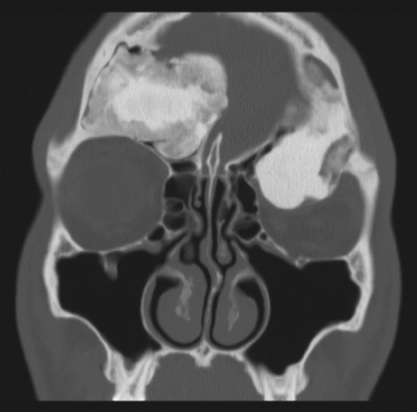
A 31-year-old man with a history of chronic frontal sinusitis presented to our university department with a painless, bony midline forehead swelling (7 cm × 7 cm) of 11 years' duration (Figure 1). In 2000, he underwent surgical intervention in Nigeria to remove a subperiosteal abscess following an episode of acute sinusitis. For this, a Lynch–Howarth incision had been made, frontal trephine and antral washouts were performed, and a percutaneous drainage tube had been inserted into the frontal sinus. The current presentation included severe inferolateral displacement of the left globe, diplopia and epiphora. Ocular movements were reduced, particularly the upward gaze. An ophthalmic review was sought: surprisingly, visual acuity, visual fields, intraocular pressure and pupil diameter were within normal limits. A computed tomography (CT) scan revealed extensive abnormality of the frontal bone, with disease projection beyond the roof, superomedial and posterior aspects of the left orbit, and multiple mucoceles in the frontal sinus component (Figure 2). The ‘ground glass’ appearance was pathognomonic of fibrous dysplasia alternating with hyperdense areas and suggestive of osteoma.
Photograph showing gross frontal deformity and left proptosis caused by massive frontal sinus osteoma.

Coronal computed tomography scan showing extensive bony change with concurrent ‘ground glass’ appearance and hyperdense areas of the frontal sinus and left orbit.
As a consequence of the extensive bony involvement, joint rhinological, oculoplastic and neurosurgical input was sought with a view to surgical management. Simultaneous en bloc resection of dysplastic bone and reconstruction using a custom-made orbital roof prosthesis and bifrontal titanium cranioplasty was planned. Both implants were computer generated and fabricated (Cavendish Implants, London, UK) prior to surgery, based on the anticipated bony defect (Figure 3).
Photographs of the titanium cranial plate (a) and orbital roof prosthesis (b).
Three doses of prophylactic cefuroxime were administered during the procedure. A bicoronal incision was made extending to the root of the zygoma bilaterally. The scalp flap was raised to the level of the frontonasal suture in the midline, exposing the supra-orbital rims. A separate pedicled pericranial flap was raised anteriorly, revealing abnormal bone in the left frontal region and left orbital roof. The titanium cranioplasty was used as a template to excise the diseased bone en bloc using a craniotome and burr. The dissection was extradural. The right supra-orbital rim was re-contoured and left in situ. The left orbital dysplasia was thinned prior to removal from the superior aspect of the supra-orbital nerve. The globe rapidly returned to a good position. Further abnormal bone was removed by burring from within the frontal sinus, frontonasal recess and anterior ethmoids. The skull base defect was repaired using the pericranial flap. Obex Neurofilm (Medtronic, Jacksonville, USA) was placed over small dural lacerations in the right frontal region.
The titanium orbital roof and cranial prostheses were fixed using titanium matrix screws (Figure 4), and a surgical drain was placed prior to soft tissue closure. Post-operative recovery was uneventful, with excellent early cosmetic result. The patient maintained normal visual acuity and normal forehead sensation.
Photograph showing the titanium cranial plate in situ.
Histological analysis of the resected specimen revealed dense cortical-type bone containing fibrous and osseous components (Figure 5). Margins were reported as clear and without evidence of malignancy. These findings were suggestive of fibrous dysplasia. However, following histological review by an expert pathologist at the University of Manchester, the diagnosis was revised to multiple frontal osteomas.
Photomicrograph showing irregular trabecular bone with active re-modelling. (H&E; ×10)
Diplopia is now limited to vertical eye movements and the patient is delighted with the surgical result. No signs of recurrence were seen at the 12-month follow up (Figure 6).
Photograph showing the post-operative surgical appearance of the patient after 12 months, with the left globe returning to a near normal position and full extirpation of the frontal bony defect.
Search strategy
To identify reports of similar cases, a search of OVID Medline was conducted in 2012. Search terms were ‘frontal sinus(es)’ or ‘frontal bone’ or ‘paranasal sinus(es)’ or ‘paranasal’ and ‘large’, ‘massive’, ‘huge’, ‘ultra-large’, or ‘osteoma(s)’ ‘fibrous dysplasia’ and titanium or cranioplasty. The search was restricted to articles published in English and reports in humans. This strategy retrieved 10 articles, of which 8 met the selection criteria for inclusion in the review.
Discussion
Numerous classification systems exist for bone tumours; which represent a clinically diverse group of disorders with similar histopathological features.Reference Brannon and Fowler4 Histopathological classification is being continually refined. Moreover, diagnosis of individual cases can often be challenging. A definitive diagnosis requires correlations of patient history, clinical findings, imaging and operative findings with histological analysis. Histologically similar findings are seen in all benign fibro-osseous lesions.Reference Brannon and Fowler4 Radiological analysis of frontal sinus tumours can be indiscriminate between osteoma, fibrous osteoma, fibrous dysplasia and ossifying fibroma. Therefore, the differential diagnosis of craniofacial bony lesions includes these pathologies.
Osteomas comprise dense focal sclerosis within bone and display unlimited growth potential. The overall incidence of osteoma is 0.14–0.43 per cent and of osteoma of the skull 0.4 per cent.Reference Patel and Borah5, Reference Childrey6 Classically, osteomas occur within the craniomaxillofacial skeleton, skull and paranasal sinuses.Reference Seiden and El Hefny7 The fronto-ethmoid region is involved in 80 per cent of cases, followed by the maxillary and sphenoid regions in decreasing frequency.Reference Onishi, Maruyama and Sawaizumi8 Giant disfiguring osteomas of the frontal sinuses are rare, as established by our literature review, but can have serious complications such as frontal lobe abscess.Reference Panagiotopoulos, Tzortzidis, Partheni, Iliadis and Fratzoglou9 Presentation is variable, but may include headache, proptosis, dizziness, facial deformity, diplopia and sinusitis, as in our patient. Serious complications, although rare, result from intracranial extension and include a mass effect on the brain and orbit. Intraparenchymal tension pneumatocele, pneumocephalus with pneumococcal meningitis, and cerebrospinal fluid (CSF) leakage have been reported.Reference Leiberman and Tovi10 Histological classification describes three basic types: ivory (or eburnated), cancellous (or spongious) and mixed (Table I).Reference Summers, Mascott, Tompkins and Richardson11 Ivory (or eburnated) osteomas comprise hard bone with thick matrix containing minimal fibrous tissue, cancellous osteomas comprise hard bone and mixed osteomas have characteristics of both. In the case of multiple osteomas, the possibility of Gardner's syndrome, an autosomal dominant disorder, should be considered.
Histological features of osteoma subtypes
Fibrous dysplasia is a rare benign tumour in which normal bone is replaced with connective tissue containing immature non-lamellar bone.Reference DiCaprio and Enneking12 The disease has a comparable propensity for occurrence within the craniofacial skeleton, which is involved in 10–25 per cent of cases.Reference Som, Brandwein, Som and Curtin13 Surgical indications are analogous to those for osteoma. In equivocal cases such as ours, a mutation-specific screening technique may be used to detect GNAS1 mutations in fibrous dyplasia.Reference Idowu, Al-Adnani, O'Donnell, Yu, Odell and Diss14 This technique has 93 per cent sensitivity and may improve an uncertain histopathological diagnosis. Unfortunately, the technique could not be performed in our case because of cost and a lack of specialist expertise.
Osteomas largely appear radiopaque upon X-ray imaging.Reference Muto, Michiya, Kanazawa and Sato15 Plain CT frequently shows a dense homogeneous mass with well-circumscribed margins, and contrast is unnecessary.Reference Del Balso and Werning16 Computed tomography scanning using a bone algorithm is the ‘gold standard’ imaging modality for most frontal osteomas. However, some authors advocate magnetic resonance imaging.Reference Shibata, Yoshii, Tsukada and Nose17
Significant growth or symptoms warrant diagnostic evaluation and intervention, usually surgical. Established indications for surgery include symptomatic osteomas, facial or forehead deformity, osteomas located at the nasofrontal duct, or osteomas occupying more than half the volume of the frontal sinus.Reference Haddad, Hadded and Zaatari18 En bloc removal of the osteoma is preferred because 10 per cent of partially resected osteomas recur.
Currently, three operative approaches to frontal sinus osteomas are commonly used: the endoscopic endonasal, supraciliary and osteoplastic flap techniques.Reference Savastano, Guarda-Nardini, Marioni and Staffieri19, Reference Goodale and Montgomery20 Endoscopic-assisted osteoma surgery theoretically reduces some of the complications associated with other surgical approaches by providing better visualisation, an absence of scarring, reduced post-operative morbidity and a shortened hospital stay. However, bleeding and inadequate resection margins remain a challenge. Although the use of endoscopic resection is now widely described for fronto-ethmoid and sphenoid sinus osteomas, an obliterated frontal sinus is surgically unsuitable. Reference Chiu, Schipor, Cohen, Kennedy and Palmer21 The supraciliary approach is suitable for small osteomas located on the anterior wall of the frontal sinus but, unfortunately, confers a high recurrence rate.
• Benign bony tumours can be difficult to diagnose radiologically and histopathologically
• Giant frontal sinus osteomas are rare
• Single-stage excision and reconstruction is desirable
• A custom-made titanium prosthesis gives excellent cosmetic and functional outcome
• Multidisciplinary input is essential for a successful outcome
In recent decades, the osteoplastic flap has been the gold standard surgical treatment for most frontal sinus disease. Associated risks are infrequent and include cosmetic deformity, dural damage, CSF leakage, forehead numbness, damage to the frontal branch of the facial nerve, orbital or intracranial damage, and headache.Reference Hardy and Montgomery22 Despite increased morbidity compared with an endoscopic approach, this technique provides superior surgical access for drilling ‘large’ or ‘giant’ osteomas (those greater than 3 cm in diameter).Reference Fobe, Melo and Fechner RE, Mills23 Only a few reports have described the use of this approach for giant frontal sinus osteomas, and all used the osteoplastic flap approach.Reference Panagiotopoulos, Tzortzidis, Partheni, Iliadis and Fratzoglou9, Reference Gossios, Bai and Psilas24, Reference Koyuncu, Belet and Sesen25 ‘Ultra-large lesions’ of more than 6 cm in diameter are even rarer.Reference Ataman, Ayas and Gursel26, Reference Bushan, Watal, Ahmed, Saxena, Goswami and Pathania27 Our case documents an osteoma of 7 cm × 7 cm in size and is the first reported case of primary reconstruction (in which the defect was simultaneously created and repaired) and of reconstruction using a custom-made titanium implant.
With significant resection of the skull comes the challenge of reconstruction. In the past, the best surgical option was considered to be an osteoplastic flap with frontal sinus obliteration.Reference Goodale and Montgomery20 For smaller lesions, pericranial bone grafts are useful local options that allow the forehead and supra-orbital areas to be reconstructed aesthetically.Reference Ozerdem, Sen, Anlatici, Erdogan and Aydin28 For larger defects, repair options include an osteogaleal flap, autogenous bone (split calvarial bone grafts, rib autograft or demineralised bone matrix), alloplastic materials (methyl methacrylate, hydroxyapatite cement, titanium mesh or acrylic implants) and, more recently, titanium.Reference Musliston and Stafford29–Reference O'Broin, Morrin, Breathnach, Allcutt and Earley36 A recent case report documenting osteoma recurrence within a bone cement and titanium mesh cranioplasty in a seven-year-old boy indicates that this technique is not without limitation.Reference Hayden, Guzman, Dulai, Mobley and Edwards37 Computer-generated titanium cranioplasty has the advantage of producing a prosthesis with an accurate fit to the defect and enables minimum insertion time, the use of a one-stage procedure, and osseointegration.Reference Joffe, McDermott, Linney, Mosse and Harris38 We believe this is a valid and valuable adjunct for the neurorhinologist when treating substantial lesions of the frontal sinus that involve extensive reconstruction following extirpation.
Conclusion
This case highlights the radiological and histological difficulties involved in benign bony lesion diagnosis. Our literature review showed this to be the first case of an ultra-large frontal sinus osteoma in which the size of the bony defect following resection rendered common reconstruction techniques unsuitable and necessitated dual titanium implants in a single-stage procedure. We found titanium cranioplasty to provide optimal reconstruction of the forehead and orbital contours. In our opinion, the successful management of these rare and challenging cases can only be achieved by a collaborative approach involving ENT expertise, neurosurgery, oculoplastics, histopathology and radiology.
Acknowledgements
We wish to thank Dr P Pal, consultant neuropathologist, Salford Royal Hospital, UK; Mr S Ataullah, consultant oculoplastic surgeon, Manchester Royal Eye Hospital, UK; and Prof A J Freemont, consultant pathologist, University of Manchester, UK, for helpful comments and suggestions for this study.