Introduction
Perforation in the posterior or superior canal wall may be created inadvertently during surgery for chronic ear pathologies like canal wall up mastoidectomy or during the facial recess approach for cochlear implantation, especially when round window visualisation is difficult. There are other causes, such as trauma, keratosis, cholesteatomas or tumours, which could lead to focal posterior canal wall defects.
Focal defects in the posterior canal wall are unacceptable. They can result in retraction pockets and cholesteatomas. There are multiple techniques and materials used for closure of focal defects in the posterior canal wall. Alloplastic materials used for closure of these defects are silicon,Reference Gluth and Sunde1 titaniumReference Sudhoff, Brors, Al-Lawati, Gimenez, Dazert and Hildmann2 and hydroxyapatite,Reference Grote and van Blitterswijk3 and natural materials used are tragal cartilageReference Parelkar, Shere and Shah4 and bone pâté.Reference Althaus5 Each of these materials has their drawbacks. In addition, none of these materials have the natural quality and curvature of the bony external auditory canal. The anatomy and curvature of the bony external auditory canal is extremely variable from case to case and within the same case from region to region. A material with similar consistency, structure and curvature of the canal that can fit into the defect to close it can greatly enhance the outcome of the surgery. We describe the use of bony sigmoid sinus plate for closure of meato-mastoid fistulae. The bony sigmoid sinus plate has similar consistency, structure and curvature as that of posterior canal wall. We can customise the thin bony plate as per need. It provides a robust repair and creates a near normal external auditory canal, without significantly narrowing the canal because of the bulk of the graft material.
Materials and methods
We reviewed all cases operated on at our centre between January 2013 and December 2019. Inclusion criteria were: (1) cases in which the posterior or superior canal walls were focally defective, either pre-operatively or intra-operatively and (2) cases where these canal wall defects were repaired by using bony sigmoid sinus plate. We had 13 cases that fulfilled these criteria.
Complete clearance of pathology was understandably a primary requirement to consider the presently described technique of repair. In patients assessed for repair, we performed a thorough examination of the fistula with a probe and angled endoscope along with cross-sectional imaging. This was because, in our experience, patients with a large fistula (greater than two thirds of the posterior canal wall) will not benefit from this repair technique. Patients with such large posterior canal wall defects were excluded from this study.
Patients were informed and signed a written consent form. All procedures contributing to this work complied with the ethical standards of the relevant national and institutional guidelines and with the Helsinki Declaration of 1975, as revised in 2008.
Technique
Disease clearance and delineation
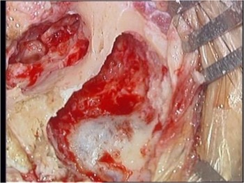
We made a post-aural incision with the patient under general anaesthesia. Routine cortical mastoidectomy was performed. An incision was made on the posterior and superior canal wall skin, parallel to the annulus and just medial to the hairy to non-hairy junctional line. Deep posterior and superior canal wall skin was elevated. After clearing all the pathology in the mastoid, the middle ear and the canal, a good haemostasis was achieved. The focal meato-mastoid fistula was defined by removing all pathologies, such as granulations, squamous epithelium and remnant tumour tissues. Uneven bony margins were drilled with diamond rose burr, thereby delineating meato-mastoid fistulae (Figure 1).
Meato-mastoid fistulae delineated and cleared of all pathology. Sigmoid plate delineated as part of disease clearance procedure.

Copying meato-mastoid fistulae defect
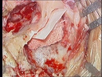
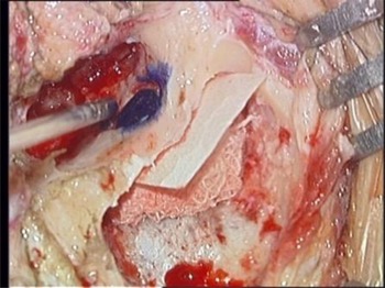
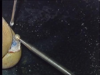
We placed a filter paper posterior (or superior) to the canal wall in the mastoid cavity. The filter paper was supported with a piece of dry gauze so that the filter paper was temporarily stabilised with respect to the canal wall (Figure 2). The filter paper was visible through the canal and the meato-mastoid fistula. We painted the filter paper through the defect of meato-mastoid fistulae with a tiny (approximately 2 mm) cotton ball. This was achieved by dipping the cotton ball in methylene blue and gently squeezing off the excess dye. The cotton ball was held in crocodile forceps for this purpose (Figure 3). The gauze and filter paper were retrieved.
Filter paper placed in the mastoid cavity on the posterior surface of the posterior canal wall. The medial end of the filter paper was supported with dry gauze.

The painted filter paper as seen through the canal.

Delineation of sigmoid plate
If the sigmoid plate was not delineated during mastoid disease clearance, the sigmoid plate was delineated at this point (Figure 1). The site selected for delineation of the sigmoid sinus plate was chosen to be where the natural curvature of the sigmoid plate and the natural curvature of the canal wall where the meato-mastoid fistula was situated matched as closely as possible. This is possible because the external auditory canal and bony sigmoid sinus plate are both oval in cross-section and thus have different curvatures in different regions.
Harvesting the sigmoid plate
Using a diamond burr and continuous irrigation, deep cuts were made to detach a piece of sigmoid plate larger than the defect size by at least 2 mm in diameter. The detached sigmoid plate was gently lifted off the sigmoid sinus.
Filter paper cutout
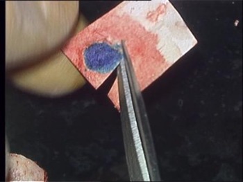
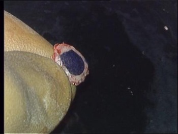
The methylene blue painted area of the filter paper was then cut (Figure 4). This painted cutout of the filter paper exactly represented meato-mastoid fistulae defect in size, shape and orientation. The cutout was dipped in methylene blue and made dripping wet. Excess methylene blue was allowed to run off. We placed this fully wet (in methylene blue) cutout of the filter paper on the convex surface of the sigmoid plate and pressed the cutout with dry gauze. The impression of the cutout was then transferred onto the sigmoid plate (Figure 5). It was necessary to place the cutout on the sigmoid plate, keeping in view the orientation of meato-mastoid fistulae with respect to sigmoid plate and canal curvatures.
The painted part of the filter paper being cutout.

Methylene blue impression copied onto the convex surface of the sigmoid plate.

Trimming excess bony sigmoid plate
Keeping a margin of 1 mm around the methylene blue mark on the sigmoid plate, excess bone was removed.
Making a step on the outer margin of sigmoid plate
At 1 mm beyond the impression of the cutout, the bony graft was drilled with diamond burr to make it 0.5 mm thick (Figure 6). This meant we now had a bony plate that had a thickness at the centre that exactly matches the size and shape of the bony defect to be closed and had a 0.5 mm thick and 1 mm wide marginal rim that will sit snugly on the bony canal wall.
The 1 mm outer margin is drilled to make the margin 0.5 mm thick.

Placing bony sigmoid sinus graft
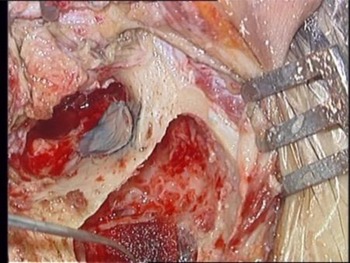
The bony sigmoid sinus graft was then placed in its final destination in proper orientation (Figure 7). The bony graft generally fits very well.
The sigmoid plate graft is placed in its final destination, closing the meato-mastoid fistula as seen through the meatus. There is a perfect curvature match of the bony graft and the posterior canal wall.

Covering the graft
The well adapted bony graft is now covered with fascia graft and remnant meatal skin.
Results
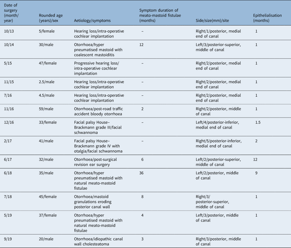
Table 1 describes aetiology, defect size and position and other clinical parameters as well as the outcome of our cases. In all cases, the bony sigmoid sinus plate was integrated. In our series of 13 cases, the age group varied from 2.5 years to 59 years. Four patients had iatrogenic meato-mastoid fistulae during facial recess approach for cochlear implantation. These patients had a defect of size 1.0 to 2.0 mm located near the medial end of the posterior canal wall. For cases where the defect size was 2 mm or less, we used bony sigmoid sinus plate without sizing it, as the defect was too small. In addition, we found it difficult to sculpt small sized bony sigmoid sinus plate, although it was not necessary for a proper fit. We simply placed a bony sigmoid sinus plate larger than the meato-mastoid fistula, anterior to the meato-mastoid fistula.
Clinical details and duration for epithelialisation over the graft

Mean follow period = 37.6 months
There were two cases of facial schwannomas, where the tumour had created a relatively larger meato-mastoid fistula (4 mm and 5 mm). Three patients had mastoiditis with a hyper-pneumatised mastoid and a meato-mastoid fistula. One case had excessive mastoid granulations, which had eroded the posterior canal wall. One case had a post-traumatic (road traffic accident) meato-mastoid fistula. This case had hyper-pneumatised mastoid and no fracture of temporal bone or posterior canal wall. It is likely that the patient had had an asymptomatic meato-mastoid fistula for some time, and the meato-mastoid fistula caused symptoms after ear trauma.
We had one case of external auditory canal cholesteatoma and one case of canal wall up cortical mastoidectomy with meato-mastoid fistulae. We used the bony sigmoid sinus plate in all our cases for repair of meato-mastoid fistulae. The outcome of the repair was satisfactory in all our cases in terms of viability, stability, epithelialisation and relief of symptoms. Two cases had delayed epithelialisation up to 9 months and 12 months after surgery. In the long-term, a mean period of 37.6 months, the posterior or superior canal wall appeared normal. High-resolution computed tomography showed no fistula in the posterior canal wall. We felt that any focal defect from 2 mm to 8 mm could be treated with this technique.
Discussion
Meato-mastoid fistulae are focal defects in the posterior or superior bony external auditory canal. They can be iatrogenic, post-traumatic or caused by certain known pathologies like external auditory canal cholesteatomas, keratosis obturans, florid granulations in the mastoid and tumours. They can sometimes be idiopathic. The occurrence of posterior canal wall mastoid cavity fistulae in external auditory canal cholesteatoma is rare.Reference Cho6 According to Persaud et al., osteo-necrosis or bony sequestra and focal loss of epithelium are typical of external auditory canal cholesteatoma.Reference Persaud, Hajioff, Thevasagayam, Wareing and Wright7 Schwannomas of the facial nerve or the chorda tympaniReference Pou and Chambers8 are rare too;Reference Kirazli, Oner, Bilgen, Ovül and Midilli9 they can cause gradual erosion of the posterior canal wall leading to meato-mastoid fistulae. Meato-mastoid fistulae with iatrogenic causes can occur during simple cortical mastoidectomy or during a facial recess approach. We must specifically look for meato-mastoid fistulae at the lateral end of the bony canal in cases with previous history of canal wall up mastoid surgery and that have post-operative otorrhoea with a normal appearing tympanic membrane. Often these patients have a not easily visible defect in the lateral part of the posterior or superior bony canal. We had one case of post-operative canal wall up surgery in our study with meato-mastoid fistulae. In these types of cases, meato-mastoid fistulae can be asymptomatic or can present with otorrhoea. Two patients with facial nerve schwannoma in our study had facial palsy and tumours presenting in the posterior-inferior canal. It is essential to remove tumours in totality and to restore the anatomy of the external auditory canal to relieve the patient of otorrhoea, preserve or restore hearing, and avoid retraction pockets or cholesteatoma formation in the future.
Cho reported a case of a meato-mastoid fistula.Reference Cho6 This study used a bone-wax template to intra-operatively size meato-mastoid fistulae defects. This is certainly a brilliant proposal for sizing the posterior canal wall defect. However, we find that the bone wax is distorted during retrieval, particularly if the mastoid cavity is small; instead, we used blotting paper and methylene blue dye to size the posterior canal wall defect. In addition, Cho used cortical bone for repair of meato-mastoid fistulae. Cortical bone lacks the natural curvature of the posterior external auditory canal. An ideal repair material for meato-mastoid fistulae repair should have an inherent consistency, structure and curvature similar to the posterior canal wall. It should be available in a size larger than the defect size and preferably be locally available. Donor site morbidity should be kept to a minimum. The bony sigmoid sinus plate fulfils these requirements. We therefore find it to be an ideal graft material for the repair of meato-mastoid fistulae.
Alloplastic materials like silastic, hydroxyapatite and titanium have been used. They have the advantage of having ready availability, no donor site morbidity, and come in all sizes and shapes. These implants have been used successfully for reconstruction of the posterior canal wall. However, they hinder vascularisation, and biofilm sometimes forms around them, ultimately causing extrusion.Reference Anand, Latif and Smith10 Repair of focal canal wall defects with cartilage has also been described.Reference Parelkar, Shere and Shah4 Cartilage does not have the natural consistency and structure as that of the posterior canal wall. In addition, it has a recoil memory and can undergo a variable amount of resorption over time, which can significantly alter the long-term outcome.
Compared with these alloplastic materials and cartilage, the bony sigmoid sinus plate has several advantages. This bone is easily available in all cases undergoing cortical mastoidectomy, which is a part of the primary procedure in most cases. Adequate size of bone can be harvested, and it can be customised as per requirement. It has a natural two-dimensional curvature that matches the natural two-dimensional curvature of the bony posterior canal wall, providing the site of harvest of bony sigmoid sinus plate is appropriately chosen. The technique of harvest and adaptation of the sigmoid plate to the posterior canal wall is not complicated and is easily repeatable. There are reports of dehiscent sigmoid sinus or sigmoid sinus diverticulum causing pulsatile tinnitus;Reference Eisenman, Raghavan, Hertzano and Morales11 however, none of our patients complained of pulsatile tinnitus. Venous bleeding can occur while lifting the bony plate over the sinus. This can be avoided by gentle and precise dissection. The bleeding can be controlled easily with pressure using Gelfoam® or Surgicel®. In addition, exposing the sigmoid sinus is a primary step in numerous lateral skull base procedures. It rarely leads to troublesome complications.
Since both external auditory canal and the sigmoid plate are ovoid in cross-section, the appropriate region of the sigmoid sinus plate can be selected to match the curvature of the region of interest of the external auditory canal.
In revision cases, the canal wall skin may take longer to epithelialise and cover the bony graft. Inadequate saline irrigation during delineation or harvest of the bony plate may cause delayed epithelialisation or even rejection because of thermal damage to the graft.
• Meato-mastoid fistulae are focal defects in the posterior or superior bony external auditory canal
• These defects can be iatrogenic, post-traumatic or pathological
• Various techniques of repair with alloplastic materials, cartilage or bone have been described
• The technique of repair using bony sigmoid sinus plate is simple, repeatable and provides an effective physiological reconstruction of the posterior canal wall
• Bony sigmoid sinus plate has a curvature, consistency and structure that match well with that of the posterior or superior canal wall
Meato-mastoid fistulae can be divided into the categories of small and large. Meato-mastoid fistulae that are smaller than 2 mm in diameter can be closed with bony sigmoid sinus plate from any region without concern about curvature. The small meato-mastoid fistulae may not need to be sized. A small piece of bony sigmoid sinus plate from the presenting sigmoid plate can be harvested very easily without undue risk to the sigmoid sinus. However, when it comes to large meato-mastoid fistulae, such as those created by facial schwannomas, bony sigmoid sinus plate must be harvested from the appropriate site, keeping its curvature and size in mind. The harvest of large bony sigmoid sinus plate from the appropriate site needs to be performed with care to avoid puncturing the sigmoid sinus and also meato-mastoid fistulae must be sized to get a perfectly fitting implant. A perfect fit will help with quick integration and avoid canal narrowing.
The bony sigmoid sinus plate is not recommended in the following situations. (1) In cases of post-radiation meato-mastoid fistulae, it is not recommended because surrounding healthy vascular tissue is essential for a good outcome. In addition, the bony sigmoid sinus plate itself may show radiation necrosis over a period. (2) This technique is not good in laterally located defects, which lack circumferential bony margins because the sigmoid plate may not fit snugly and may remain immobilised during the integration phase. (3) It is also not good for large defects (more than two thirds of the posterior canal wall defect).
Conclusion
The senior author's (IS) technique described above for repair of focal posterior canal wall defects causing meato-mastoid fistulae is simple and repeatable and has very encouraging outcomes as our patients were symptom free. Mean follow-up period showed normal posterior canal wall, and high-resolution computed tomography of temporal bone showed no bony defect in posterior canal wall. We did not have donor site morbidity. The technique needs validation by repeating it at other centres and with other surgeons, after understanding its nuances.
Competing interests
None declared