Implications
In ruminants, adequate development of foetal brown or post-natal white adipose tissues relative to muscle development is a major challenge to promote metabolic adaptation both at birth for neonate survival, especially in sheep, and in adult life for productive efficiency during the gestation-lactation cycle of dairy females or for carcass yield and quality of meat animals. In addition, better knowledge of brown adipogenesis could help to prevent excessive fat development in ruminants and other species.
Introduction
Producing meat animals with a desirable lean-to-fat ratio (i.e. relative amounts of muscle and adipose tissue) is an economic challenge for the beef industry. The lean-to-fat ratio is the result of a dynamic balance between the number and size of muscle and adipose cells. There is striking evidence for developmental and functional links between muscle and adipose tissue (AT): the successive waves of growth of muscle and AT suggest a priority for muscle growth, and in double-muscled Belgian blue cattle, increased muscular development is concomitant with a decrease in the carcass and muscular AT (Bellinge et al., Reference Bellinge, Liberles, Iaschi, O’Brien and Tay2005). Thus, adipose and muscle cells seem to be linked by competition or prioritization in their commitment, differentiation, and/or uptake and metabolism of nutrients, all of which determine their number and size. Yet few studies have directly considered the reciprocal links between muscle and AT (Argiles et al., Reference Argiles, Lopez-Soriano, Almendro, Busquets and Lopez-Soriano2005; Dyck et al., Reference Dyck, Heigenhauser and Bruce2006; Eckardt et al., Reference Eckardt, Sell and Eckel2008). Indirect identification of the developmental and functional links between muscle and AT is made possible by current knowledge of the cellular and molecular processes involved in their respective development and/or related to marked variations in the lean-to-fat ratio in ruminants. This review examines (i) the processes and regulation of myogenesis and adipogenesis in cattle, (ii) the variation in the masses of muscle and/or AT, and the related cellular and molecular adaptations induced by factors that cause a wide variability in lean-to-fat ratio, namely age, genotype, sex and nutrition, and (iii) current knowledge of mechanisms likely to underlie the ‘adipose–muscular cross-talk’ suggested above. We focus on current available data obtained from cattle, and also from other species when relevant.
Dynamics of muscle growth
In mammals, skeletal muscle comprises about 55% of the body. The muscle mass is largely determined by the number and size of muscle fibres. Muscle fibres are heterogeneous due to different properties, both morphological (size) and physiological (contractile and metabolic). This heterogeneity is not restricted to post-natal life, but originates in foetal life in species that are relatively mature at birth such as cattle (Picard et al., Reference Picard, Lefaucheur, Berri and Duclos2002).
Cellular events triggering myogenesis
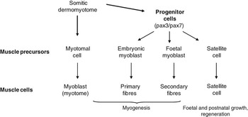
Myogenesis is the developmental process leading to muscle formation, extensively described in rodents and chicks. The trunk and head musculature derive, respectively, from the paraxial mesoderm in the trunk and the cranial paraxial mesoderm located anterior to the somites (Tzahor, Reference Tzahor2009). Early in the embryonic development of vertebrates, paraxial mesodermal cells differentiate into skeletal muscle progenitors. Shortly after somitogenesis, some myogenic progenitors derived from the dermomyotome give rise to mononuclear myoblasts. This initial phase generates the early muscle, called the primary myotome (Figure 1) located beneath the dermomyotome. The myoblasts proliferate, become post-mitotic, elongate and fuse together to form myotubes, which further develop into the mature striated muscle fibres (the basic units of skeletal muscle). Formation of the skeletal muscle proceeds in successive distinct, but overlapping steps involving different types of myoblasts that result in formation of myotomal cells, embryonic and foetal myoblasts, and satellite cells (Figure 1). This results in muscle heterogeneity and diversification. In addition, satellite cells found adjacent to the myofibres are detected as soon as 2 months in bovine foetuses (Russell and Oteruelo, Reference Russell and Oteruelo1981) and 85 days in sheep foetuses (Greenwood et al., Reference Greenwood, Slepetis, Hermanson and Bell1999) and contribute to muscle growth and regeneration (Figure 1). The different generations of myofibres are installed in close relation to the growth factor environment of muscle progenitors (Biressi et al., Reference Biressi, Molinaro and Cossu2007). During primary myogenesis, embryonic myoblasts fuse into primary myofibres, whereas differentiation of foetal and satellite cells is inhibited by transforming growth factor-β (TGF-β) and bone morphogenetic protein (BMP, (Cusella de Angelis et al., Reference Cusella de Angelis, Molinari, Ledonne, Coletta, Vivarelli, Bouche, Molinaro, Ferrari and Cossu1994)). Once formed, primary myofibres may induce a new wave of proliferation in foetal myoblasts, possibly through secretion of mitogens such as fibroblast growth factor (FGF). At the foetal stage, decreased TGF-β and BMP levels may be responsible for the differentiation of foetal myoblasts into secondary myofibres, while platelet-derived growth factor (PDGF) may keep satellite cells undifferentiated (Biressi et al., Reference Biressi, Molinaro and Cossu2007).
Developmental cascade and cellular events leading to myogenesis in vertebrates. Successive waves of muscle precursors contribute to muscle formation and diversification. The different myoblast generations, expressing the paired box transcription factors Pax3/Pax7, derive from the dorsal compartment of somites (dermomyotome) and lead to the different types of myofibres in skeletal muscle (adapted from Tajbakhsh (Reference Tajbakhsh2005) and Biressi et al. (Reference Biressi, Molinaro and Cossu2007)).

During myogenesis, myofibres acquire their specific contractile and metabolic properties driven, respectively, by their myosin heavy chain (MyHC) isoform content and development of energy metabolism enzymatic pathways. They can be classified into slow (type I) and fast (IIA, IIX, IIB) myofibres.
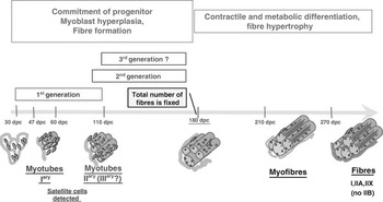
In cattle, as in other species the properties of myofibres are acquired in a three-stage process (embryonic, foetal and post-natal; (Picard et al., Reference Picard, Lefaucheur, Berri and Duclos2002); Figure 2). A primary or embryonic generation is observed from 30 days post-conception (dpc) onwards and is completely differentiated by the end of the second trimester (around 180 dpc). These primary myofibres mature into slow twitch type I myofibres in most muscles except in exclusively fast adult muscles, in which they are converted into fast myofibres (Picard et al., Reference Picard, Robelin, Pons and Geay1994). A secondary or foetal generation is observed from the end of the first trimester onwards, and mostly matures into fast IIX myofibres. The secondary myofibres are converted after birth into fast or slow myofibres depending on the muscle (Picard et al., Reference Picard, Lefaucheur, Berri and Duclos2002). Although controversial, the existence of a third generation has been proposed based on different anti-MyHC antibodies. It was observed from 40% of the gestation period onwards gives rise to fast IIA, slow I and IIC myofibres. This last myofibre type is undifferentiated at birth, and expresses both fast and slow MyHC isoforms. The analysis of several differentiation markers shows that contractile and metabolic maturation occurs during the last trimester (Picard et al., Reference Picard, Lefaucheur, Berri and Duclos2002). Expressions of the developmental MyHC isoforms (embryonic, foetal, α-cardiac, etc.) decrease during this period (Gagnière et al., Reference Gagnière, Picard and Geay1999a; Picard et al., Reference Picard, Jurie, Duris and Renand2006). They are progressively replaced by the adult fast MyHCs. The slow isoform of MyHC is expressed earliest; it is observed in myotubes from 30 dpc, but only in the first generation from 180 dpc. Later, this isoform is also detected in myofibres of the second and third generations, which give rise to type I myofibres in the adult muscle. Three weeks after birth, cattle muscles contain only adult MyHC isoforms I, IIa and IIx (Picard et al., Reference Picard, Jurie, Duris and Renand2006). They will not express the IIb isoform, except in some muscles of some cattle (Picard and Cassar-Malek, Reference Picard and Cassar-Malek2009). Consequently, the contractile maturation of muscles in cattle, like in sheep (Maier et al., Reference Maier, McEwan, Dodds, Fischman, Fitzsimons and Harris1992) and humans (Shrager et al., Reference Shrager, Desjardins, Burkman, Konig, Stewart, Su, Shah, Bricklin, Tewari, Hoffman, Rickels, Jullian, Rubinstein and Stedman2000), but unlike in rodents (Cho et al., Reference Cho, Hughes, Karschmizrachi, Travis, Leinwand and Blau1994), is particularly advanced at birth. During the last trimester, there is also a marked increase in the activities of enzymes from glycolytic and oxidative metabolism in cattle muscles (Gagnière et al., Reference Gagnière, Picard, Jurie and Geay1999b). In cattle, all future type I myofibres exhibit an oxidative metabolism from 210 dpc. For IIA myofibres, the oxidative metabolism increases during the last trimester and all of them have an oxidative metabolism from birth onwards (Picard et al., Reference Picard, Jurie, Duris and Renand2006). Activities of enzymes involved in the glycolytic metabolism also increase during this period in relation to changes in isoform expressions, for example, lactate dehydrogenase (LDH) isoforms are converted from cardiac to skeletal isoform (Picard et al., Reference Picard, Jurie, Duris and Renand2006).
Main steps in bovine myogenesis. At least two generations of myoblasts proliferate during the first two trimesters of foetal life. Primary myotubes are present before 47 days (Russell and Oteruelo, Reference Russell and Oteruelo1981; Picard et al. Reference Picard, Lefaucheur, Berri and Duclos2002). The total number of myofibres is fixed from approximately 180 days onwards. From this stage, the muscle tissue is morphologically well organized, the cells have a myofibre shape and no longer a myotube shape, also demonstrating the intense morphological differentiation between 110 and 180 dpc. The last trimester is characterized by intensive contractile and metabolic maturation of myofibres (adapted from Picard et al. (Reference Picard, Lefaucheur, Berri and Duclos2002)). Satellite cells are detectable since 63 days of gestation (Russell and Oteruelo, Reference Russell and Oteruelo1981).

During the development, the process of muscle growth is continuous. At the cellular level, muscle accretion can be defined as a combined increase in the number of myofibres (hyperplasia) and in their diameter and length (hypertrophy). During foetal development, muscle growth occurs mainly through hyperplasia. Different studies conducted in cattle foetuses have shown that the total number of myofibres is set by the end of the second trimester of gestation (180 dpc; see (Picard et al., Reference Picard, Lefaucheur, Berri and Duclos2002)). The size of myofibres increases by hypertrophy from this stage as observed also in sheep (Greenwood et al., Reference Greenwood, Slepetis, Hermanson and Bell1999; Greenwood et al., Reference Greenwood, Slepetis and Bell2000) and continues during the perinatal and post-natal periods (Brandstetter et al., Reference Brandstetter, Picard and Geay1998; Jurie et al., Reference Jurie, Picard and Geay1999). This process involves the fusion of satellite cells to existing myofibres as described in other vertebrate species (Biressi et al., Reference Biressi, Molinaro and Cossu2007).
Molecular events during myogenesis
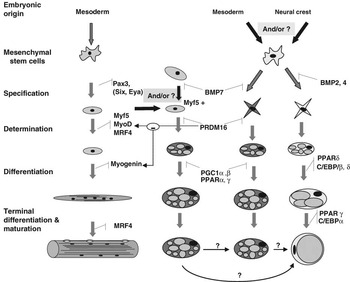
The myogenic pathways and signalling have been described mainly in rodents. Networks of transcription factors are required for molecular specification and myogenic determination and differentiation (Figure 3). The myogenic regulatory factors (MRFs), including MyoD (Davis et al., Reference Davis, Weintraub and Lassar1987), Myf5 (Braun et al., Reference Braun, Buschhausendenker, Bober, Tannich and Arnold1989), myogenin (Wright et al., Reference Wright, Sassoon and Lin1989) and MRF4 (Rhodes and Konieczny, Reference Rhodes and Konieczny1989) are specific myogenic transcription factors. They act in concert with associated co-factors and transcription factors including E-proteins and members of the myocyte enhancer factor 2 family to control cell identity and differentiation (Bryson-Richardson and Currie, Reference Bryson-Richardson and Currie2008). MRFs activate transcription through heterodimerization with MRFs or with ubiquitously expressed E-proteins and binding to E-box consensus sites in the promoters of muscle-specific genes (Lassar et al., Reference Lassar, Davis, Wright, Kadesch, Murre, Voronova, Baltimore and Weintraub1991).
Transcription factors controlling the specification, determination and differentiation of the myogenic and adipogenic lineages. Myf5, MyoD and myogenic regulatory factor 4 (MRF4) are required for myogenic determination, whereas differentiation depends on myogenin and MRF4, as demonstrated by knock-out experiments (adapted from Rudnicki et al. (Reference Rudnicki, Schnegelsberg, Stead, Braun, Arnold and Jaenisch1993), Kassar-Duchossoy et al. (Reference Kassar-Duchossoy, Gayraud-Morel, Gomes, Rocancourt, Buckingham, Shinin and Tajbakhsh2004), Buckingham (Reference Buckingham2007), Buckingham and Relaix (Reference Buckingham and Relaix2007)). The embryonic origin of white adipocytes remain to be clarified (Billon et al., Reference Billon, Monteiro and Dani2008). Bone morphogenetic protein 7 (BMP7) drives the brown determination (Tseng et al., Reference Tseng, Kokkotou, Schulz, Huang, Winnay, Taniguchi, Tran, Suzuki, Espinoza, Yamamoto, Ahrens, Dudley, Norris, Kulkarni and Kahn2008) and PRDM16 activates the brown differentiation program at least in part through peroxisome proliferator-activated receptor-γ (PPARγ) binding and coactivation, and blocks the expression of Myf5 and MyoD (Seale et al., Reference Seale, Bjork, Yang, Kajimura, Chin, Kuang, Scime, Devarakonda, Conroe, Erdjument-Bromage, Tempst, Rudnicki, Beier and Spiegelman2008 and Reference Seale, Kajimura and Spiegelman2009). BMP2 and 4 drives the white determination (Tseng et al., Reference Tseng, Kokkotou, Schulz, Huang, Winnay, Taniguchi, Tran, Suzuki, Espinoza, Yamamoto, Ahrens, Dudley, Norris, Kulkarni and Kahn2008), and PPARγ and CCAAT enhancer-binding proteins (C/EBPs) are pivotal to driving the differentiation of white pre-adipocytes (Gregoire et al., Reference Gregoire, Smas and Sul1998). Whether a single Myf5+ cell gives rise to both brown adipose tissues (BAT) and muscle, whether there are distinct populations of Myf5+ progenitors, or whether brown adipocytes are transformed into true white or white-like adipocytes, remains unknown.

MRFs are expressed in a highly regulated spatial and temporal fashion. Skeletal myogenesis is initiated by the activation of Myf5 and MyoD expressions (Emerson, Reference Emerson1990; Rudnicki et al., Reference Rudnicki, Schnegelsberg, Stead, Braun, Arnold and Jaenisch1993) under the control of wingless-integration site (Wnt) and Sonic Hedgehog signalling pathways (Bryson-Richardson and Currie, Reference Bryson-Richardson and Currie2008). More specifically, Myf5, MyoD and MRF4 act as determination genes (Figure 3), since all skeletal muscles and myoblasts can be eliminated by inactivating these three genes in triple mutant mice (Kassar-Duchossoy et al., Reference Kassar-Duchossoy, Gayraud-Morel, Gomes, Rocancourt, Buckingham, Shinin and Tajbakhsh2004). Gene targeting experiments have shown that Myf5 and MyoD have distinct regulatory and partially redundant functions (Bryson-Richardson and Currie, Reference Bryson-Richardson and Currie2008); data indicate that epaxial musculature (deep muscles of the back) depends on Myf5 expression, whereas MyoD is required for hypaxial muscle formation (body wall and limb muscles, diaphragm, hypoglossal and tongue muscle). At least one of these MRFs is required for activation of myogenin and MRF4 (Rudnicki et al., Reference Rudnicki, Schnegelsberg, Stead, Braun, Arnold and Jaenisch1993; Valdez et al., Reference Valdez, Richardson, Klein and Olson2000). MRF4 has a biphasic expression pattern in the myotome, but not in the limb and is the predominant MRF expressed in the adult (Rhodes and Konieczny, Reference Rhodes and Konieczny1989). Gene targeting and knocked-in experiments have shown that myogenin and MRF4 are, respectively, crucial for the differentiation and the maintenance of the terminally differentiated state (Figure 3). Interestingly, MRF4 can compensate for the absence of Myf5 and MyoD for trunk muscle formation (Kassar-Duchossoy et al., Reference Kassar-Duchossoy, Gayraud-Morel, Gomes, Rocancourt, Buckingham, Shinin and Tajbakhsh2004).
Pax3, a member of the paired box family of transcription factors (Buckingham and Relaix, Reference Buckingham and Relaix2007) is an upstream regulator of the myogenic cascade required for primary myogenesis (Tajbakhsh et al., Reference Tajbakhsh, Rocancourt, Cossu and Buckingham1997) and trunk muscle development. It may activate Myf5 directly (Bajard et al., Reference Bajard, Relaix, Lagha, Rocancourt, Daubas and Buckingham2006). Pax3 probably also controls myogenesis indirectly via other transcription factors such as members of the six family and their co-factors Eya and Dach (Buckingham and Relaix, Reference Buckingham and Relaix2007). A Pax3- and a Myf5-dependent myogenic pathway is required for early myogenesis in the hypaxial and epaxial muscle, respectively. There is some evidence that distinct networks operate for specification of the muscle lineages in the head (Tzahor, Reference Tzahor2009). In post-natal muscle, Pax3 and Pax7 regulate the entry of satellite cells into the myogenic program via activation of MyoD (Relaix et al., Reference Relaix, Montarras, Zaffran, Gayraud-Morel, Rocancourt, Tajbakhsh, Mansouri, Cumano and Buckingham2006). More specifically, Pax7 is required for the specification of satellite cells (Seale et al., Reference Seale, Sabourin, Girgis-Gabardo, Mansouri, Gruss and Rudnicki2000) and ensures satellite cell survival (Relaix et al., Reference Relaix, Montarras, Zaffran, Gayraud-Morel, Rocancourt, Tajbakhsh, Mansouri, Cumano and Buckingham2006). However in Pax7 mutants, some satellite cells are still detectable (Oustanina et al., Reference Oustanina, Hause and Braun2004) and recent data suggest that Pax7 would not be required for satellite cell myogenesis in adulthood (Lepper et al., Reference Lepper, Conway and Fan2009). These molecular events observed in rodents are likely to apply for late foetal and post-natal muscle growth in ruminants as indicated by on-going molecular profiling during bovine myogenesis (Sudre et al., Reference Sudre, Leroux, Pietu, Cassar-Malek, Petit, Listrat, Auffray, Picard, Martin and Hocquette2003; Lehnert et al., Reference Lehnert, Reverter, Byrne, Wang, Nattrass, Hudson and Greenwood2007; Chaze et al., Reference Chaze, Meunier, Chambon, Jurie and Picard2008 and Reference Chaze, Meunier, Chambon, Jurie and Picard2009).
Dynamics of AT growth
AT is a diffuse organ that comprises 5% to 35% of cattle body mass, depending on age, genotype and nutrition. Development of AT occurs in multiple, anatomically distinct sites. Three major locations; visceral, subcutaneous and muscular, are further subdivided into smaller depots defined by anatomical location (e.g. perirenal v. omental). In cattle as in other mammals, AT has been classified into two distinct types: white AT (WAT), the primary site of energy storage, and brown AT (BAT) specialized for energy expenditure. Brown adipocytes are smaller than white adipocytes, are usually multi-locular (in comparison with white adipocytes with a single lipid droplet) and contain relatively large numbers of mitochondria. Brown adipocytes may be characterized by the expression of uncoupling protein-1 in the inner mitochondrial membrane, which allows energy expenditure through thermogenesis (Vernon, Reference Vernon1986; Cannon and Nedergaard, Reference Cannon and Nedergaard2004) as well as by the expression of proteins involved in mitochondrial biogenesis and oxidative activity (Fruhbeck et al., Reference Fruhbeck, Sesma and Burrell2009). Growth of BAT and WAT results from increases in adipocyte number (hyperplasia) and/or volume (hypertrophy).
Chronology of AT growth: anatomical location specificity
The chronology of ATs appearance depends on both species and anatomical location. In cattle, the perirenal AT appears in foetuses at 80 dpc, followed by subcutaneous and intermuscular AT from 180 dpc onwards, while intramuscular AT is dissectible only after birth (Vernon, Reference Vernon1986). In the last 20 days before birth most of the AT is abdominal and intermuscular, with little subcutaneous AT (Robelin et al., Reference Robelin, Barboiron and Jailler1985). In cattle and sheep at birth, the different AT depots, except for the subcutaneous depot in some bovine breeds (Smith et al., Reference Smith, Carstens, Randel, Mersmann and Lunt2004), can be considered as brown due to the high expression of UCP1, cytochrome c oxidase and ADP/ATP carrier protein (Casteilla et al., Reference Casteilla, Forest, Robelin, Ricquier, Lombet and Ailhaud1987; Casteilla et al., Reference Casteilla, Champigny, Bouillaud, Robelin and Ricquier1989). UCP1 mRNA reaches its highest level in the various BAT at birth and is no longer detectable 2 days later in cattle (Casteilla et al., Reference Casteilla, Forest, Robelin, Ricquier, Lombet and Ailhaud1987), and sheep, probably as the result of a sharp decline in the expression of peroxisome proliferator-activated receptor-α (PPARα) and peroxisome proliferator-activated receptor-c coactivator-1α (PGC-1α) (Lomax et al., Reference Lomax, Sadiq, Karamanlidis, Karamitri, Trayhurn and Hazlerigg2007). In cattle, since the estimated number of cells remains constant during the first week following birth, it has been thought that a transdifferentiation of brown into white adipocytes occurs. In cattle, 2 to 3 weeks after birth, all AT are currently considered white. Whether brown adipocytes can be transformed into white-like adipocytes with different properties or into true white adipocytes (Cinti, Reference Cinti2009) remains to be clarified. The existence of separate, distinct populations of precursor cells for brown and white adipocytes (see later below), and the presence of brown adipocytes in adult humans (Nedergaard et al., Reference Nedergaard, Bengtsson and Cannon2007), suggest that BAT and WAT are two distinct forms of AT that probably participate jointly in the development and plasticity of AT throughout life, at least in humans (Casteilla and Dani, Reference Casteilla and Dani2006).
The relative contributions of hyperplasia and hypertrophy to the growth of AT vary among tissue locations and age (Cianzio et al., Reference Cianzio, Topel, Whitehurst, Beitz and Self1985, Vernon, Reference Vernon1986). Hyperplasia mainly occurs during foetal and/or early post-natal life, but it can also arise during later stages or in adult life in cattle; the number of total adipocytes has been estimated at around 19 billion at the end of foetal life, and then increases sixfold after birth to reach an estimated 124 billion in adults (Robelin and Casteilla, Reference Robelin and Casteilla1990). Post-natal growth through hyperplasia strongly depends on the anatomical location of AT (being nil in perirenal AT and higher in intermuscular than omental or subcutaneous AT (Vernon, Reference Vernon1986)) and on the average diameter of existing adipocytes. If an average diameter of approximately 90 μM (which seems to an upper limit of size) is reached, hyperplasia contributes to AT growth in fattening growing cattle (Garbutt et al., Reference Garbutt, Anthony, Walker and Mcguire1979; Robelin, Reference Robelin1981; Schoonmaker et al., Reference Schoonmaker, Fluharty and Loerch2004). Furthermore, approximately 10% of the body’s fat cells are regenerated each year in humans (Spalding et al., Reference Spalding, Arner, Westermark, Bernard, Buchholz, Bergmann, Blomqvist, Hoffstedt, Naslund, Britton, Concha, Hassan, Ryden, Frisen and Arner2008). However, mature adipocytes are post-mitotic, which implies that proliferative adipocyte precursor cells must exist and be poised to respond to metabolic demands to maintain adipose dynamics in adults. Adipocyte precursors have been isolated from the stromal vascular fraction of bovine WAT (Hausman et al., Reference Hausman, Dodson, Ajuwon, Azain, Barnes, Guan, Jiang, Poulos, Sainz, Smith, Spurlock, Novakofski, Fernyhough and Bergen2009).
Hypertrophy begins during foetal life and is the main mechanism by which AT grows after birth. The volume of the adipocytes increases considerably (more than 100-fold) in perirenal WAT, and to a lesser extent (4- to 10-fold) in subcutaneous, intermuscular and omental bovine WAT between 150 and 600 days of age (Vernon, Reference Vernon1986).
Adipocytes develop from adipocyte precursor cells (pre-adipocytes), which in turn derive from progenitors that are not yet well characterized, through processes of adipose differentiation and growth, termed adipogenesis (Figure 3).
Cellular and molecular events triggering the adipose fate: from an embryonic cell to a pre-adipocyte
The developmental origin of AT, unlike that of muscle, has received little attention. Recent evidence suggests that white adipocytes may have different origins along the anterior-posterior axis: neural crest in the cephalic region, and putatively mesoderm in the trunk (Billon et al., Reference Billon, Monteiro and Dani2008). In addition, at least some precursors of white fat cells may derive from mural cells associated with blood vessels in mice (Tang et al., Reference Tang, Zeve, Suh, Bosnakovski, Kyba, Hammer, Tallquist and Graff2008).
Adipocytes are generally described as derived from a mesenchymal stem cell (MSC) in both cattle (Bosnakovski et al., Reference Bosnakovski, Mizuno, Kim, Takagi, Okumura and Fujinaga2005) and humans (Gregoire et al., Reference Gregoire, Smas and Sul1998). The prevailing idea that a common MSC gives rise to BAT and WAT has been challenged by the characterization of distinct precursor cells that are committed in either brown or white adipogenesis (Seale et al., Reference Seale, Bjork, Yang, Kajimura, Chin, Kuang, Scime, Devarakonda, Conroe, Erdjument-Bromage, Tempst, Rudnicki, Beier and Spiegelman2008). BMPs have recently been proposed as powerful regulators of brown and white adipogenesis both in vitro and in vivo in mice (Tseng et al., Reference Tseng, Kokkotou, Schulz, Huang, Winnay, Taniguchi, Tran, Suzuki, Espinoza, Yamamoto, Ahrens, Dudley, Norris, Kulkarni and Kahn2008). In particular, BMP2 and BMP4 have been shown to promote the commitment of precursors in the white adipogenic pathway, whereas BMP7 selectively stimulates the commitment of brown precursors. BMP7 then induces the expression of transcriptional regulator PRDM16 (PR domain zinc finger protein 16) required for brown fat determination (Seale et al., Reference Seale, Kajimura, Yang, Chin, Rohas, Uldry, Tavernier, Langin and Spiegelman2007; Tseng et al., Reference Tseng, Kokkotou, Schulz, Huang, Winnay, Taniguchi, Tran, Suzuki, Espinoza, Yamamoto, Ahrens, Dudley, Norris, Kulkarni and Kahn2008), while suppressing the expression of genes specific for white fat cells (Kajimura et al., Reference Kajimura, Seale, Tomaru, Erdjument-Bromage, Cooper, Ruas, Chin, Tempst, Lazar and Spiegelman2008). However, the possibility that WAT and BAT may originate from different cell progenitors remains open. In mice, brown adipocytes that develop within WAT in response to β-adrenergic stimulation and brown adipocytes in BAT depot do not derive from the same progenitor (Seale et al., Reference Seale, Bjork, Yang, Kajimura, Chin, Kuang, Scime, Devarakonda, Conroe, Erdjument-Bromage, Tempst, Rudnicki, Beier and Spiegelman2008) and they differentiate through different programs (Xue et al., Reference Xue, Rim, Hogan, Coulter, Koza and Kozak2007). Thus, it is reasonable to suppose that there might be a common progenitor for white/brown adipocytes in some or all WAT locations. Overall, current data highlight the complexity of adipogenesis and suggest that the cellular and functional heterogeneity of ATs may have a developmental origin, as previously described for muscle. Whether mice data can be transposed to other species remains to be studied, given that the developmental chronology and the relative proportion of BAT and WAT throughout life are very different between rodents and humans or ruminants.
The ‘committed’ cells are adipoblasts that proliferate in vitro until they reach confluence and become growth-arrested at the G1/S phase of the cell cycle. Growth arrest (not cell confluence per se) appears to be required for final differentiation. At that stage, cells become pre-adipocytes expressing early markers of adipogenesis (Boone et al., Reference Boone, Mourot, Gregoire and Remacle2000).
Cellular and molecular events controlling adipocyte differentiation: from pre-adipocyte to mature lipid-filled adipocyte
The second phase of adipogenesis is the differentiation of pre-adipocytes into mature adipocytes, that is, permanently cell cycle-arrested, spherical, lipid-filled cells. The cellular events involve three (in vivo) to four stages (in vitro): the growth arrest described above, clonal expansion (only in cell lines), and early and terminal differentiation. These stages are orchestrated by a tightly regulated transcriptional cascade involving nuclear receptors, as deciphered by extensive studies in cell culture models, in particular mouse 3T3-L1 and 3T3-F442A cell lines and immortalized brown pre-adipocyte cell lines (Gregoire et al., Reference Gregoire, Smas and Sul1998; Boone et al., Reference Boone, Mourot, Gregoire and Remacle2000; Lefterova and Lazar, Reference Lefterova and Lazar2009; Seale et al., Reference Seale, Kajimura and Spiegelman2009). CCAAT/enhancer-binding protein (CEBP-β and -δ, and mainly -α) and PPARγ, are central transcriptional regulators of adipogenesis, through an induction of the expression of many downstream target genes involved in the lipid metabolism and other WAT genes. However, PPARγ has emerged as the master gene of WAT terminal differentiation, and to a lesser extent of BAT. PRDM16 binds to PPARγ (Seale et al., Reference Seale, Bjork, Yang, Kajimura, Chin, Kuang, Scime, Devarakonda, Conroe, Erdjument-Bromage, Tempst, Rudnicki, Beier and Spiegelman2008) and co-activates its transcriptional function, leading to the emergence of brown adipocytes expressing UCP1 and other BAT genes (Timmons et al., Reference Timmons, Wennmalm, Larsson, Walden, Lassmann, Petrovic, Hamilton, Gimeno, Wahlestedt, Baar, Nedergaard and Cannon2007). PRDM16 also co-activates PGC1-α and -β through direct physical interactions. Interestingly, PGC1-α has emerged as a dominant regulator of mitochondrial biogenesis and oxidative metabolic pathways. In addition to these central players, the adipogenic program involves many proteins, among which retinoid X receptor, sterol regulatory element binding protein-1c, Forkhead box C2, and twist-1 have been reported to promote WAT and/or BAT differentiation, while Wnt-10b and PRDM16, respectively, inhibit differentiation of both WAT and BAT, and of WAT only (Lefterova and Lazar, Reference Lefterova and Lazar2009; Seale et al., Reference Seale, Kajimura and Spiegelman2009).
The conservation of these cellular and molecular events across the cell culture models and in vivo in monogastric species suggests that they should also drive the terminal differentiation of ruminant adipocytes. This is supported by the central role of PPARγ in the control of the terminal differentiation of pre-adipocytes isolated from the stromal-vascular fraction in bovine WAT (Ohyama et al., Reference Ohyama, Matsuda, Torii, Matsui, Yano, Kawada and Ishihara1998; Torii et al., Reference Torii, Ohyama, Matsui and Yano1998) and ovine BAT (Soret et al., Reference Soret, Lee, Finley, Lee and Vernon1999). However, CEBP-α expression was either not detected (Taniguchi et al., Reference Taniguchi, Leluo, Bing, Dodson, Okine and Moore2008) or did not parallel the adipogenic process (Ohsaki et al., Reference Ohsaki, Sawa, Sasazaki, Kano, Taniguchi, Mukai and Mannen2007) in cattle.
During white adipogenesis pre-adipocytes also undergo a dramatic change in cell shape, from fusiform to spherical. Cytoskeletal remodelling and extracellular matrix alterations seem to be prerequisites for terminal differentiation, probably by modulating the interaction of cells with their environment (both cell–cell and cell–growth factor interactions). Briefly, the extracellular matrix is converted from a fibronectin-rich stroma to a laminin-rich basal lamina (Gregoire et al., Reference Gregoire, Smas and Sul1998). Modifications of extracellular matrix components during adipogenesis depend on a balance between component deposition and degradation orchestrated by matrix metalloproteinase and their inhibitors in both cattle (Tan et al., Reference Tan, Reverter, Wang, Byrne, McWilliam and Lehnert2006) and monogastric species (Gregoire et al., Reference Gregoire, Smas and Sul1998).
The hormones, growth factors and nutrients that regulate the terminal adipocyte differentiation in vitro have been extensively reviewed in monogastrics (Gregoire et al., Reference Gregoire, Smas and Sul1998; Boone et al., Reference Boone, Mourot, Gregoire and Remacle2000; Avram et al., Reference Avram, Avram and James2007) and more recently in ruminants (Hausman et al., Reference Hausman, Dodson, Ajuwon, Azain, Barnes, Guan, Jiang, Poulos, Sainz, Smith, Spurlock, Novakofski, Fernyhough and Bergen2009).
Cellular and molecular events controlling adipocyte hypertrophy: a balance between lipogenesis, lipolysis and fatty acid oxidation
The size of adipocytes results mainly from the size of the lipid droplet in WAT and from the size and number of lipid droplets in BAT. The main constituents of lipid droplets are triacylglycerols (TAG). The rate of TAG deposition and hence of hypertrophy depends on the relative rates of lipogenesis, lipolysis, and fatty acid (FA) oxidation.
Three main lipogenic pathways are involved in TAG deposition in cattle as in other mammals (Vernon, Reference Vernon1980; Chilliard, Reference Chilliard1993). FA are synthesized de novo, mainly in WAT from acetate and to a lesser extent from lactate, or arise from hydrolysis of plasma TAG by lipoprotein lipase (LPL). Fatty acids are subsequently esterified and stored as TAG in the lipid droplet. Available data on genes and enzymes of the main lipogenic pathways in ruminants has been recently reviewed (Bernard et al., Reference Bernard, Leroux and Chilliard2008). Similar precursors and lipogenic pathways are probably involved in TAG deposition in perirenal BAT of ovine (Vernon, Reference Vernon1986) and bovine (Smith et al., Reference Smith, Carstens, Randel, Mersmann and Lunt2004; Hocquette et al., Reference Hocquette, Sauerwein, Higashiyama, Picard and Abe2006) foetuses.
Within WAT, stored TAG can be rapidly mobilized by the hydrolytic action of lipases, resulting in the release of non-esterified (NEFA), which are used by other tissues, for example, during energy deprivation. Until recently, hormone-sensitive lipase (HSL) was considered to be the enzyme responsible for the first step of TAG hydrolysis in mammals. However, this concept was challenged recently by (i) studies on hormone-sensitive lipase-null mice maintaining a residual fat cell lipolysis, (ii) the identification of a novel adipocyte TAG lipase named adipose triglyceride lipase (ATGL), desnutrin or phospho-lipase A2 (Lafontan and Langin, Reference Lafontan and Langin2009). The current understanding of the lipolysis steps is that at least three major lipases cooperate to release three FA molecules and one glycerol molecule from each TAG molecule (Lafontan and Langin, Reference Lafontan and Langin2009; Zechner et al., Reference Zechner, Kienesberger, Haemmerle, Zimmermann and Lass2009). Similarly in BAT, lipases promote the hydrolysis of stored TAG, leading to the mobilization of FAs as fuel for thermogenesis. The high ATGL activity in BAT together with the impaired thermogenesis in ATGL-null mice, while HSL-null mice exhibited normal thermogenesis, suggest that ATGL is essential for the provision of FA as substrate for BAT thermogenesis (Zechner et al., Reference Zechner, Kienesberger, Haemmerle, Zimmermann and Lass2009). To date, only HSL has been identified in bovine WAT through its mRNA abundance (Bonnet et al., Reference Bonnet, Faulconnier, Flechet, Hocquette, Leroux, Langin, Martin and Chilliard1998; Sumner and Mcnamara, Reference Sumner and Mcnamara2007; Xu et al., Reference Xu, Oh, Lee, Kim, Li, Yin, Jin, Jin, Kim, Kim, Yeo and Choi2008) and its activity (Sprinkle et al., Reference Sprinkle, Ferrell, Holloway, Warrington, Greene, Wu and Stuth1998; Kazala et al., Reference Kazala, Petrak, Lozeman, Mir, Laroche, Deng and Weselake2003). The thermogenic protein UCP1, together with enzymes involved in the citric acid cycle and the respiratory chain, are expressed in bovine foetal BAT, allowing FA oxidation and heat production (Casteilla et al., Reference Casteilla, Champigny, Bouillaud, Robelin and Ricquier1989; Smith et al., Reference Smith, Carstens, Randel, Mersmann and Lunt2004; Hocquette et al., Reference Hocquette, Sauerwein, Higashiyama, Picard and Abe2006).
The balance between the storage and mobilization of TAG is under the control of multiple hormones and nutrients, as previously reviewed (Vernon, Reference Vernon1980; Chilliard, Reference Chilliard1993; Hausman et al., Reference Hausman, Dodson, Ajuwon, Azain, Barnes, Guan, Jiang, Poulos, Sainz, Smith, Spurlock, Novakofski, Fernyhough and Bergen2009), and adipokines. In ruminants, leptin is the best studied adipokine (Chilliard et al., Reference Chilliard, Delavaud and Bonnet2005), but its direct effects on bovine WAT lipolysis and lipogenesis remain unclear (Newby et al., Reference Newby, Gertler and Vernon2001).
Nutritional and physiological control of muscular and AT growth
The number of muscle fibres is set at birth, implying that any events that trigger a regulation or balance between the number of muscular and adipose cells must occur and be regulated during foetal life. Cell hypertrophy is thought to be the main mechanism, whereby muscle and fat masses increase in post-natal life, thanks to increased protein deposition and nutrient storage. A simultaneous increase in adipocyte numbers may also occur, essentially in the late-developing subcutaneous and muscular WAT.
Programming of foetal lean-to-fat ratio by maternal nutrition during pregnancy
The relationships between maternal nutrient intake during pregnancy and the growth of the foetus help to ensure successful pregnancy and the life-long health and productivity of offspring, partly as the result of controlled lean-to-fat ratio (Wu et al., Reference Wu, Bazer, Wallace and Spencer2006; Cottrell and Ozanne, Reference Cottrell and Ozanne2008; Dulloo, Reference Dulloo2008). In ruminants, several strategies have been used to study the effects of maternal nutrition: (i) changes in maternal energy intake to modify the nutrient availability for the foetus, (ii) experimental restriction of nutrient delivery through placental carunclectomy, and (iii) reduction in the uterine blood flow to induce small birth weight as a model for human intrauterine growth retardation (Redmer et al., Reference Redmer, Wallace and Reynolds2004; Cottrell and Ozanne, Reference Cottrell and Ozanne2008; Forhead and Fowden, Reference Forhead and Fowden2009). The effects of manipulating maternal nutrition on the muscle and AT growth have been studied in sheep but rarely in cattle. Whatever the species, it clearly appears that the effects of maternal nutrition depend on the stage of gestation (Redmer et al., Reference Redmer, Wallace and Reynolds2004; Symonds et al., Reference Symonds, Stephenson, Gardner and Budge2007).
Effects of maternal undernutrition (50% of control intake) on muscle development were addressed at three different periods of pregnancy in ewes around the primary myofibre formation, the secondary myofibre formation and the post-myofibre formation. The most critical period was before the peak in secondary myofibre formation in ewes (30 to 70 days). It resulted in increased diameter of fast myofibres, no change in diameter of slow myofibres and decreased fast-to-slow myofibre ratio per unit area (Zhu et al., Reference Zhu, Ford, Means, Hess, Nathanielsz and Du2006; Brameld and Daniel, Reference Brameld and Daniel2008). Elsewhere, undernutrition in ewes between 85 to 115 days of gestation, which encompasses the period of cessation of myofibre formation, had no effect on the number of myofibres in the newborn lambs, but decreased the weight of muscles in lambs (Fahey et al., Reference Fahey, Brameld, Parr and Buttery2005).
Maternal undernutrition (approximately 50% requirement of controls) had opposite effects according to the stage of pregnancy evaluated relative to BAT development, which appears around 50 dpc (Vernon, Reference Vernon1986). Maternal undernutrition during early to mid-gestation (28 to 80 days of gestation (Bispham et al., Reference Bispham, Gopalakrishnan, Dandrea, Wilson, Budge, Keisler, Broughton Pipkin, Stephenson and Symonds2003 and Reference Bispham, Gardner, Gnanalingham, Stephenson, Symonds and Budge2005)) increased the mass of perirenal BAT (representing 95% of the total foetal fat mass at 141 dpc) in near-term foetuses, whereas it decreased it if undernutrition occurred during late gestation (115 days until term; (Budge et al., Reference Budge, Edwards, McMillen, Bryce, Warnes, Pearce, Stephenson and Symonds2004)). The increased adiposity induced by early maternal undernutrition was greater in lambs born from mothers fed to requirement (100% maintenace energy requirement (MER)) than to appetite (150% MER) in late gestation. This adaptation was accompanied in the perirenal BAT of the fattest lambs by increases in mRNA abundance of UCP2, PPARα, leptin, IGF-I and IGF-II (Insulin-like growth factor I and II) receptors, but not of UCP1, PPARγ, leptin receptor, IGF-I and IGF-II (Bispham et al., Reference Bispham, Gopalakrishnan, Dandrea, Wilson, Budge, Keisler, Broughton Pipkin, Stephenson and Symonds2003 and Reference Bispham, Gardner, Gnanalingham, Stephenson, Symonds and Budge2005). Whether these metabolic adaptations enhance hyperplasia and/or hypertrophy of adipocytes remains to be determined. In calves born from mothers fed a restricted level of protein (−4% of dry matter compared with control, isoenergetic), the weight of perirenal BAT, adipocyte size and lipogenic activities were the same as in control calves at birth (Martin et al., Reference Martin, Carstens, Taylor, Sweatt, Eli, Lunt and Smith1997).
Maternal overfeeding (approximately +55% of control) during mid- to late gestation did not affect the weight of perirenal BAT or the adipocyte size of the near-term foetuses, but increased glycaemia, insulinaemia and mRNA abundance of PPARγ, leptin, adiponectin, LPL in perirenal BAT (Muhlhausler et al., Reference Muhlhausler, Duffield and Mcmillen2007).
Whether the maternal nutrition affects adipose and muscular growth in cattle to the same extent as in sheep is still to be determined. Species specificities in nutritional foetal programming have been observed between sheep and humans (Symonds et al., Reference Symonds, Stephenson, Gardner and Budge2007) and hypothesized between sheep and cattle, due to differences in the timing of placental and foetal growths (Greenwood and Cafe, Reference Greenwood and Cafe2007). Nevertheless, do these adaptations have post-natal consequences for lean-to-fat ratio?
Consequences of foetal nutritional programming on post-natal lean-to-fat ratio
The post-natal consequences of maternal nutrition have mainly been addressed in lambs and calves born with different birth weights as a model of foetal undernutrition (Greenwood et al., Reference Greenwood, Hunt, Hermanson and Bell1998; Greenwood and Cafe, Reference Greenwood and Cafe2007) or born from underfed mothers (Tudor et al., Reference Tudor, Utting and O’Rourke1980; Gardner et al., Reference Gardner, Tingey, Van Bon, Ozanne, Wilson, Dandrea, Keisler, Stephenson and Symonds2005; Daniel et al., Reference Daniel, Brameld, Craigon, Scollan and Buttery2007; Ford et al., Reference Ford, Hess, Schwope, Nijland, Gilbert, Vonnahme, Means, Han and Nathanielsz2007). Maternal undernutrition during early pregnancy increased backfat thickness and leptinemia at age 4 months (Ford et al., Reference Ford, Hess, Schwope, Nijland, Gilbert, Vonnahme, Means, Han and Nathanielsz2007), and perirenal adipocyte size (Daniel et al., Reference Daniel, Brameld, Craigon, Scollan and Buttery2007) and absolute and relative visceral fat mass (Gardner et al., Reference Gardner, Tingey, Van Bon, Ozanne, Wilson, Dandrea, Keisler, Stephenson and Symonds2005; Ford et al., Reference Ford, Hess, Schwope, Nijland, Gilbert, Vonnahme, Means, Han and Nathanielsz2007) at age 6 to 12 months in lambs. At slaughter higher masses of AT were recorded in lambs as a result of foetal programming in response to ewe undernutrition (Daniel et al., Reference Daniel, Brameld, Craigon, Scollan and Buttery2007; Ford et al., Reference Ford, Hess, Schwope, Nijland, Gilbert, Vonnahme, Means, Han and Nathanielsz2007). In contrast to AT, differences in myofibre proportions and characteristics induced by maternal early undernutrition were no longer observed in lambs at age 6 months (Daniel et al., Reference Daniel, Brameld, Craigon, Scollan and Buttery2007) or in calves at weaning or when grown to 30 months of age (Greenwood and Cafe, Reference Greenwood and Cafe2007). As long-term consequences for ruminant production, at slaughter, higher masses of AT were recorded when the quality of the post-natal nutrition allowed a high growth rate either to compensate for large differences in birth weight in lambs (Greenwood et al., Reference Greenwood, Hunt, Hermanson and Bell1998), but not in steers or heifers grown to 400 or 370 kg live weight (Tudor et al., Reference Tudor, Utting and O’Rourke1980) or to an average of 675 kg live weight (380 kg carcass weight) at 30 months of age (Greenwood et al., Reference Greenwood, Cafe, Hearnshaw, Hennessy, Thompson and Morris2006; Greenwood and Cafe, Reference Greenwood and Cafe2007). To date scarce experiments suggest that the lean-to-fat ratio is not adversely affected in the long term by growth in utero, although the feeding level during the post-natal period may be important in determining subsequent higher carcass adiposity. However, foetal metabolic adaptations to maternal undernutrition exist at least in sheep that will favour a disproportionately higher rate of fat gain relative to muscle gain in a post-natal environment providing plentiful nutrition. These adaptations, also called ‘thrifty mechanism’ (Cottrell and Ozanne, Reference Cottrell and Ozanne2008; Dulloo, Reference Dulloo2008), result partly from cross-talk between muscle and WAT (see ‘Interactions’ below).
Post-natal regulation of the lean-to-fat ratio by nutrition
Depositions of muscle and AT result from a constant turnover in biosynthesis and degradation of proteins and lipids. Nutrition has a major impact on the rate of accretion of proteins (Jones et al., Reference Jones, Starkey, Calkins and Crouse1990; Therkildsen, Reference Therkildsen2005) and lipids (Vernon, Reference Vernon1980; Hausman et al., Reference Hausman, Dodson, Ajuwon, Azain, Barnes, Guan, Jiang, Poulos, Sainz, Smith, Spurlock, Novakofski, Fernyhough and Bergen2009). During the post-natal growth of cattle, protein and fat accretions are concomitant, with a preferential protein accretion during early growth, and of fat during later growth. The switch occurs when empty body weight reaches 300 kg in concentrate-fed steers and bulls (Owens et al., Reference Owens, Gill, Secrist and Coleman1995). The energy level or composition of the diet have been shown to affect the lean-to-fat ratio differently according to the age and thus the respective growth rates of muscle and AT (Berge et al., Reference Berge, Geay and Micol1991; Greenwood and Cafe, Reference Greenwood and Cafe2007).
In calves, the lean-to-fat ratio at weaning depends on the level of milk intake. Milk intake reduction (−40%) produced a 122% increase in the lean-to-fat ratio as the result of a strong decrease in lipid deposition (−63%) while protein deposition fell slightly (−20%) at age 95 days (Robelin and Chilliard, Reference Robelin and Chilliard1989). This was accompanied by a reduced adipocyte hypertrophy with no effect on their number, and a decreased lipogenesis in WAT. In muscle, energy restriction from birth to weaning decreased cross-section area and increased the proportion of fast glycolytic fibres, which were not detectable at slaughter (Picard et al., Reference Picard, Gagnière, Robelin and Geay1995). Although the increase in lean-to-fat ratio at weaning is spectacular, it is generally less marked when slaughter occurs later, depending on the feeding following weaning (Berge et al., Reference Berge, Geay and Micol1991; Greenwood and Cafe, Reference Greenwood and Cafe2007). Cattle restricted during the milk feeding period then fed at pasture for a prolonged period and feedlot finished (Greenwood et al., Reference Greenwood, Cafe, Hearnshaw, Hennessy, Thompson and Morris2006; Greenwood and Cafe, Reference Greenwood and Cafe2007) have carcasses that are of similar composition (when slaughtered at equivalent carcass weight) or leaner (when slaughtered at equivalent age) than cattle that were better nourished during the milk feeding period. In contrast, when concentrate-based diets are fed from weaning period, the carcass of cattle restricted during the milk feeding period may be fatter (at equivalent weight) than their well-grown counterparts (Tudor et al., Reference Tudor, Utting and O’Rourke1980).
After weaning and until slaughter, a long-term reduction in the level of intake increases the carcass lean-to-fat ratio (at similar live weight). This results from a marked decrease in fat deposition as shown in cattle with differing propensity to fatten (old v. young, fat v. lean breed (Geay and Robelin, Reference Geay and Robelin1979; Hornick et al., Reference Hornick, Van Eenaeme, Gqrard, Dufrasne and Istasse2000)). The decreased fat deposition results preferentially from a decrease in adipocyte size because of decreased lipogenesis and increased lipolysis (Vernon, Reference Vernon1980; Bonnet et al., Reference Bonnet, Faulconnier, Flechet, Hocquette, Leroux, Langin, Martin and Chilliard1998). In contrast, a long-term high level of intake decreases the lean-to-fat ratio in carcasses of similar weight because of excess energy deposited as fat (Tudor et al., Reference Tudor, Utting and O’Rourke1980). This results mainly from an increase in adipocyte size and lipogenesis (Vernon, Reference Vernon1980) and occasionally to a concomitant increase in adipocyte numbers when the average cell size reached about 90 μM (Garbutt et al., Reference Garbutt, Anthony, Walker and Mcguire1979; Schoonmaker et al., Reference Schoonmaker, Fluharty and Loerch2004). Short-term but severe feed restriction has controversial effects on lean-to-fat ratio depending on the severity and length of restriction, age and breed (Berge et al., Reference Berge, Geay and Micol1991; Hornick et al., Reference Hornick, Van Eenaeme, Gqrard, Dufrasne and Istasse2000). Restricted cattle then refed exhibited compensatory growth to achieve leaner, fatter or similar carcasses compared with well-nourished cattle. If refeeding occurred when muscle growth is limited (in steers, fat breeds or adults compared with bulls, lean breeds or youngs) and when fat deposition predominated, this resulted in a fatter carcass (Hornick et al., Reference Hornick, Van Eenaeme, Gqrard, Dufrasne and Istasse2000). In muscle, mild restriction followed by ad libitum feeding had little effect on myofibre size, but increased the frequency of hybrid IIC myofibres and the oxidative metabolism, with differences depending on muscles, suggesting their differential nutritional sensitivity (Cassar-Malek et al., Reference Cassar-Malek, Hocquette, Jurie, Listrat, Jailler, Bauchart, Briand and Picard2004). After severe nutritional restriction immediately post-weaning, the size of the more glycolytic fibre types was more adversely affected than the more oxidative types, resulting in an increased relative area of type I, slow oxidative myofibres. However, adequate time and nutrient at pasture enabled the recovery in myofibre (Greenwood et al., Reference Greenwood, Tomkins, Hunter, Allingham, Harden and Harper2009).
The composition of the diet has been shown to affect protein and fat deposition. Growing cattle fed grass silage had a lower lean-to-fat ratio than those fed an isoenergetic dried grass diet, as a result of a limited rate of protein accretion without variations in fat accretion (Greathead et al., Reference Greathead, Dawson, Craigon, Sessions, Scollan and Buttery2006). Simultaneously, grass silage tended to increase the weight of internal WAT and increased lipogenic activities in perirenal AT. Long-term consumption of maize silage compared with cut or pasture grass increased lipogenic activities in perirenal, intermuscular and subcutaneous WAT, with no variation in adiposity in growing steers conducted at similar weight gain (Faulconnier et al., Reference Faulconnier, Ortigues-Marty, Delavaud, Dozias, Jailler, Micol and Chilliard2007). A review of data (Chilliard, Reference Chilliard1993) indicated that most dietary fats tended to increase body fat deposition in growing cattle if the supplementation did not consistently decrease dietary dry matter intake (DMI). Isoenergetic but low protein diets fed to growing cattle reduced the deposition of protein relative to fat, and hence the lean-to-fat ratio (Hornick et al., Reference Hornick, Van Eenaeme, Gqrard, Dufrasne and Istasse2000).
Taken together, these data emphasize previous conclusions on the priorities for growth (Palsson, Reference Palsson1955). They show that post-natal muscle and WAT growth are closely related, mainly through prioritization or competition for hypertrophy of their cells, and thus for nutrient partitioning, with a major effect on WAT.
Variation in lean-to-fat ratio among cattle types: sex and genotype
Cattle are classified as early-maturing (fat breeds, female) v. late-maturing (lean breed, male) depending on the onset and early maximal rate of WAT growth relative to muscle growth (Pethick et al., Reference Pethick, Barendse, Hocquette, Thompson and Wang2007). The lean-to-fat ratio thus varies strongly with cattle maturity, for example with age, sex and genotype. Sixteen-month-old steers and heifers have lean-to-fat ratios 20% and 45% lower than bulls (Gettys et al., Reference Gettys, Henricks and Schanbacher1988). This may partly result from the greater amount of fat, larger adipocytes and in general greater lipogenic activities in females than in males, both in meat and dairy breeds, compared at similar adiposity (Eguinoa et al., Reference Eguinoa, Brocklehurst, Arana, Mendizabal, Vernon and Purroy2003).
Among the studied bovine extreme genotypes, Belgian Blue and Holstein Friesian have 10- and 2.5-fold higher lean-to-fat ratios than Japanese Black steers at 24 to 26 months of age (however, these figures were maximized by feeding low v. high concentrate diets, respectively, (Gotoh et al., Reference Gotoh, Albrecht, Teuscher, Kawabata, Sakashita, Iwamoto and Wegner2009)). Muscle hypertrophy (20% higher muscle mass on average than other cattle breeds) concomitant to adipose atrophy in the double-muscled Belgian Blue (Bellinge et al., Reference Bellinge, Liberles, Iaschi, O’Brien and Tay2005) contributes to its extremely high lean-to-fat ratio, while the reverse is observed in the Japanese Black (Zembayashi, Reference Zembayashi1994). Muscles from double-muscled cattle are hypertrophied consequently to myostatin (a member of the TGF-β family, which negatively controls the muscle mass (McPherron et al., Reference McPherron, Lawler and Lee1997)) mutation resulting in non-functional myostatin. Other genes than myostatin have been associated with muscle hypertrophy phenotype in sheep (callipyge gene, and nearby gene called Dlk1, reviewed by (Freking et al., Reference Freking, Smith and Leymaster2004)) and mice (follistatin gene encoding a inhibitor of myostatin function, (Nakatani et al., Reference Nakatani, Takehara, Sugino, Matsumoto, Hashimoto, Hasegawa, Murakami, Uezumi, Takeda, Noji, Sunada and Tsuchida2008)) but their relevance remains to be studied in cattle. Differences in muscle and adipose cell number and/or size among genotypes explain differences in lean-to-fat ratio.
Muscle fibre number is higher in double-muscled cattle as the result of a greater proliferation of secondary cell generation during foetal life (Picard et al., Reference Picard, Depreux and Geay1998; Deveaux et al., Reference Deveaux, Cassar-Malek and Picard2001) and a delay in the differentiation phase (Gagnière et al., Reference Gagnière, Ménissier, Geay and Picard2000). The degree of hypertrophy is variable among muscles, some superficial muscles could contain twice as many myofibres per muscle as other breeds with normal musculature (Ashmore et al., Reference Ashmore, Parker, Stokes and Doerr1974; Wegner et al., Reference Wegner, Albrecht, Fiedler, Teuscher, Papstein and Ender2000). Transcriptomic studies have revealed molecular signatures of the double-muscled phenotype and emphasized specific features in connective tissue growth, muscle energy and lipid metabolism (Cassar-Malek et al., Reference Cassar-Malek, Passelaigue, Bernard, Leger and Hocquette2007), and in apoptotic processes (Chelh et al., Reference Chelh, Meunier, Picard, Reecy, Chevalier, Hocquette and Cassar-Malek2009). Similarly, Charolais cattle selected for high v. low muscle growth capacity have a higher total number of myofibres (indicating hyperplasia), starting during foetal life, and a higher proliferation of myoblasts in vitro (Duris et al., Reference Duris, Renand and Picard1999). From the last trimester of gestation onwards, the muscles of high growth capacity Charolais foetuses display a higher proportion of IIX myofibres (fast glycolytic), as shown in double-muscled foetuses (Picard et al., Reference Picard, Jurie, Duris and Renand2006). This feature is associated with delayed physiological maturity of muscle, inducing a delay in the shift in myofibre types after birth. In WAT, the number of adipocytes per depot was higher in the fatter than in the leaner cattle at similar age or weight: in Hereford cross Angus v. Holstein (Hood and Allen, Reference Hood and Allen1973), in Angus v. Santa Gertrudis (Miller et al., Reference Miller, Cross, Lunt and Smith1991), and in hardy v. meat Spanish crossbreeds (Alzón et al., Reference Alzón, Mendizabal, Arana, Albertí and Purroy2007). However, differences in adipocyte number varied more in the late-maturing subcutaneous, inter- and intra-muscular WAT than in the earlier-maturing omental and perirenal WAT. This is consistent with the low hyperplasia ability of perirenal WAT after birth (see above).
Muscle fibre size, especially that of the fast glycolytic fibres, is greater in Charolais than in Holstein steers at the age of 18 months (Bellmann et al., Reference Bellmann, Wegner, Teuscher, Schneider and Ender2004). In addition, double-muscled compared with conventional cattle and Charolais selected for high v. low muscle growth capacity have muscles with a higher proportion of fast glycolytic fibres (Cassar-Malek et al., Reference Cassar-Malek, Ueda, Bernard, Jurie, Sudre, Listrat, Barnola, Gentes, Leroux, Renand, Martin and Hocquette2005; Picard et al., Reference Picard, Jurie, Duris and Renand2006) and a lower intramuscular fat content (Gotoh et al., Reference Gotoh, Albrecht, Teuscher, Kawabata, Sakashita, Iwamoto and Wegner2009), as observed between genotypes with a high v. low lean-to-fat ratio (May et al., Reference May, Savell, Lunt, Wilson, Laurenz and Smith1994; Bellmann et al., Reference Bellmann, Wegner, Teuscher, Schneider and Ender2004). This is accompanied by a reduced expression of proteins related to oxidative and lipid metabolism in muscle (Bouley et al., Reference Bouley, Meunier, Chambon, De Smet, Hocquette and Picard2005; Wang et al., Reference Wang, Byrne, Reverter, Harper, Taniguchi, McWilliam, Mannen, Oyama and Lehnert2005; Bonnet et al., Reference Bonnet, Faulconnier, Leroux, Jurie, Cassar-Malek, Bauchart, Boulesteix, Pethick, Hocquette and Chilliard2007; Jurie et al., Reference Jurie, Cassar-Malek, Bonnet, Leroux, Bauchart, Boulesteix, Pethick and Hocquette2007; Graugnard et al., Reference Graugnard, Piantoni, Bionaz, Berger, Faulkner and Loor2009). In several studies comparing pure or crossbred Pirenaican, Limousin, Holstein, Wagyu, Santa Gertrudis, Angus, and Japanese Black growing cattle it was repeatedly observed that the leaner breeds had lower adipocyte size in carcass and muscular WAT (Miller et al., Reference Miller, Cross, Lunt and Smith1991; May et al., Reference May, Savell, Lunt, Wilson, Laurenz and Smith1994; Eguinoa et al., Reference Eguinoa, Brocklehurst, Arana, Mendizabal, Vernon and Purroy2003), concomitant with lower gene expression of C/EBPs (Yamada et al., Reference Yamada, Kawakami and Nakanishi2009) and PPARγ (Bonnet et al., Reference Bonnet, Faulconnier, Leroux, Jurie, Cassar-Malek, Bauchart, Boulesteix, Pethick, Hocquette and Chilliard2007), lipogenic activities (Hood and Allen, Reference Hood and Allen1973; Miller et al., Reference Miller, Cross, Lunt and Smith1991; Eguinoa et al., Reference Eguinoa, Brocklehurst, Arana, Mendizabal, Vernon and Purroy2003; Bonnet et al., Reference Bonnet, Faulconnier, Leroux, Jurie, Cassar-Malek, Bauchart, Boulesteix, Pethick, Hocquette and Chilliard2007), leptin gene expression (Chilliard et al., Reference Chilliard, Delavaud and Bonnet2005; Bonnet et al., Reference Bonnet, Faulconnier, Leroux, Jurie, Cassar-Malek, Bauchart, Boulesteix, Pethick, Hocquette and Chilliard2007), and higher resistin (Komatsu et al., Reference Komatsu, Itoh, Hodate, Hazegawa, Obara and Kushibiki2005) when slaughtered at similar age. However, when growing animals were slaughtered at different ages, but similar carcass adiposities, no differences between breeds persisted for gene expression of C/EBPs (Xu et al., Reference Xu, Albrecht, Viergutz, Nurnberg, Zhao and Wegner2009; Yamada et al., Reference Yamada, Kawakami and Nakanishi2009), lipogenic activities (Chakrabarty and Romans, Reference Chakrabarty and Romans1972; Eguinoa et al., Reference Eguinoa, Brocklehurst, Arana, Mendizabal, Vernon and Purroy2003) or leptin gene expression (Chilliard et al., Reference Chilliard, Delavaud and Bonnet2005). This shows that the lower adiposity repeatedly observed in lean genotypes partly results from a delay in adipocyte hypertrophy relative to fatter genotypes, and thus to their lower maturity at any given age or weight.
Overall, data from several cattle types suggest that differences in lean-to-fat ratio result from differences in the size and metabolic properties of muscle fibres and adipocytes, combined with differences in the number of adipocytes.
Cellular and molecular interactions between adipose and muscular tissues that help to control lean-to-fat ratio
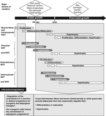
As we have seen, muscle and AT both display cellular and functional heterogeneity that varies along their respective growth phases. Current knowledge, reviewed above, highlights the cellular and molecular networks involved in muscular and adipose ontogenesis, and their different growth timelines. However, variations in lean-to-fat ratio in response to the developmental and nutritional cues confirm that events that control muscle and AT cells size and number are closely related. Current data, mainly available in monogastrics, suggest that molecular links and/or cross-talk occur between muscle and AT that may reciprocally act on their cell proliferation and/or differentiation, substrate metabolism, and functioning, depending on the stage of development (Figure 4).
Depiction of the cellular events involved in the ontogenesis of muscle fibres (light grey) brown adipose tissues (BAT; dark grey) and white adipose tissues (WAT; white) adipose tissues in cattle. Major variation factors and putative interactions between tissues and/or regulation are indicated.

Molecular control of the cell lineage fate
The decrease in the number of secondary myofibres and the increased adiposity in offspring born from mothers underfed during early pregnancy, together with the reciprocal variations observed in cattle harbouring a high growth potential, suggest that a balance occurs in the commitment of a common progenitor cell into the myogenic or brown adipogenic lineages. However, neither simultaneous regulation of different progenitors nor cross-talk between the different cell types can be ruled out. Interestingly, recent results (Figure 3) showed that BAT cells, but not WAT cells arose from Myf-5 expressing progenitors committed into the brown adipogenic or muscular programme, depending on the expression of PRDM16 (Seale et al., Reference Seale, Bjork, Yang, Kajimura, Chin, Kuang, Scime, Devarakonda, Conroe, Erdjument-Bromage, Tempst, Rudnicki, Beier and Spiegelman2008). Loss of PRDM16 in primary brown fat precursors caused myogenic gene expression and muscle differentiation; in vitro PRDM16 expression in committed myoblasts, both primary and immortalized, changed their phenotype into brown adipocytes (Seale et al., Reference Seale, Bjork, Yang, Kajimura, Chin, Kuang, Scime, Devarakonda, Conroe, Erdjument-Bromage, Tempst, Rudnicki, Beier and Spiegelman2008). In addition, consistent with a common muscle and BAT progenitor, other groups have reported that (i) BAT and skeletal muscle are derived from a common progenitor in the dermomyotome (Atit et al., Reference Atit, Sgaier, Mohamed, Taketo, Dufort, Joyner, Niswander and Conlon2006), (ii) myogenin-deficient neonatal mice not only have impaired muscle development but also exhibit an accumulation of brown adipocytes in the anatomical location of muscles (Hasty et al., Reference Hasty, Bradley, Morris, Edmondson, Venuti, Olson and Klein1993) and (iii) brown but not white adipose precursors express many genes and microRNA specific to muscle precursors (Timmons et al., Reference Timmons, Wennmalm, Larsson, Walden, Lassmann, Petrovic, Hamilton, Gimeno, Wahlestedt, Baar, Nedergaard and Cannon2007). Finally, adult skeletal muscle has been shown to be a ‘reservoir’ of brown adipocyte progenitors in humans (Crisan et al., Reference Crisan, Casteilla, Lehr, Carmona, Paoloni-Giacobino, Yap, Sun, Leger, Logar, Penicaud, Schrauwen, Cameron-Smith, Russell, Peault and Giacobino2008) and mice (Almind et al., Reference Almind, Manieri, Sivitz, Cinti and Kahn2007). Whether a single Myf5+ cell gives rise to both BAT and muscle, or whether there are distinct populations of Myf5+ progenitors remains unknown.
These results challenge the prevailing model whereby a common MSC gives rise to muscle, WAT and BAT (and also bone) in response to appropriate developmental cues (Seale et al., Reference Seale, Kajimura and Spiegelman2009). However, brown adipocytes arise from Myf-5-expressing progenitors only in the BAT and not in WAT in mice (see discussion of AT growth above). This raises the question of the origin of WAT and its putative link with muscle. Muscle cells and some white adipocytes have been described as originating from the mesoderm and from a common MSC, whose cell fate is under the control of molecular pathways that are beginning to be identified. The canonical Wnt promotes myogenesis while suppressing adipogenesis in mice MSC (Seale et al., Reference Seale, Kajimura and Spiegelman2009) and bovine bone marrow stromal cells (Tan et al., Reference Tan, Reverter, Wang, Byrne, McWilliam and Lehnert2006). Wnt inhibits both brown and white adipogenesis by blocking the induction of key transcription factors for adipogenesis (PPARγ and C/EBPα in WAT and PGC1α in BAT (Seale et al., Reference Seale, Kajimura and Spiegelman2009)). Myostatin inhibits white adipogenesis through the activation of Wnt signalling, which in turn inhibits the expression of PPARγ in human MSC and primary white pre-adipocytes (Guo et al., Reference Guo, Flanagan, Jasuja, Kirkland, Jiang and Bhasin2008). Conversely, myostatin promotes the commitment and differentiation of multi-potent mesenchymal cells lines into the adipogenic lineage and inhibits their differentiation into the myogenic lineage (Artaza et al., Reference Artaza, Bhasin, Magee, Reisz-Porszasz, Shen, Groome, Fareez and Gonzalez-Cadavid2005). This shows that myostatin effects on in vitro white adipogenesis seem to be cell-type specific. Whether myostatin similarly regulates the commitment of both BAT and WAT progenitors remains unknown, as does the hierarchization in, or cooperation between, PRDM16, β-catenin and myostatin signalling. It may, however, be hypothesized that proteins such as PRDM16 and Wnt regulate the commitment and early differentiation of (i) a common progenitor that gives rise to either muscle and BAT, or muscle and WAT, depending on anatomical site, or (ii) different progenitors that give rise to muscle, BAT and WAT. To date, none of these hypotheses can be evaluated due to lack of knowledge on the developmental origin of AT. In addition, whatever their origin, the precocial differentiated myotubes, and especially secondary myotubes, may affect the commitment and differentiation of adipose progenitors through the secretion of myokines such as myostatin.
Reciprocal regulation of AT and muscle cell differentiation in vitro
Once committed, the adipose and muscular cells undergo differentiation. In addition to the metabolic and endocrine regulations specific to each tissue (see above), cross-talk between adipose and muscle cells leading to the reciprocal regulation of their differentiation has been deciphered by in vitro studies. This cross-talk mainly concerns myogenic cells and brown or white fat cells during the foetal or post-natal periods, respectively. We used models of primary co-culture and conditioned medium to study the direct interactions that occur during myogenesis and adipogenesis in bovine foetuses. In preliminary results, we observed that myogenesis was impaired by proliferative pre-adipocyte-conditioned media, as shown by a low fusion index, an indicator of myoblast fusion (Cassar-Malek et al., Reference Cassar-Malek, Bonnet, Chilliard and Picard2006). Conversely, myogenesis was enhanced by adipocyte-conditioned medium, through an increase in the myotube area. Elsewhere, the differentiation of foetal pre-adipocytes was enhanced by co-culture with primary bovine myoblasts (Bonnet et al., Reference Bonnet, Cassar-Malek, Delavaud, Tourret, Chilliard and Picard2008). This may result from a regulation by paracrine signals and/or from physical cellular interactions as already described between white adipocytes and endothelial cells. The paracrine signals involved in this cross-talk between foetal cells remain unknown, while some have been identified in the cross-talk between white adipocytes and muscular cells; leptin increased the fusion index (+28%, P < 0.02, Bonnet et al., unpublished results) and stimulated glucose uptake in C2C12 myogenic cells (Berti and Gammeltoft, Reference Berti and Gammeltoft1999). Myostatin has been shown to impair differentiation of bovine white pre-adipocytes by inhibiting the expression of PPARγ and C/EBPα (Hirai et al., Reference Hirai, Matsumoto, Hino, Kawachi, Matsui and Yano2007). These few data highlight the complexity of the cross-talk between muscle and BAT or WAT, which involves both secreted factors and cell–cell interactions, and concern different cell types depending on the growth timelines of muscles and ATs.
Reciprocal regulation between WAT and muscle of nutrient metabolism and tissue function
Competition or prioritization between adipose and muscle cells for the uptake and metabolism of nutrients is suggested by the inhibited or delayed AT growth in feed-restricted cattle and in bovine genotypes exhibiting high levels of muscle development (see above). The dynamic control of nutrient partitioning and storage depends on balancing energy demand of the tissue with energy supply. In ruminants this balance is maintained through the integration of many different signals that communicate the nutrient status of the organism to the periphery, including insulin, growth hormone (GH), cortisol, catecholamine, NEFA, glucose and adipokines such as leptin (Chilliard et al., Reference Chilliard, Bocquier and Doreau1998; Chilliard et al., Reference Chilliard, Delavaud and Bonnet2005). Insulin and GH are deeply involved in the regulation of nutrient partitioning. Interestingly, the GH/insulin ratio was higher in steers than in heifers, concomitantly to higher lean-to-fat ratio (Gettys et al., Reference Gettys, Henricks and Schanbacher1988), during the initial phase of growth recovery (Hornick et al., Reference Hornick, Van Eenaeme, Gqrard, Dufrasne and Istasse2000), and higher in Holstein than Japanese Black (Shingu et al., Reference Shingu, Hodate, Kushibiki, Ueda, Watanabe, Shinoda and Matsumoto2001). This is in agreement with the well-known proteogenic and antilipogenic effects of GH and the lipogenic and proteogenic effects of insulin. Concomitantly, an additional signal that may regulate nutrient partitioning during growth is the plasma level of leptin. Leptin, whose secretion by WAT increases as body fatness increases and/or energy intake increases, stimulates energy expenditure and FA oxidation (through stimulation of AMP-activated protein kinase (AMPK) activity) in muscle, liver and WAT, inhibits WAT lipogenesis and decreases appetite. Plasma leptin is low during the first year of life in calves (Blum et al., Reference Blum, Zbinden, Hammon and Chilliard2005), is lower in steers than in heifers (Brandt et al., Reference Brandt, Keisler, Meyer, Schmidt and Berg2007) and in lean than in fat breeds (Chilliard et al., Reference Chilliard, Delavaud and Bonnet2005), which may contribute to favouring muscle growth in early life and in late-maturing cattle. Lean steers compared with fat steers have a decreased oxidative metabolism (Jurie et al., Reference Jurie, Cassar-Malek, Bonnet, Leroux, Bauchart, Boulesteix, Pethick and Hocquette2007), an increased AMPK activity (Underwood et al., Reference Underwood, Means, Zhu, Ford, Hess and Du2008), in addition to decreased leptinemia and leptin gene expression (Chilliard et al., Reference Chilliard, Delavaud and Bonnet2005; Bonnet et al., Reference Bonnet, Faulconnier, Leroux, Jurie, Cassar-Malek, Bauchart, Boulesteix, Pethick, Hocquette and Chilliard2007). Thus leptin, by activating AMPK, may favour nutrient oxidation in muscle over nutrient storage in WAT.
It has also been shown that preferential growth of AT may occur in growing animals fed a high concentrate diet after a period of foetal or post-natal restriction (see above). In monogatrics, a ‘thrifty mechanism’ was proposed to sustain the compensatory growth and preferential deposition of WAT in the post-natal period (Dulloo, Reference Dulloo2008). A decrease in plasma level of leptin or other adipose-specific signal(s) that sense the state of AT store depletion leads to a decrease in PI3K- and AMPK-stimulated energy expenditure in muscle. These muscular adaptations result in reduced glucose utilization, which during re-feeding will lead to concomitant insulin and leptin resistance. The resulting hyperinsulinaemia serves to redirect the glucose spared from oxidation in muscle towards de novo lipogenesis and fat storage in WAT, in line with the preferential growth of AT demonstrated after growth retardation in ruminants (see above). Evidence in favour of the existence of a ‘thrifty mechanism’ in ruminants comes first from the observation that leptin, through its muscle receptors (Chelikani et al., Reference Chelikani, Glimm and Kennelly2003), disrupts the insulin-signalling pathway through the IRS-1-PI3K pathway in bovine myogenic cells (Lulu Strat et al., Reference Lulu Strat, Kokta, Dodson, Gertler, Wu and Hill2005). Further evidence comes from metabolic adaptations to nutritional foetal programming in sheep. Activities of IRS-1-PI3K and AMPK were decreased in semitendinosus muscle of overweight, hyperglycaemic and hyperinsulinemic foetuses from overfed ewes compared with controls (Zhu et al., Reference Zhu, Han, Tong, Ma, Kimzey, Underwood, Xiao, Hess, Ford, Nathanielsz and Du2008). In addition, the post-natal higher adiposity induced by maternal undernutrition is accompanied by glucose intolerance and insulin resistance (Gardner et al., Reference Gardner, Tingey, Van Bon, Ozanne, Wilson, Dandrea, Keisler, Stephenson and Symonds2005). In this study, hyperleptinemia was not observed, but a rapid rise in leptinemia was reported in pre-term calves during the first week of life up to levels measured in full-term calves (Blum et al., Reference Blum, Zbinden, Hammon and Chilliard2005). The ‘thrifty mechanism’ has also been associated with decreased energy expenditure, which would allow fat mass recovery. The higher adiposity of low-birth-weight lambs has been attributed to a 30% lower energy requirement for maintenance, coupled with a higher relative intake (Greenwood et al., Reference Greenwood, Hunt, Hermanson and Bell1998). Whether the ‘thrifty mechanism’ in cattle, a species that is adapted to low glucose availability, affects adipose and muscular growth in cattle as in humans, and concomitantly affects volatile fatty acid together with glucose metabolism, remains to be determined.
To date, few data address the role of adipokines on the regulation of protein metabolism in muscle (Argiles et al., Reference Argiles, Lopez-Soriano, Almendro, Busquets and Lopez-Soriano2005). Likewise, the effects of myokines on AT metabolism have never been addressed in ruminants and scant results in monogastrics report, for example, that interleukins 6 and 15 affect AT growth through decreased lipogenesis, and for interleukin 6 through increased lipolysis (Argiles et al., Reference Argiles, Lopez-Soriano, Almendro, Busquets and Lopez-Soriano2005).
Concluding remarks and future trends
Muscle growth results from an increase in the number of myofibres during the first two thirds of pregnancy and thereafter exclusively from an increase in fibre size. Before 180 dpc, muscle plasticity mainly arises from variability in the number of secondary myofibres, which are sensitive to foetal programming by early severe maternal nutrition in sheep or by factors related to the bovine genotype. The number of primary myofibres is controlled by factors intrinsic to the animal and is resistant to manipulation. After 180 dpc and throughout the post-natal period, muscle plasticity derives from the variation in myofibre hypertrophy, which allows cattle to adapt to their environment.
AT grows by the successive growth of foetal brown and post-natal white AT. Unlike muscle cells the number of adipocytes is never set. In addition, the timing of the balance between increase in size and increase in number of adipocytes differs depending on the anatomical site of AT: end of the foetal growth for perirenal, around age 1 year for subcutaneous and intermuscular AT and probably later for the late-maturing intramuscular AT. To date, few data are available on the plasticity of foetal BAT in ruminants. However, the perirenal BAT growth seems to be sensitive to foetal programming by early maternal nutrition with an impact on post-natal perirenal WAT growth in sheep. Post-natal plasticity of WAT growth results mainly from variations in the size of existing adipocytes. However, when an average upper limit size of adipocytes is reached, AT also grows by an increase in adipocyte number. This shows that the number of white adipocytes is determined early in life. However, the developmental origin of white adipocytes remains to be clarified. Do white adipocytes arise from a transdifferentiation of brown adipocytes or do they come from the same or different progenitors, which develop in the perinatal period and progressively replace brown adipocytes? Cattle may offer a model of brown adipogenesis that could be relevant to human health outcomes; some therapeutic strategies under investigation advocate inducing the development of small amounts of BAT in obese humans.
Interactions between myofibres and brown adipocytes remain largely unexplored, but may modify both foetal and post-natal growth. Does the plasticity in the numbers of secondary myofibres and in adiposity before 180 dpc result from the regulation of the commitment of a single common progenitor or of two distinct ones? Interactions between myofibres and white adipocytes are beginning to be described and the underlying pathways mainly concern a regulation of nutrient partitioning. The variations in lean-to-fat ratio result from variations in adipocyte number and size and to a lesser extent in myofibre size, which suggests that metabolic and endocrine adaptations regulating nutrient partitioning protect muscle growth thanks to the plasticity of WAT. The mediators involved in the reciprocal regulation between muscle and AT for nutrient partitioning remains largely unknown due to lack of knowledge on myokine secretion and effect on AT, and on effects of adipokines, other than leptin, on muscle. The understanding of these interactions is a prerequisite to a possible manipulation of the lean-to-fat ratio.
Lastly, the understanding of the regulation of the balance between adipogenesis and myogenesis may become more complex because of signalling coming from bone growth. Higher bone mass in bovine genotype harbouring a high muscle and a low AT growth, together with the identification of common cell progenitors for the three tissues, suggest that osteogenesis may interact with adipogenesis and/or myogenesis.