Atrial septal defect is one common CHD that makes up a significant proportion of cases needing closure, as irreversible pulmonary vascular changes occur if not repaired on time.Reference Geva, Martins and Wald1 Ostium secundum defects which are either large or with flimsy and inadequate rims usually undergo surgical patch closure. Recurrent defects after surgical atrial septal defect closure are uncommon and have been reported with a varying incidence of 2–7%. Reference Meijboom, Hess and Szatmari2,Reference Valdes-Cruz, Pieroni and Jones3 We present a case of a 16-year-old young male with symptomatic recurrent atrial septal defect secondary to patch degradation after 10 years of initial surgical closure. Very few reports of device closure of recurrent atrial septal defects are cited in literature. Our aim is to highlight importance of long-term echocardiographic surveillance in surgical patch closure patients which in turn will enhance the understanding of the concepts related to recurrent defects of CHDs.
Case report
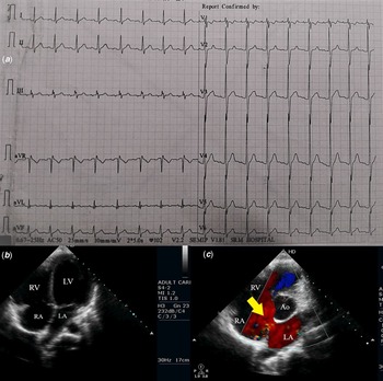
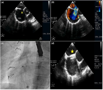
A 16-year-old boy presented with palpitations, exertional fatigue, and NYHA class II dyspnoea for last 6 months. He had undergone successful surgical patch closure using autologous pericardial patch for 28 mm ostium secundum atrial septal defect with deficient inferoposterior rims, 10 years back. Auscultation showed a wide fixed split S2 and grade 2/6 systolic murmur in the upper left sternal border. Electrocardiogram showed sinus tachycardia, Right bundle branch block, notching in the QRS complex of lead 3 (Fig 1A). Transthoracic echocardiogram showed a dilated right atrium and ventricle (Fig 1B), with a doubtful jet into the right atrium across the interatrial septum (Fig 1C). Transesophageal echocardiogram showed a new eccentric, anterosuperior defect of 11 mm size in the pericardial patch with a left to right shunt. The rims formed by the patch were found to be adequate except in the retro-aortic region (Fig 2A, B) (Video 1). Cardiac catheterisation showed a Qp:Qs ratio of 1.4:1. There was no documented evidence of any residual shunting in the immediate post-operative period. Considering his age, the recent development of symptoms, and the change in functional status, it was decided to close the defect percutaneously. Amplatzer 14 mm atrial septal occluder was successfully deployed across the defect after confirming the stability (Fig 2C) (Video 2). Post-procedural transesophageal echocardiogram showed a stable device position (Fig 2D). The patient’s symptoms improved and is on regular follow-up.
(a) Electrocardiogram on admission having sinus tachycardia, right bundle branch block, and prominent RV forces. ( b) 2D transthoracic echocardiogram apical 4 chamber view showing dilated RA, RV. ( c) Modified 5- chamber view showing doubtful jet across the surgical patch (yellow arrow).

(a, b) TEE images showing 11 mm recurrent ASD (yellow arrowhead) in the pericardial patch with an absent aortic rim and with a left to right shunt on colour flow doppler. ( c ) Fluoroscopic image after successful Amplatzer ASO 14 mm device deployment. Sternal sutures suggestive of previous surgery can be seen. ( d ) TEE image indicating stable device position (yellow star) across the pericardial patch. ASD – Atrial septal defect; RA – right atrium; LA – Left atrium; RV - right ventricle; LV – Left ventricle; Ao- Aorta; TEE – Transoesophageal echocardiography; ASO – Atrial septal occluder.

Discussion
In the current transcatheter device closure era for ostium secundum defects, surgical patch closure is done mainly for large defects or defects with inadequate rims and other complex CHDs. Patch closure is done using pericardium, either autologous or bovine and prosthetic material. Prosthetic materials like Dacron and Polytetrafluoroethylene carry a risk of endocarditis and thrombus formation. Reference Okwulehie, Dharmapuram, Swain, Ramdoss, Sundararaghavan and Kona4 Autologous pericardial patch being easily available, sterile, and non-immunoreactive is the most commonly preferred material. Reference Okwulehie, Dharmapuram, Swain, Ramdoss, Sundararaghavan and Kona4 However, it has a rough surface with a tendency to curl at the edges, leading to difficult handling and suturing. Treatment with 0.6% glutaraldehyde improves its handling qualities by cross-linking collagen, thereby reducing elasticity and enhancing shape fixation. Hence, glutaraldehyde fixed autologous pericardium is preferred, more so for repair of complex heart defects. Reference Liava'a and Kalfa5
The presence of a shunt across the patch of a surgically closed atrial septal defect is uncommon. There may be a persistent defect at the end of surgery termed as residual defect. Defects developing later in time after initial successful surgical closure are termed as recurrent defects. Evidence has been sparse in definition, diagnosis, and management of these conditions. They have mainly been attributed to two pathological processes namely suture dehiscence and patch degeneration. Suture dehiscence which usually occurs in the early postoperative period is seen at the suture site and is caused by excessive tension, trauma, and kinking stresses at knots. Suture dehiscence is commonly associated with closures involving complex CHDs with abnormal geometry of the defect or associated structures. In contrast, patch degeneration which usually occurs a few years later refers to shrinkage, retraction, calcification, aneurysmal dilatation, or degradation of the patch. Glutaraldehyde treatment of autologous pericardium has been shown to accelerate degenerative process. Unlike dehiscence which occurs primarily at suture lines, patch degradation may be located anywhere leading to eccentric shunts. Reference Atik, Afiune and Caneo6,Reference Mohanty and Kumar7 The precise timing of development of recurrent ASD in our patient is difficult to predict, and could have variably progressed anywhere from immediate post-operative period to 6 months prior to index hospitalisation. Both possibilities of patch degeneration and late suture dehiscence were considered. A consensus of possible patch degeneration was arrived at after the heart team discussion, considering the slow progressive dyspnoea, absence of dehiscence flap near the retro aortic region, and the rarity of late suture dehiscence presenting 10 years after the initial surgery. Further imaging was limited due to logistic reasons. Obviously, this consensus represents an educated speculation.
Device closure of recurrent defects follow the same technique and selection criteria as for native secundum atrial septal defects. TEE has been the conventional method to assess the adequacy of the patch rims and device sizing. In future, newer modalities like intracardiac echocardiography or 3D printing may yield more information regarding patch thickness in this particular subset of patients. Percutaneous closure is less invasive, cost-effective with lesser complication rates compared to redo-surgery. Reference Mohanty and Kumar7–Reference Chessa, Butera, Giamberti, Bini and Carminati10 This case highlights the importance of long-term clinical and echocardiographic surveillance in patients after surgical atrial septal defect closure. There should be a low threshold for transesophageal echocardiogram in the event of any suspicious findings on transthoracic echocardiogram along with a change in the patient’s clinical status.
Transcatheter closure is an excellent, feasible treatment modality compared to redo surgery is select cases of recurrent atrial septal defect.
Supplementary material
To view supplementary material for this article, please visit https://doi.org/10.1017/S1047951122001962
Acknowledgements
None.
Financial support
This research received no specific grant from any funding agency, commercial, or not-for-profit sectors.
Conflicts of interest
None.