

Significant outcomes
• Wild-type mice upregulate proteasomal activity in neural tissues in response to 1-methyl-4-phenyl-1,2,3,6-tetrahydropyridine (MPTP) exposure in vivo, whereas Fas-deficient mice do not.
• Agonistic stimulation of the Fas receptor induces significant upregulation of chymotrypsin-like and caspase-like proteasomal activities in Fas-expressing SH-SY5Y neuroblastoma cells.
• Data suggest that Fas can promote proteasomal activity, which may reduce protein aggregation observed in PD and other neurodegenerative disorders. This may begin to provide a mechanism by which Fas exerts neuroprotective effects in the MPTP model of PD.
Limitations
• In this investigation, a limited amount of data is presented to explore a potentially complicated system. More experiments are necessary in order to further validate and understand the relationship between Fas and the ubiquitin proteasome system (UPS) in the normal and diseased brain.
• Only acute responses are observed, and the addition of later time points after MPTP treatment would shed light on the chronic effects of MPTP on the induction of proteasomal activity in wild-type and Fas-deficient mice.
• Fas is likely acting through multiple mechanisms, not limited to the induction of proteasomal activity. Fas has been shown previously to induce phagolysosomal activity and neurite growth.
Introduction
Fas, a member of the tumour necrosis factor receptor superfamily, has been extensively studied as an apoptosis-inducing receptor (Reference Nagata1). In the nervous system, it can induce neuronal apoptosis (Reference Martin-Villalba, Herr and Jeremias2–Reference Raoul, Henderson and Pettmann4); however, Fas can also mediate numerous other functions (Reference Peter, Budd and Desbarats5). It is upregulated in neurons and glial cells in response to cellular stress or injury (Reference Choi and Benveniste6), and can promote recovery after sciatic nerve crush and induce neurite outgrowth from dorsal root ganglion cells (Reference Desbarats, Birge, Mimouni-Rongy, Weinstein, Palerme and Newell7). In a previous study, we investigated the role of Fas signalling in the 1-methyl-4-phenyl-1,2,3,6-tetrahydropyridine (MPTP)-induced mouse model of Parkinson's Disease (PD) (Reference Landau, Luk and Jones8). Fas-deficient lymphoproliferative (lpr) mice developed a phenotype resembling clinical PD, characterised by extensive nigrostriatal degeneration accompanied by tremor, hypokinesia, and loss of motor coordination, at a dose of MPTP which caused neither neural degeneration nor behavioural impairment in wild-type (wt) mice. Thus, deficient Fas expression increased susceptibility of dopaminergic neurons to MPTP toxicity in vivo, supporting a neuroprotective role for Fas (Reference Landau, Luk and Jones8).
PD, a progressive neurodegenerative disease, is the second most common neurodegenerative disease after Alzheimer's disease. Symptoms of PD include bradykinesia, tremor, postural instability and impaired motor coordination. PD results from the selective degeneration of dopamine neurons in the substantia nigra of the brain. The remaining neurons contain Lewy bodies, which are protein aggregates composed of α-synuclein, ubiquitin, and parkin. The aggregation of modified proteins is a pathological hallmark of most neurodegenerative diseases (Reference Ciechanover and Brundin9).
The ubiquitin proteasome system (UPS) is an intracellular pathway that targets damaged proteins for degradation. A series of enzyme-mediated reactions occur in which several ubiquitin molecules are ligated to the substrate to be degraded as a targeting signal to the proteasome. The proteasome degrades the ubiquitin-tagged substrate proteins into small peptides and ensures the release of ubiquitin molecules with the help of deubiquitinating enzymes.
Defects in protein degradation by the UPS have been reported in PD (Reference McNaught, Jackson, JnoBaptiste, Kapustin and Olanow10). In familial PD, rare mutations in parkin, an ubiquitin-activating enzyme, and ubiquitin C-terminal hydrolase 1, a deubiquitinating enzyme, are implicated in dysfunction of the UPS (Reference Shimura, Schlossmacher and Hattori11,Reference Leroy, Boyer and Auburger12). Selective impairment of proteasomal activity and reduced expression of proteasomal subunits have been reported in postmortem tissue from the substantia nigra of patients with sporadic PD (Reference McNaught, Belizaire, Isacson, Jenner and Olanow13,Reference McNaught, Belizaire, Jenner, Olanow and Isacson14). Results from rat studies indicate a reduced expression of proteasome activators in brain regions involved in PD compared with other brain areas, and in dopaminergic cells compared with other cell types (Reference McNaught, Jnobaptiste, Jackson and Jengelley15). Proteasome inhibition has been used as a tool to induce experimental PD in animal models (Reference McNaught, Perl, Brownell and Olanow16–Reference Sun, Anantharam, Zhang, Latchoumycandane, Kanthasamy and Kanthasamy18). Aggregated α-synuclein, the main component of Lewy bodies, inhibits proteasomal activity, and proteasomal activity is decreased in cells overexpressing α-synuclein (Reference Fujita, Sugama and Nakai19,Reference Chu, Dodiya, Aebischer, Olanow and Kordower20). A hypodysfunctional UPS may underlie abnormal protein accumulation, thereby facilitating the formation of toxic protein aggregates and increasing the vulnerability of nigral dopaminergic neurons to degeneration in sporadic PD.
Aim of the study
The aim of this study was to determine whether Fas expression and stimulation modified the activity of the UPS in neural tissue, in particular in the context of the MPTP model of PD. We measured proteasome enzyme activities in the midbrain of wt and Fas-deficient lpr mice, treated with MPTP or saline as a control. We aimed to determine whether Fas expression and engagement could promote proteasome enzyme activity, which may in part account for its neuroprotective role in MPTP-induced parkinsonism in mice.
Materials and methods
Mice
We purchased wt C57BL/6 mice from Charles River Canada, and we bred B6.MRL-lpr mice in our facility. We followed Canadian Council on Animal Care ethical guidelines, and the McGill University Animal Care Committee approved all animal experiments. All efforts were made to minimise animal suffering and reduce the number of animals used.
MPTP treatment and midbrain dissection
We injected wt and Fas-deficient lpr mice twice with saline or 30 mg/kg MPTP-HCl (Sigma, Aldrich, St. Louis, MO, USA) at 24-h intervals. We euthanised mice by cervical dislocation 2 h after the second injection and dissected the midbrain (Reference Smeyne and Smeyne21) for the proteasome enzyme assays.
Proteasome enzyme assays
We lysed midbrain tissue or SH-SY5Y cells in Tris/ethylenediaminetetraacetic acid (EDTA) by 100 strokes of manual homogenisation on ice, and cleared the lysate by centrifugation. We adjusted the protein concentration of the supernatant to 0.1 mg/ml and plated quadruplicates of each sample. We plated two extra replicates with the proteasomal inhibitor MG132 (15 mM). We added fluorescent substrates (2.5 mM) in proteasome assay buffer (8 mM DTT, 10 mM MgCl2, 4 mM ATP) immediately prior to the first reading on an Analyst Fluorescence plate reader (Molecular Devices/LJL Biosystems, Sunnyvale, CA, USA). We used substrates Z-LLE-AMC for caspase-like activity, Suc-LLVY-AMC for chymotrypsin-like activity and Boc-LRR-AMC for trypsin-like activity. We measured fluorescence at 30 min or 1 h intervals for 4–5 h for chymotrypsin and caspase activities, and for 1 h for trypsin activity, and maintained plates at 37 °C/5% CO2 protected from light between readings.
Cell lines and antibody stimulation
We obtained SH-SY5Y cells from the American Type Culture Collection (ATCC) and maintained them in Dulbecco's Modified Eagle Medium (DMEM) supplemented with 10% fetal calf serum (FCS). Fas-positive cells were selected by three cycles of fluorescence activated cell sorting (FACS) for the 5% of cells with highest Fas expression. Cells were allowed to expand in culture between each sort. For assays, the SH-SY5Y cells were grown to 80% confluence in 6-well plates then stimulated for 18 h with 10 ng/ml agonistic anti-human Fas antibody (clone CH11) or isotype-matched control antibody. Cells were harvested using cell scrapers, without trypsin.
Cell viability assay
We plated Fas-positive SH-SY5Y and Jurkat cells in phenol-red-free DMEM and treated them with 20 ng/ml agonistic anti-human Fas antibody (clone CH11) or isotype-matched control antibody. The SH-SY5Y cells were incubated for 4 h and the Jurkat cells for 40 h at 37 °C/5% CO2. We assayed cell viability using WST-1 reagent (Roche Diagnostics, Quebec, Canada) according to the manufacturer's instructions.
Flow cytometry
To sort the SH-SY5Y cells and to determine their levels of Fas expression, we stained the cells with phycoerythrin (PE)-conjugated anti-human Fas antibody (clone DX2) and sorted and analysed them using a FACSAria cell sorter (Becton Dickinson, San Jose, CA, USA).
Statistics
We calculated standard error of the mean (SEM) and derived p values using Student's t-tests.
Results
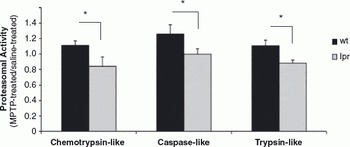
In a previous study, we had found that deficient Fas expression increased the susceptibility of mice to MPTP-induced Parkinsonism (Reference Landau, Luk and Jones8). To determine whether proteasomal activity correlates with Fas-induced neuroprotection in this model, we measured proteasome enzyme activities in the midbrain of wt and Fas-deficient lpr mice, treated with MPTP or saline as a control. Midbrain neural tissue from wt and lpr mice displayed similar baseline proteasomal activities. However, in wt mice, MPTP exposure resulted in upregulation of proteasome function, whereas in lpr mice, MPTP treatment caused a decrease in proteasomal activities (Fig. 1).
Fas-deficient lpr mice treated with MPTP show decreased proteasomal activities. Mice were injected with 30 mg/kg MPTP or with saline as a control, twice at 24 h intervals. Proteasome activities in midbrain lysates of wt and lpr mice were measured fluorometrically 2 h after the second injection. Data are expressed as a ratio of the activity in MPTP-treated versus saline-treated mice for each strain. Averages and SEM of three independent experiments are shown (n = 6 mice per group, *p < 0.05).

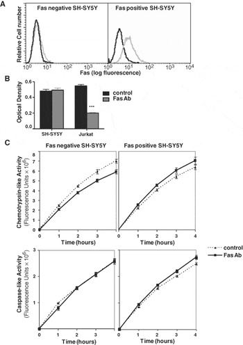
Changes in midbrain proteasomal activity may reflect glial and neuronal proteasome function. We tested the direct consequence of Fas stimulation on neuronal proteasomal activity in vitro using the SH-SY5Y neuroblastoma cell line. We treated SH-SY5Y cells that were negative or very low for Fas expression, and SH-SY5Y cells selected for higher levels of Fas expression (Fig. 2a) with an agonistic stimulatory antibody to Fas or an isotype-matched control antibody. We have previously shown that Fas ligation is not toxic to SH-SY5Y cells (Reference Landau, Luk and Jones8), and we show here that the viability of SH-SY5Y cells is unchanged upon exposure to anti-Fas antibody, at a dose toxic to over 60% of Jurkat cells (Fig. 2b). We found that Fas-engagement-induced significant upregulation of chymotrypsin-like and caspase-like proteasomal activities in Fas-expressing SH-SY5Y cells, but had no effect or decreased proteasomal activity in Fas-negative cells (Fig. 2c). These results suggest that signalling through Fas can enhance proteasomal activity in neuronal cells directly.
Fas engagement upregulates proteasome activities in a neuroblastoma cell line. (a) Flow cytometric profiles of Fas-negative (left) and Fas-positive (right) SH-SY5Y cells. Black line, isotype control staining; grey line, Fas staining. (b) Optical density (OD) as a measure of viability in SH-SY5Y and Jurkat cells treated with 20 ng/ml CH-11 anti-Fas antibody (grey bars) or control antibody (black bars). (c) Graphs show the rate of proteasomal activities in control-antibody-treated or agonistic Fas-antibody-treated SH-SY5Y cells. Increases in chemotrypsin-like activity at 1, 2 and 3 h and in caspase-like activity at 3 and 4 h in response to Fas stimulation in Fas-positive SH-SY5Y cells are significant (p < 0.05). The proteasome inhibitor MG132 completely suppressed all measured activities. Averages and SEM from four measurements are shown, and the data are representatives of three independent experiments.

Discussion
Summary of main findings
In this article, we show that Fas expression and/or signalling promotes proteasomal activity. Fas-deficient mice, unlike wt mice, do not upregulate proteasome function in response to MPTP in vivo. Furthermore, using SH-SY5Y neuroblastoma cells, a cell type not vulnerable to Fas-induced apoptosis, we find that Fas engagement with an activating antibody induces significant upregulation of proteasomal activities in Fas-expressing SH-SY5Y cells, but has no effect in Fas-negative cells.
Relevance to existing work
These preliminary data suggest a mechanistic explanation for the neuroprotective role of Fas that we have previously described in both SH-SY5Y cells and mice exposed to MPTP (Reference Landau, Luk and Jones8). All three proteasomal activities were increased in wt, but not in Fas-deficient mice in response to MPTP. Furthermore, when Fas-positive SH-SY5Y cells were stimulated with an activating antibody to Fas, chemotrypsin-like and caspase-like proteasomal activities were increased. In addition to these data, we have found that the U937 macrophage cell line upregulates all three proteasomal activities in response to Fas engagement, to a greater extent than SH-SY5Y cells (data not shown), possibly because U937 cells express higher levels of Fas than SH-SY5Y cells. It therefore appears that multiple cell types expressing Fas may have the ability to upregulate UPS activity in response to Fas stimulation. Furthermore, the human Fas-associated factor 1 (hFAF1) has been proposed to serve as a regulatory protein in the UPS (Reference Song, Yim, Kim, Kim and Lee22), which further supports the potential of Fas to induce proteasomal activity.
In patients with PD, Fas and Fas ligand expressions are reduced in the neurons of the substantia nigra (Reference Ferrer, Blanco, Cutillas and Ambrosio23). Concomitantly, the soluble form of Fas is increased in tissue from the nigrostriatal region of PD brains (Reference Mogi, Harada and Kondo24). Soluble Fas can act as a decoy receptor and can block Fas ligand binding to cell surface Fas. Thus, Fas signalling in patients with PD may be diminished both by decreased cell surface Fas expression and by the presence of soluble Fas. We have previously found that patients with PD manifest a defect in inducible Fas expression (Reference Landau, Luk and Jones8). As mentioned above, selective impairment of proteasomal activity and reduced expression of proteasomal subunits have been reported in postmortem tissues from the substantia nigra of patients with sporadic PD (Reference McNaught, Belizaire, Isacson, Jenner and Olanow13,Reference McNaught, Belizaire, Jenner, Olanow and Isacson14). On the basis of our present data, we speculate that decreased Fas expression and signalling in the PD brain could result in decreased activity of the UPS, and consequently inadequate degradation of proteins, culminating in oxidative stress and neuronal death.
Strengths and limitations of this study
Our findings describe a novel potential function of Fas as an inducer of proteasomal activity in neural tissue. The increase in Fas-mediated proteasomal activity may contribute to dopaminergic neuron survival after exposure to PD-inducing agents by slowing the accumulation of damaged proteins which would normally be processed through the UPS. This protection may be common to other neurodegenerative diseases featuring the accumulation of proteins as a pathological hallmark.
In this investigation, limited data are presented to explore a potentially complicated system. More experiments are necessary in order to further validate and understand the relationship between Fas and the UPS in the normal and diseased brain. Fas expression can be manipulated using overexpression or RNAi, or Fas signalling induced with Fas ligand or agonistic antibodies, to further explore Fas-dependent UPS activation. Although we have shown increases in proteasomal activities, we have not measured actual protein aggregation of ubiquitin, parkin or α–synuclein, which could significantly strengthen this work. Finally, proteasomal activity was measured 2 h after the last MPTP injection in mice. Kinetic studies would determine whether Fas expression exerts transient, reversible, or long-term effects on MPTP-induced UPS activity.
PD is a heterogeneous and debilitating disease. Treatment can slow progression of the disease, but eventual deterioration is inevitable, and at present there is no cure. Data from patients with PD and mice-bearing mutations in Fas suggest that susceptibility to PD neurodegeneration is increased by a decrease in Fas expression, and therefore that Fas is neuroprotective in PD. Our current data suggest a novel role of Fas in enhancing protein degradation through the UPS, and potentially attenuating harmful protein aggregation, which may partially explain its neuroprotective properties. Further work will be required to elucidate the pathway leading from Fas signalling to activation of the UPS. Previous work has shown that hFAF1 is a candidate molecular link between Fas and the UPS (Reference Song, Yim, Kim, Kim and Lee22). However, other pathways may also be implicated, including upregulation of proteasomal activity by Fas-induced cell stress. Studies of Fas as a neuroprotective factor could lead to treatments that promote survival of dopamine neurons. Future extensive studies are necessary to further explore the relationship between Fas, proteasome function and neuroprotection.
Acknowledgements
This research was funded by a Parkinson Society Canada New Investigator Award (J. D.) and by the Canadian Institutes of Health Research (CIHR; 53337). J. D. was supported by a CIHR New Investigator Salary Award and A. M. L. was the recipient of a CIHR Canada Graduate Scholarship.