Introduction
Setaria digitata belongs to the superfamily Filarioidea, the family Setariidae, and the genus Setaria. The life cycle involves ungulates as definitive hosts and mosquitoes as vectors. The first stage larvae are known as microfilariae, often sheathed and released into the bloodstream by the adult female worm. While taking a blood meal, mosquitoes take up the infective microfilariae, which are transmitted to the next natural host while feeding. The adult worm resides in the peritoneal cavity of cattle and buffalo and is often non-pathogenic, although in some cases, it causes mild peritonitis (Solusby Reference Soulsby1982). The aberrant migration of third stage (L3) larvae occurs in hosts like goats, sheep, and horses (Soulsby Reference Soulsby1982; Mohan et al. Reference Mohan, Ananda, Shridhar, Puttalakshmamma and Placid2009). Ophthalmic migration usually occurs in horses, and cerebrospinal migration occurs in horses, sheep, and goats, causing enzootic cerebrospinal nematodiasis/lumbar paralysis (Soulsby Reference Soulsby1982; Maharana et al. Reference Maharana, Potliya, Ganguly, Bisla, Mishra and Ganguly2020). The heterotrophic migration and swirling movement of the worm in the anterior chamber of the eye of horses, cattle, and goats can cause pathogenic changes to the cornea leading to corneal opacity and blindness (Mohan et al. Reference Mohan, Ananda, Shridhar, Puttalakshmamma and Placid2009; Gautam et al. Reference Gautam, Verma, Shah, Raghuvanshi, Nehra and Kumar2018). Also, there are reports of erratic migration of larvae of Setaria sp. to the urinary bladder, oviduct, liver, and heart (Yoshikawa et al. Reference Yoshikawa, Oyamada and Yoshikawa1976; Fujita et al. Reference Fujita, Imai, Ishii, Nunoya, Takahashi, Tomita and Oikawa1985; Sundar et al. Reference Sundar, D’souza and Jagannath2005). There are 43 species of Setaria reported worldwide, so accurate identification of the parasite with simple morphological features is insufficient (Wijesundera Reference Wijesundera2001). Therefore, molecular techniques, sequence data, and phylogenetic analysis will help in the confirmation of the parasite at the species level and also help in understanding the evolutionary pattern, relationship of species, and parasite biology (Senanayake et al. Reference Senanayake, Söderberg, Põlajev, Malmberg, Karunanayake, Tennekoon and Niazi2020). Tamilmahan et al. (Reference Tamilmahan, Zama, Pathak, Muneeswaran and Karthik2013) reported an incidence of 57.02% of equine ocular setariasis between 2002–2011. Here, we report the first case of ocular setariasis in a buffalo. Further, the parasite was confirmed as S. digitata by amplifying the 12S rRNA region, sequencing, and its phylogenetic analysis.
Materials and methods
The clinical case was presented at the referral veterinary polyclinic, Indian Veterinary Research Institute (IVRI). The parasite sample was analysed at the division of parasitology, IVRI, Izatnagar, Uttar Pradesh, India.
Case report
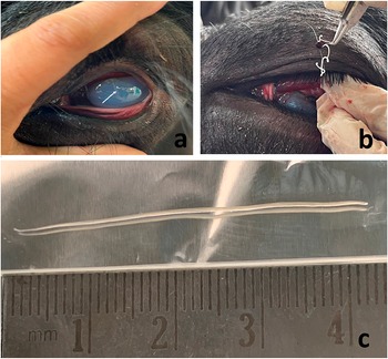
An adult Indian buffalo (Bubalus bubalis) presented with a history of restlessness, excessive lacrimation, and leukomatous cornea. On gross clinical examination, corneal opacity, blepharospasm, and discharge from the eye were observed (Figure 1a). A thin, milky white, thread-like, cylindrical, swirling worm was observed in the anterior chamber of the left eye. The blood sample was collected for wet film examination, peripheral blood smear, and complete blood count (CBC). No microfilariae could be detected in both wet film/blood smear, and CBC was also normal. Surgical intervention was preferred over medication. Proper restraining and irrigation of the eye with normal saline was done, and then auriculopalpebral and Peterson nerve block was given (Rafee Reference Rafee and Amarpal2016). A 5 mm incision was made at the 6 o’clock position at the limbus of the eye, and the worm was removed with the help of ocular forceps (Figure 1b). The surgical site was left to heal by administration of post-surgical antibiotic and anti-inflammatory drops.
(a) Whitish, thread-like worm (arrow) present in the anterior chamber of left eye with leukomatous cornea; (b) Retrieval of worm with ocular forceps; (c) Gross morphology of worm retrieved measuring 43 mm long.

Morphological identification
For morphological identification, the retrieved worm was washed thrice with phosphate-buffered saline (PBS), and both the anterior and posterior ends were observed under the light microscope. The identification was made based on criteria by Rhee et al. (Reference Rhee, Choi, Park and Jang1994).
Molecular Identification
Genomic DNA was isolated for molecular analysis using the phenol-chloroform-isoamyl alcohol method (Sambrook & Russel Reference Sambrook and Russell2006). The concentration of DNA was measured using a nano-spectrometer (Qiagen, Hilden, Germany) and subsequently stored at -20°C for further molecular work. Polymerase chain reaction (PCR) was performed with the extracted genomic DNA using the primers: SDF: 5' -AGT CCT CCC TTG TTG CTG GT-3' and SDR: 5'-GGG TGG TTT GTA CCC CTC CG-3' (Peng et al. Reference Peng, Armiladiana, Ruhil, Maizan and Choong2019). The amplification was done in a 50 uL reaction volume by adding 25 uL of 2X PCR master mix (Sapphire Amp Fast PCR Master Mix, TaKaRa, Shiga, Japan), 10 pmol each of forward primer and reverse primer, and 40 ng of template. The reaction conditions were: 94°C for 4 min, followed by 39 cycles of 94°C for 80 sec, 46°C for 80 sec, 72°C for 60 sec, and 72°C for 10 min (Yu et al. Reference Yu, Liu, Chen, Yi, Liu, Zhu and Li2021). The amplicons were gel purified and custom sequenced. The sequences were analysed with published sequences available in the NCBI data bank. The ClustalW program (https://www.genome.jp/tools-bin/clustalw) was used to obtain multiple sequence alignment, and then phylogenetic analysis was done using MEGA11 software (MEGA11: Molecular Evolutionary Genetics Analysis version 11 (Tamura et al. Reference Tamura, Stecher and Kumar2021)). A Tamura Nei substitution model was used to construct the distance matrix, and the phylogenetic tree was constructed using the maximum likelihood approach with a 500-bootstrap value (Yang Reference Yang2007).
Results and discussion
Parasite
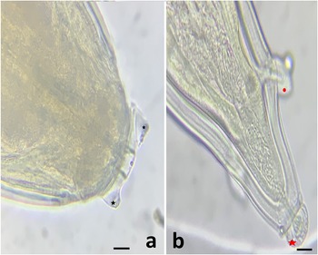
In gross examination: a 43 mm long, both-ends tapered, thread-like, milky white worm was observed (Figure 1c). The anterior end is round with two projections in the peri buccal area (Figure 2a), and the posterior end with a spherical bulb and lateral appendage is characteristic of female Seteria digitata (Figure 2b).
(a) Anterior end showing the presence of two tuberences (black star; dorsal projection and ventral projection); (b) Posterior end showing spherical bulb (red star) and lateral appendages (red round). Scale bar = 100 μm.

Seteria genus includes S.digitata, S.labitopapilosa, S. equina, and S.cervi, which are similar in morphology. Therefore, PCR was used to further confirm species by amplifying the 12S rRNA gene. After gel electrophoresis, the amplicons were purified and sequenced. The sequence result showed a 243 bp band, which confirmed the worm isolated was S. digitata by Basic Local Alignment Search Tool (BLAST) analysis and comparison with other sequences from the National Center for Biotechnology Information (NCBI).
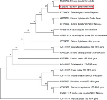
After BLAST analysis, the query sequence showed more than 99% similarity to the S. digitata mitochondrion complete genome (KY284626.1) and S. digitata Thailand isolates (OP895162.1), which confirmed the sequence was of S. digitata. Phylogenetic analysis showed a major clade formed by the genus Setaria and another by the genus Brugia + Wuchereria + Onchocerca, and an outgroup was formed by Thelazia gulosa (Figure 3).
Phylogenetic tree constructed in MEGA 11 software with related Setaria species and Thalezia gulosa as an outgroup, using maximum likelihood method after Tamura Nei substitution model and 500 bootstrap values. The generated sequence is represented in the red rectangle and other species’ accession numbers are also mentioned.

Occular setariasis caused by S. digitata mainly causes its pathogenic effect in equines. However, there are reports of ocular setariasis in goats and cattle (Mohan et al. Reference Mohan, Ananda, Shridhar, Puttalakshmamma and Placid2009; Gautam et al. Reference Gautam, Verma, Shah, Raghuvanshi, Nehra and Kumar2018; Shin et al. Reference Shin, Cho and Wee2002) but not in buffalo. Sundar & D’Souza (Reference Sundar and D’Souza2015) reported that the length of an adult male and female worm can reach up to 82 mm and 156 mm, respectively, and the length of fourth-stage larvae can be 22–23 mm (Tung et al. Reference Tung, Lai, Ooi, Yang and Wang2003). Here the retrieved worm was 45 mm long, suggesting it was an immature worm. The microscopic examination of the worm revealed ventral and dorsal protuberances at the anterior end and a smooth knob at the posterior end, suggestive of a female S. digitata worm. A surgical procedure was chosen over medication because of the lower risk and faster corneal opacity improvement. The presence of a dead worm in the eye can cause immunological reactions leading to further complications like degeneration of structures in the eye, enhancing intraocular pressure and eyeball rupture (Peng et al. Reference Peng, Armiladiana, Ruhil, Maizan and Choong2019). Ocular setariasis is caused mainly by S. digitata, but morphological characters cannot differentiate S. digitata from its congeners (Jayasinghee et al. Reference Jayasinghe and Wijesundera2003). Thus, for molecular confirmation, the 12S rRNA gene is targeted, which is an evolutionary marker (Yatawara et al. Reference Yatawara, Wickramasinghe, Nagataki, Rajapakse and Agatsuma2007; Junsiri et al. Reference Junsiri, Kamkong, Chinkangsadarn, Ouisuwan and Taweethavonsawat2023). Earlier, the sequence data analysis of the S. digitata showed 243 bp (Peng et al. Reference Peng, Armiladiana, Ruhil, Maizan and Choong2019; Yu et al. Reference Yu, Liu, Chen, Yi, Liu, Zhu and Li2021). Some potential mosquito vectors for the transmission of filarial nematodes are Anopheles, Mansonia, Culex, Armigeres, Aedes, and Ochlerotatus. Anopheles sp. and Armigeres sp. are mainly responsible for transmitting Setaria sp. (Siriyasatien et al. Reference Siriyasatien, Intayot, Sawaswong, Preativatanyou, Wacharapluesadee, Boonserm, Sor-Suwan, Ayuyoe, Cantos-Barreda and Phumee2023). The area where this case was reported has a prevalence of 63 species of mosquitoes, which include 15 species of Anopheles and Aedes, 24 species of Culex, 3 species of Mansonia, and 2 species each of Verrallina, Mimomyia, and Ochlerotatus (Kanojia & Geevarghese Reference Kanojia and Geevarghese2005). In Asia, the occurrence of S. digitata is high, and there are many reports of aberrant ophthalmic migration in equines (Shin et al. Reference Shin, Ahn, Suh, Kim, Jeong, Kim and Shin2017). However, this migration pattern is rare in buffalo; this the first report of an unusual occurrence of ocular setariasis in buffalo, which might be due to the high prevalence of vectors for S.digitata in the summer season. The accidental case may have occurred because of the vector’s high prevalence in the summer (Tung et al. Reference Tung, Cheng, Lai, Wang, Wang and Lee2004).
Acknowledgements
The authors express their sincere gratitude to the Head of Division, Division of Parasitology, for providing research facilities.
Financial support
None.
Competing interest
None.
Ethical standard
All the authors here by declare that they have followed the ethical standard for this experiment.