Introduction
Progressive facial hemiatrophy, generally referred to as Parry–Romberg syndrome, is a rare condition of unknown aetiology characterised by slow, progressive, unilateral facial atrophy that affects the skin, subcutaneous tissue, muscle and bone.Reference Singh and Bajpai1–Reference Mazzeo, Fisher, Mayer and Mathieu3
Various theories of pathogenesis have been proposed, including infection,Reference Abele, Bedingfield, Chandler and Given4 cervical sympathetic loss or imbalance,Reference Moss and Crikelair5, Reference Scope, Barzilai, Trau, Orenstein, Winkler and Haik6 trigeminal peripheral neuritis,Reference Pensler, Murphy and Mulliken2 lesions in the central nervous system,Reference Carreno, Donaire, Barcelo, Rumia, Falip and Agudo7 and scleroderma.Reference Sakuraoka, Tajima and Nishikawa8 In recent years, several articles have addressed this syndrome, but mainly from a plastic surgery, rather than an aetiological, perspective.Reference Moore, Wong, Proudman and David9, Reference Guerrerosantos, Guerrerosantos and Orozco10
In this article, we present a case of progressive facial hemiatrophy with an associated osseous lesion involving the left hemimandible, diagnosed using three-dimensional computed tomography (CT) and thermographic imaging. Clinical findings indicated that sympathetic hyperactivity may have contributed to the pathogenesis of this patient's progressive facial hemiatrophy. These investigative findings, based on modern imaging modalities, were not available in the 1800s and early 1900s, when the pathogenesis of this rare syndrome was extensively discussed.
In addition to cosmetic problems, this illness causes several functional problems due to loss of facial symmetry and associated identity features. Affected patients require a multidisciplinary approach to treatment, which can include physicians, dentists, plastic surgeons and otolaryngologists. Although progressive facial hemiatrophy is a rare syndrome, otolaryngologists should be aware of its symptoms, aetiology and treatment.
Case report
A 65-year-old woman with no significant medical history visited our out-patient clinic in 2008 complaining of left subaural pain, as a referral from a dermatologist. Since 2001, a purplish pigmentation had developed in her left submandibular region; cutaneous biopsy revealed that the pigmentation was due to an old haemorrhage. The dermatologist had detected facial hemiatrophy without morphea, and referred her to our department. The patient stated that she had been having difficulty opening her mouth since 2003, but that she had not detected the atrophy on the left side of her face. We surmised that the difficulty in mouth opening may have resulted from malinterdigitation because of mandibular atrophy.
Serological analyses for antinuclear antibody, anti-DNA antibody, complement immunoglobulins and anti Borrelia burgdorferi antibody were either normal or negative.
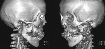
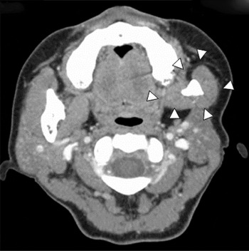
Three-dimensional CT revealed resorption of the left mandible (Figure 1). A facial CT revealed atrophy of the masseter, pterygoids, and the ramus and angle of the mandible (Figure 2).
Three-dimensional computed tomography reconstruction images showing resorption of the left mandibular ramus and condylar process. H = head; P = posterior; R = right; A = anterior; F = foot; L = left

Axial facial computed tomography image showing atrophy of the mandible, masseter muscle and pterygoideus muscles (arrowheads).

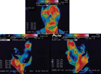
Thermographic examination showed that the affected side of the face was somewhat warmer than the unaffected side (Figure 3).
Thermographic images showing that the affected (left) side of the face is slightly warmer than the unaffected (right) side. The ratio of the area presumed to have a skin temperature of more than 36°C on the affected side versus the unaffected side was 1.7:1.

Hyperhidrosis was identified on the left side using Minor's iodine starch test. Ophthalmological examination revealed no abnormal findings (including no abnormal pupillary size). There was no neurological deficit, based on the patient's history, physical examination and magnetic resonance imaging (MRI).
From these findings, stimulation of the cervical sympathetic system was indicated as the aetiology. Thus, linear polarised light irradiation, using the Super Lizer system (Super Lizer HA-550; Tokyo Iken, Tokyo, Japan) was applied near the stellate ganglion to decrease the sympathetic output. An image analysis was performed using Adobe Photoshop and Microanalyser software (Nihon Poladigital KK, Tokyo, Japan); this indicated that, before treatment, the ratio between the area presumed to have a skin temperature of more than 36°C on the affected side versus the unaffected side was 1.7:1. After the treatment, this ratio had decreased to 1.5:1. A reference ratio of 1.2:1 was obtained from one of the authors (HM) (thermographic examination not shown).
After two years, the patient's hemiatrophy had not progressed, and her spontaneous temporomandibular joint pain had resolved.
Discussion
Progressive facial hemiatrophy, or Parry–Romberg syndrome, was first described by Parry in 1825 and later elaborated clinically by Romberg in 1846.
The underlying pathogenesis remains elusive, and aetiological theories include infection by Borrelia burgdorferi,Reference Abele, Bedingfield, Chandler and Given4 cervical sympathetic loss or imbalance,Reference Moss and Crikelair5, Reference Scope, Barzilai, Trau, Orenstein, Winkler and Haik6 trigeminal peripheral neuritis,Reference Pensler, Murphy and Mulliken2 lesions in the central nervous system,Reference Carreno, Donaire, Barcelo, Rumia, Falip and Agudo7 and scleroderma.Reference Sakuraoka, Tajima and Nishikawa8 Although these theories lack conclusive evidence, each is accompanied by convincing clinical reasoning.
ReesReference Rees and Facial11 suggested that Parry–Romberg disease represents a syndrome rather than a specific disease entity. This syndrome characteristically occurs during the first or second decade of life as localised, progressive atrophy of the skin and subcutaneous tissue within the dermatome of one or more branches of the trigeminal nerve supplying one side of the face. However, the involvement of the trigeminal nerve may be a secondary occurrence, and the distribution of lesions along its branches and the common manifestations of trigeminal neuralgia may result from the involvement of sympathetic fibres in the nerve, or from nerve compression by narrowed bone canals.Reference Jablonska and Jablonska12 Furthermore, trophic changes in the face are mediated by the sensory root of the trigeminal nerve, which is derived primarily from the cervical sympathetic trunk and not the trigeminus.Reference Wartenberg13 Conceivably, higher autonomic centres may be damaged, resulting in disorders of the blood supply to the cerebral blood vessels and secondary neurological manifestations.
• Progressive facial hemiatrophy (Parry–Romberg syndrome) is a rare cause of slowly progressive, unilateral facial atrophy
• Three-dimensional computed tomography and thermographic imaging are useful
• Cervical sympathetic hyperactivity may be present, and may be a cause
• The presented case had stellate ganglion blockade using linear polarised near-infrared light
• Such treatment is especially suited to cases with mild deformity
Our patient did not have B burgdorferi antibody, nor any antecedent traumatic event which may have acted as a trigger. In addition, she did not have scleroderma or any associated cutaneous findings characteristic of morphea, and her brain MRI findings were normal. Therefore, the above-mentioned theories involving infection, central nervous system lesions and scleroderma were not applicable.
On the other hand, high temperatures (as seen on the thermographic examination) and hyperhidrosis on the affected side indicated unilateral stimulation of the cervical sympathetic chain as a possible underlying cause.
This condition can cause loss of subcutaneous adipose tissue and even bone, through an increase in bone resorption,Reference Togari and Arai14 leading to facial hemiatrophy. However, whether stimulation of the cervical sympathetic chain will increase or decrease skin temperature is uncertain, because both effects can occur in this syndrome.Reference Kawano, Araki, Arakawa, Matsumono, Yamada and Kira15 Several interesting studies have reported this point. Progressive facial hemiatrophy may also accompany Horner's syndrome, and in rare cases it is observed with Pourfour du Petit syndrome,Reference Wartenberg13, Reference Kawano, Araki, Arakawa, Matsumono, Yamada and Kira15 the opposite of Horner's syndrome. The aetiology of Pourfour du Petit syndrome is similar to that of Horner's syndrome, and Pourfour du Petit syndrome may precede Horner's syndrome.Reference Aouba, Der Agopian, Genty-Le Goff, Mutschler, Janin and Patri16, Reference Ségura, Speeg-Schatz, Wagner and Kern17 Therefore, progressive facial hemiatrophy may manifest itself as a state of paralysis after chronic stimulation of the sympathetic cervical chain. In other words, progressive facial hemiatrophy can present as hyperfunction or hypofunction of the sympathetic cervical chain, depending on its stage of progression.
In our case, stimulation of the cervical sympathetic chain was indicated as a possible underlying cause. Thus, linear polarised near-infrared light, produced using a linear polarised near-infrared light therapy instrument (the Super Lizer HA-550), was applied near the stellate ganglion to create a stellate ganglion block. This therapy has been previously used in Japan for the treatment of various painful diseases.Reference Yokoyama and Sugiyama18 In brief, the linear polarised near-infrared light therapy instrument consisted of a power source, light source, light-guiding system and lens unit. The light source was a 150-W iodine lamp with an adjustable arm that allowed the position of the lens unit to be adjusted. The apparatus emitted linear polarised near-infrared light with wavelengths of 0.6–1.6 mm; the light was not a laser because it did not have a uniform wavelength.Reference Wajima, Shitara, Inoue and Ogawa19 Irradiation near the stellate ganglion using the Super Lizer instrument is an especially notable therapeutic method which is used together with, or as a substitute for, stellate ganglion blockade.
The mainstay of treatment for progressive facial hemiatrophy is plastic surgery, with lipoinjection in mildly deformed cases and cartilage and bone grafting in severe cases.Reference Guerrerosantos, Guerrerosantos and Orozco10 However, the deformity in our patient was mild and almost imperceptible to strangers, and her complaint was left subaural pain that seemed to be derived from temporomandibular joint derangement. After therapy, the spontaneous pain in the temporomandibular joint was relieved; however, whether this was because of the therapy or spontaneous remission of the syndrome is difficult to interpret, as the natural course of stabilisation is more than two to 10 years.Reference Kawano, Araki, Arakawa, Matsumono, Yamada and Kira15 However, because linear polarised light irradiation is minimally invasive, it can be considered a treatment option, particularly in cases with mild deformity.
Conclusion
We present a rare case of progressive facial hemiatrophy in which cervical sympathetic hyperactivity was indicated as the underlying aetiology, based on clinical and investigative findings. Currently, discussion about this condition mainly centres on therapeutic approaches, such as plastic surgery using lipoinjection or cartilage and bone grafting, rather than on the underlying aetiology.
However, the underlying pathogenesis of this syndrome could be elucidated by the presented evidence that sympathetic hyperactivity might also be present, and that chronic stimulation of the sympathetic cervical chain might lead to paralysis. Taking this into consideration, stellate ganglion blockade should be considered as a treatment option, particularly in mildly deformed cases.