


Introduction
Brief history
The major breakthrough in nutritional research on fatty acids was described in the studies by Burr and Burr(Reference Burr and Burr1,Reference Burr and Burr2) , where the essentiality of polyunsaturated fatty acids (PUFA) was discussed, specifically linoleic acid. Until that point, these fatty acids were considered as a source of calories and, since they could be synthesised from carbohydrates, non-essential. Later, Wesson and Burr(Reference Wesson and Burr3), in a study with rats fed diets without lipid sources, observed that the inclusion of linoleic and linolenic acids led to the remission of clinical manifestations related to essential fatty acid deficiency. These findings generated numerous studies that used linoleic and alpha-linolenic acid (ALA) from the omega-6 (n-6) and omega-3 (n-3) series, respectively, in animal diets. However, other studies(Reference Hume, Nunn and Smedley-Maclean4) did not find responses with the addition of linolenic acid, and coupled with the fact that many laboratories were unable to confirm these responses, the n-3 series generated some disinterest in research until the 1970s.
Subsequently, studies on ALA have been conducted and published, and the conversion into longer-chain and more unsaturated fatty acids was identified under the action of elongase and desaturase enzymes, such as docosahexaenoic acid (DHA) and eicosapentaenoic acid (EPA)(Reference Spector and Kim5). In the 1970s, studies related to EPA and DHA identified the presence of DHA in the retinas of animals from different species and that the presence of this fatty acid increased the electrical response of photoreceptor cells to light(Reference Anderson and Maude6,Reference Benolken, Anderson and Wheeler7) . In addition, antithrombotic effects were associated with EPA, and its use as a protector against coronary thrombosis was linked to the promotion of numerous benefits(Reference Dyerberg, Bang and Stoffersen8). In summary, the physiological roles related to EPA and DHA, such as their involvement in the structure of the cell membrane and cellular responses, have effectively demonstrated that DHA plays an essential role in the development of the nervous system, while EPA is capable of modulating immune functions and the inflammatory response in animals(Reference Lenox9). Since then, the application of EPA and DHA have been under investigation in many diseases, including those mentioned in this article, but also others, such as gingivitis(Reference Lourenço, Booij-Vrieling and Vossebeld10) and keratoconjunctivitis sicca(Reference Silva, Nai and Giuffrida11).
Thus, the aim of this review was to compile information relevant to the veterinary field regarding the applicability of omega-3 fatty acids in eight areas of clinical practice – dermatology, orthopaedics, oncology, cardiology, endocrinology, nephrology, cognition and behaviour – by highlighting their indications, mechanisms of action, potential clinical justifications and possible adverse effects, ultimately providing an up-to-date resource for consultation and study.
Structure
Fatty acids can be classified according to chain length, number of double bonds and the location of the first double bond in relation to the methyl or omega group of the carbon chain(Reference Lenox9). Thus, the general structure of a fatty acid consists of a chain of carbon and hydrogen atoms with a carboxyl group at one end of the chain and a methyl group at the other. Fatty acids containing double bonds in the chain are called unsaturated, and when there are two or more double bonds, they are called PUFA, which are identified by the number and position of the first double bond counting from the chain terminal(Reference Trevizan and De Mello Kessler12). Long-chain PUFA with at least eighteen carbon atoms and one unsaturation are not synthesised by dogs and cats, and are therefore essential for these species.
These fatty acids are primarily found in the plant kingdom, with linoleic acid and ALA being the most prominent, serving as precursors to the n-6 and n-3 series, respectively(13). From the n-3 series, the main PUFA are ALA, EPA and DHA(14), which can modulate inflammatory processes, act as precursors to eicosanoids and prostaglandins, play structural roles as components of cell membranes, promote healthy growth and development, and affect the skin and coat health of dogs and cats(Reference Lenox9). ALA, an isomer of linolenic acid, consists of an eighteen-carbon chain with three double bonds at carbons 9, 12 and 15(Reference Shahidi and Ambigaipalan15). Under the action of elongase and desaturase enzymes, part of this acid can be converted into EPA and subsequently into DHA(13).
Dogs and cats do not synthesise ALA, but due to the low conversion of ALA to EPA and DHA(Reference Bauer16), the National Research Council (NRC) has listed the dietary requirements for EPA and DHA for dogs and cats, which vary among different life stages(13). EPA is a twenty-carbon acid with five double bonds at positions 5, 8, 11, 14 and 17. DHA, conversely, consists of a twenty-two-carbon chain with six double bonds in CIS configuration at positions 4, 7, 10, 13, 16 and 19 of the carbon chain(Reference Shahidi and Ambigaipalan15). The ratio of n-3 to n-6 fatty acids and the total amount of each in the diet are crucial for maintaining wellbeing. The NRC recommends a ratio of 2·6:1 to 26:1 of n-6 to n-3(13). It is worth noting that requirements vary among different life stages. Growing dogs and cats require more DHA than adult dogs and cats(13).
Sources
When the literature discusses sources of n-3, it is important to specify which fatty acid in the series is being referred to, as the majority of ALA is not converted to EPA and DHA by mammals. Sources of ALA that can be included in the diet are often in the form of flaxseed flour or oil. Canola, corn, and soyabean oils and flours can also be included, but they contain much smaller amounts of ALA compared with flaxseed(Reference Bauer17). Thus, some commercial dog and cat foods may contain sources of n-3 PUFA, primarily EPA and DHA, with marine fish oil (FO) from deep-sea fish such as salmon, tuna, menhaden, anchovies, herring and mackerel being commonly used(Reference Dahms, Bailey-Hall and Sylvester18). Dahms et al.(Reference Dahms, Bailey-Hall and Sylvester18) evaluated an algae oil as an alternative source of EPA and DHA, obtained through large-scale fermentation of microalgae from the Schizochytrium spp. genus. This oil was added to the diet of pregnant and lactating female dogs and subsequently to the puppies for 26 weeks during the growth phase, at inclusion levels of up to 3%, which is equivalent to 30 g/kg of diet on a dry matter basis. In general, it proved to be a sustainable alternative (preserving marine fish resources), highly bioavailable, and very safe even at high levels.
Inflammation, pathways and precursors
As mentioned, although mammals are unable to synthesise ALA, they are capable of metabolising it by introducing new double bonds through desaturation and elongation of the chain; thus, converting a small portion of ALA into EPA, and subsequently, a portion of EPA into DHA. Since their bioconversion is very limited in dogs and cats, these n-3 fatty acids need to be provided to the animal through their diet(Reference Bauer19). Building on this, cats exhibit inactivity or low activity of desaturase enzymes, making them inefficient at converting ALA to EPA and consequently leading to DHA deficiency. This emphasises the necessity of including these nutrients in their diet(Reference Trevizan and De Mello Kessler12). It is worth noting that in felines, due to the limited action of the desaturase enzyme (delta 6), it competes for both ALA and linoleic acid (n-6), and therefore, a diet with high levels of ALA can impair the conversion of linoleic acid to arachidonic acid (AA). Therefore, excessive amounts of n-3 fatty acids compared with n-6 fatty acids may be contraindicated in feline species(Reference Bauer20).
Conversely, dogs can convert linoleic acid to AA. These and other metabolic differences in dogs and cats form the basis for the different requirements of PUFA(Reference Hall, Jackson and Vondran21). Further, in dogs, the beneficial effects of n-3 PUFA supplementation have been associated with the reduction of cardiovascular diseases, chronic renal failure, neoplastic processes, atopic dermatitis(Reference Stoeckel, Bachmann and Dobeleit22) and degenerative joint diseases(Reference Adler, Schoeniger and Fuhrmann23). It is believed that the benefit of EPA and DHA is due to their role as precursors of potent anti-inflammatory mediators(Reference Weylandt, Chiu and Gomolka24). In addition, they have immunomodulatory and less inflammatory effects due to their competitive inhibition of the AA cascade, whose end metabolites (prostaglandins, thromboxanes, and leukotrienes) have greater pro-inflammatory activity(Reference Shahidi and Ambigaipalan15). The increase in circulating levels of EPA and DHA also results in the reduction of matrix metalloproteinase expression, interleukins (IL-1 and IL-2), cyclooxygenase-2 (COX-2) and tumour necrosis factor-alpha (TNF-alpha)(Reference Zainal, Longman and Hurst25), enhancing their immunomodulatory effect.
Nevertheless, n-3 should be administered consistently, as the effects of EPA and DHA are time-dependent, allowing them to incorporate into cell membranes. A recent study shows that EPA and DHA reach their maximum concentration within erythrocyte membranes between 30 and 60 d(Reference Combarros, Castilla-Castaño and Lecru26). Since these are anucleated cells, extrapolating these results to other tissues is challenging, as other organs may respond more rapidly and require shorter supplementation periods. Nonetheless, studies with very short supplementation durations should be interpreted with caution, as a lack of effect may indicate that omega-3 has not had sufficient time to fully integrate into the target tissue being studied.
Applications of omega-3 in chronic diseases and cognition
Omega-3 and dermatology
Dermatological alterations that affect dogs and cats can be related to infectious agents, chemicals, physical damage and immune reactions, including allergies, atopic dermatitis, infectious diseases and skin tumours(Reference Woldemeskel27). In humans, as well as in other animals, diets deficient in essential fatty acids can lead to skin and coat changes, such as scaly dermatitis, alopecia and delayed wound healing(Reference Mogensen28). Among the essential fatty acids, the prescription of n-3 series in veterinary clinical practice as an adjuvant in the treatment of dermatopathies is growing among clinicians(Reference Omega Fatty Acids29). In pruritic conditions affecting dogs, their recommendation can be a safe alternative that minimises the use of systemic glucocorticoids(Reference Logas and Kunkle30). As one of the main pruritic alterations that affect the skin of dogs and cats, atopic dermatitis is frequent in clinical practice and can be characterised by pruritus and production of immunoglobulin E, resulting from the inflammatory process related to environmental antigens(Reference Halliwell31).
The dietary addition of nutraceuticals, such as PUFA of the n-3 series, present in FO and other marine sources, has the ability to modify the inflammatory response by modulating the animal’s immune system(Reference Bauer17). In this way, the use of EPA and DHA can contribute to promoting health, either preventively or therapeutically, by reducing the use of conventional medications, which are often associated with unwanted side effects, or by reducing the response time for improvement in the treatment of various dermatopathies(Reference Omega Fatty Acids29). However, there is no consensus regarding the concentrations and doses of each component of this series, as well as the association and relationship with the n-6 series. Therefore, the pathway involved in the improvement of dermatological conditions may be strictly related to immunomodulation than incorporation into the haircoat, as it did not happen in a study by Combarros et al. (Reference Combarros, Castilla-Castaño and Lecru26).
De Santiago et al.(Reference de Santiago, Arribas and Llamas32) conducted a study for 2 months, in which they used forty dogs diagnosed with atopic dermatitis, divided into two groups. One of these groups was subjected to a control diet, and the other to a diet supplemented with antioxidants, polyphenols and PUFA of the n-3 series. After 60 d, they were able to observe that the dogs fed the enriched diet showed improvements in the clinical signs related to dermatitis, which were measured by a reduction in the extent and severity index score of canine atopic dermatitis (CADESI-4®), validated by Olivry et al.(Reference Olivry, Saridomichelakis and Nuttall33), as well as a noticeable improvement reported by the owners. The concentration of EPA in the enriched diet was 0·81 g/1000 kcal associated with 0·57 g/1000 kcal of DHA. Daily energy requirements were met as estimated by the National Research Council (95–180 × kg0·75). The total n-3 fatty acid concentration in the enriched diet was six times higher than the concentration in the control diet. The composition of EPA and DHA in the control diet was not measured, so further studies are needed to individually evaluate each nutrient and its contribution to the clinical improvement of the patient.
In the clinical practice guidelines for treating canine atopic dermatitis, Olivry et al. (Reference Olivry, DeBoer and Favrot34), highlighted that the use of PUFA is a safe therapeutic option for dogs with this condition. They noted that while PUFA alone did not appear to significantly improve patient outcomes, especially in acute cases, their long-term use in combination with specific medications led to a sparing effect on glucocorticoids(Reference Sævik, Bergvall and Holm35), antihistamines(Reference Paterson36) and immunosuppressants such as cyclosporine. In addition, this combination resulted in improvements in clinical signs related to dermatopathy(Reference Müller, Linek and Löwenstein37). This sparing effect of n-3 on steroid use was evaluated in a study conducted by Sævik et al.(Reference Sævik, Bergvall and Holm35), which lasted for 12 weeks and involved sixty dogs with atopic dermatitis. All dogs received oral prednisolone based on the level of pruritus and were randomly divided into treatment groups, consisting of a control group and a group that received a combination of borage seed oil and FO. The essential fatty acid supplement, as well as the placebo, was administered orally once a day according to the manufacturer’s recommendation. For dogs in the supplemented group weighing less than 10 kg, a recommended dosage of 190 mg of linoleic acid, 105 mg of gamma-linolenic acid (GLA), 9·9 mg of EPA and 6·6 mg of DHA was given. For dogs weighing between 10 and 25 kg, the recommended dosage was double the dose used for dogs weighing less than 10 kg, and for dogs weighing over 25 kg, triple that dose was recommended. The average dose of n-6 and n-3 fatty acids was 32 mg and 1·8 mg per kg of body weight, respectively.
In addition, studies conducted by Müller et al.(Reference Müller, Linek and Löwenstein37) observed a reduction in the use of cyclosporine in a study with thirty-six dogs diagnosed with atopic dermatitis, with sixteen dogs in the control group and twenty dogs supplemented with PUFA. The supplemented group of dogs received 3 mL of the supplement per 10 kg of body weight per day, which contained approximately 360 mg of ALA, 410 mg of EPA, 249 mg of DHA and smaller fractions of n-6 fatty acids, although not specified. The n-6 to n-3 ratio was 1:3·75, much lower than the ratio used by Sævik et al.(Reference Sævik, Bergvall and Holm35), who used an n-6 to n-3 ratio of 17·88:1. This lower 6:3 ratio could explain the cutaneous anti-inflammatory effect. Diets rich in EPA and DHA, and with low concentrations of n-6 fatty acids, especially linoleic acid (LA) and AA, have a nutritional profile with lower inflammatory potential and can act as anti-inflammatories if the concentrations of n-3 acids and gamma-linoleic acid (GLA) are high(Reference Bauer17).
To evaluate the anti-inflammatory effects of n-3 fatty acids, Logas and Kunkle(Reference Logas and Kunkle30) conducted a study on the impact of FO supplementation with high concentrations of EPA in sixteen dogs displaying signs of pruritic idiopathic dermatitis, atopy and/or atopic dermatitis due to ectoparasite bites. All dogs underwent 6 weeks of each treatment with a 3-week interval without supplementation, totalling 15 weeks of evaluation. The supplements used in the treatments were 1 mL of FO containing 180 mg EPA and 120 mg DHA per 4·5 kg of body weight, once a day for 6 weeks, and the other treatment was supplemented with 1 mL of corn oil containing 570 mg of LA and 50 mg of GLA, at the same dose, duration and frequency of administration. The dogs were evaluated for pruritus, alopecia, self-mutilation and the quality and appearance of the coat, and there were differences and improvements in the evaluated criteria when the animals were supplemented with fish oil. It is worth noting that when the authors described high doses of EPA and DHA, these were not higher than the concentration of n-6, maintaining a higher n-6 to n-3 ratio. Moreover, Abba et al.(Reference Abba, Mussa and Vercelli38) evaluated the effects on twenty-two dogs with varying degrees of atopy and supplemented them with a mixture of 17 mg EPA/kg, 5 mg DHA/kg, 35 mg GLA and an n-6 to n-3 ratio of 5·5:1. They observed improvements in dogs in the early stages of atopy, emphasising that early diagnosis and supplementation are important for clinical improvement.
There is no consensus regarding the results obtained in animals with certain pathologies when subjected to n-3 supplementation. In a review conducted by Lenox and Bauer(Reference Lenox and Bauer39), they assessed the potential adverse effects of n-3 fatty acids and considered it essential to understand the concentrations of EPA and DHA in order to recommend the appropriate amount of FO, which is used as a source of these acids. Attention should also be given to the ratio between n-6 and n-3, as high concentrations of ALA, although an n-3 fatty acid, do not have the same effects as EPA and DHA, which may be present. Similarly, GLA, despite belonging to the n-6 PUFA, does not act similarly to ALA and AA, which are pro-inflammatory mediators. Conditions such as those described by Lenox and Bauer(Reference Lenox and Bauer39) can contribute to the lack of consensus, as many studies do not clearly specify the dosages and the omega-3 and n-6 fatty acids used, as well as the ratio between the series.
n-3 fatty acids have already been studied in different conditions and for different purposes, such as poor haircoat quality(Reference Combarros, Castilla-Castaño and Lecru26). The supplementation of 16 mg/kg/d of EPA and 10 mg/kg/d of DHA reduced the seborrheic index after 60 d of supplementation. This study was recently evaluated in a systematic review where the level of bias arising from different steps of methodology was considered low(Reference Magalhães, Lourenço and Gregório40). Supplementary Table 1 lists a few studies that evaluated the use of n-3 fatty acids in small animal dermatology and their limitations. The dosages of EPA and DHA in these studies ranged from 1 to 43 mg/kg and 0·6 to 30 mg/kg, respectively. However, many of the studies had limitations, making it difficult to draw definitive conclusions about a specific effective dose. It may be more feasible to select a dosage based on those commonly used for other conditions that have demonstrated positive outcomes. This limitation, as well as the difficulty and variety of presenting the used dosages, compromises our understanding, as Magalhães et al. (Reference Magalhães, Lourenço and Gregório40) also discussed.
Omega-3 and orthopaedics
Another important clinical application of PUFA is in situations of osteoarticular alterations, specifically in animals with osteoarthritis, a clinical condition associated with chronic inflammation. Several studies have demonstrated positive effects of n-3 PUFA supplementation in dogs and cats with osteoarthritis(Reference Adler, Schoeniger and Fuhrmann23,Reference Barrouin-Melo, Anturaniemi and Sankari41–Reference Corbee, Barnier and van de Lest46) (Supplementary Table 2). In the prospective randomised controlled study by Fritsch et al. (Reference Fritsch, Allen and Dodd42), 106 client-owned dogs with stable chronic osteoarthritis were studied and divided into two groups. The control group consisted of fifty-seven dogs (diet with 0·1% total n-3), while the test group comprised fifty-two dogs (diet with 3·5% total n-3). The authors observed that after 12 weeks of study, consumption of the diet with high concentrations of n-3 allowed for a faster reduction in carprofen dosage compared with the dogs that consumed the control diet. In a randomised, double-blinded, controlled clinical study by Roush et al. (Reference Roush, Cross and Renberg43), thirty-eight dogs with osteoarthritis were divided into two groups: the control group (n = 16; diet with 0·11% total n-3) and the test group (n = 22; diet with 3·47% total n-3). After 90 d of study, the authors observed that dogs fed a diet rich in n-3 showed significant improvement in lameness and weight-bearing compared with the animals in the control group.
Conversely, the same research group conducted another randomised, double-blinded, controlled clinical trial(Reference Roush, Dodd and Fritsch44). In this study, a group of seventy-one dogs with osteoarthritis consumed a diet with high concentrations of total n-3 (3·47% dry matter (DM)). However, there were no improvements in lameness score, pain on palpation, weight-bearing grade, range of joint motion or willingness to hold the contralateral limb when compared with a group of fifty-six dogs with osteoarthritis that consumed food with thirty-one times lower concentrations of total n-3 (0·11% DM). In contrast, Moreau et al.(Reference Moreau, Troncy and del Castillo45) conducted a randomised, placebo-controlled, double-blinded trial with thirty dogs diagnosed with osteoarthritis. In this study, the dogs were divided into two groups: the control group (0·18% DM total n-3) and the test group (1·47% DM total n-3). After 7 and 13 weeks of treatment, the test group demonstrated improvements in locomotor disability and daily activity performance, suggesting a positive effect of n-3 supplementation.
Similarly, Mehler et al. (Reference Mehler, May and King47) demonstrated that a group of dogs (n = 41) with naturally acquired osteoarthritis that received n-3 fatty acid supplementation at an average dosage of 69 mg EPA + DHA/kg/d showed significant improvement in lameness, discomfort and total Joint Severity Score after 84 d of treatment, compared with thirty-three dogs that received placebo oil supplementation. However, unlike the previous studies, the diets were not standardised, which may have influenced the observed results. It is important to mention that a study conducted with cats demonstrated similar effects to those observed in dogs. In a randomised, double-blinded, placebo-controlled, crossover-designed study (10 weeks/period) with sixteen cats diagnosed with osteoarthritis, it was shown that supplementation with n-3 PUFA (1·53 g EPA/1000 kcal ME (Metabolizable energy); 0·31 g DHA/1000 kcal ME) resulted in improved activity levels, greater ability to walk up and down stairs, reduced stiffness during gait, increased interaction with the owners and higher jumping ability compared with a control diet composed of corn oil(Reference Corbee, Barnier and van de Lest46).
Several mechanisms are proposed through which PUFA help improve clinical signs in animals with osteoarthritis. It is known that PUFA act by inhibiting the secretion or expression of compounds involved in cartilage degradation, such as pro-inflammatory cytokines, prostaglandins and enzymes such as matrix metalloproteinases (MMPs) and disintegrin and metalloproteinase with thrombospondin motifs(Reference Abshirini, Ilesanmi-Oyelere and Kruger48). Fatty acids consumed through the diet are incorporated into chondrocyte membranes, where they mediate signalling pathways. Studies have suggested that the composition of dietary fat can influence inflammatory conditions as well as the progression of cartilage degradation. Therefore, it could impact the clinical signs and progression of osteoarthritis, as fatty acids have different, or even opposite, effects on joint cartilage.
In vitro and in vivo studies have demonstrated that both saturated fatty acids and n-6 PUFA result in increased pro-inflammatory and pro-apoptotic markers, while n-3 PUFA have been associated with the opposite effects, including a decrease in degradation markers in chondrocytes and synoviocytes(Reference Abshirini, Ilesanmi-Oyelere and Kruger48–Reference Loef, Schoones and Kloppenburg51). In an in vitro study using a cell culture system with bovine chondrocytes(Reference Zainal, Longman and Hurst25), it was observed that EPA, DHA and ALA reduced the gene expression of proteins involved in the aetiology of osteoarthritis (ADAMTS-4, ADAMTS-5, MMP-3, MMP-13, COX-2, IL-1α, IL-1β and TNF-α) after exposure to IL-1 in cell culture, while AA had no effect. Another in vitro study with bovine and human chondrocytes demonstrated that EPA reduced the gene expression of COX-2, as well as its concentrations, which is the opposite effect observed for AA. This effect is associated with a reduction in the inflammatory process, one of the factors that may explain the improvement in clinical signs in individuals with osteoarthritis receiving n-3 PUFA supplementation(Reference Hurst, Rees and Randerson52).
Another molecular mechanism that may explain the positive effects of n-3 PUFA in animals with osteoarthritis is the reduction in the expression of molecules associated with oxidative stress, which is related to the pathophysiology of osteoarthritis. In this regard, nitric oxide plays a crucial role in the progression of osteoarthritis through various mechanisms. It stimulates the synthesis of pro-inflammatory cytokines(Reference Abramson53), induces chondrocyte apoptosis via the interaction between inducible nitric oxide synthase (iNOS) and COX-2(Reference Notoya, Jovanovic and Reboul54), and regulates extracellular matrix degradation by increasing MMP activation(Reference Davies, Graham and MacFarlane55). In addition, PGE2 reduces the synthesis of the endogenous antagonist of the interleukin-1 receptor (IL-1Ra)(Reference Abshirini, Ilesanmi-Oyelere and Kruger48,Reference Abramson53) . In an in vitro study by Adler et al. (Reference Adler, Schoeniger and Fuhrmann23), EPA was able to reduce the gene expression of iNOS and consequently the production of nitric oxide in canine chondrocytes stimulated by interleukin-1β (IL-1β) to induce osteoarthritic changes. Moreover, in a prospective randomised, controlled, double-blinded study with dogs diagnosed with osteoarthritis (n = 77), it was shown that after 16 weeks of treatment, supplementation with n-3 PUFA (90 mg/kg body weight (BW) of EPA and 20 mg/kg BW of DHA) improved some markers of oxidative stress. However, there was no difference when this group was compared with a control group that received corn oil supplementation during the same period(Reference Barrouin-Melo, Anturaniemi and Sankari41).
Molecular mechanisms associated with oxidative stress also could explain the positive effects of n-3 PUFA in individuals with osteoarthritis. In an in vitro study with human chondrocytes, it was observed that EPA inhibited chondrocyte apoptosis induced by oxidative stress through various mechanisms, such as inhibiting caspase-3 and poly (ADP-ribose) polymerase cleavage, inhibiting p38 MAPK and p53 phosphorylation, as well as inhibiting the expression of MMP-3 and MMP-13(Reference Sakata, Hayashi and Fujishiro56). Furthermore, in an in vitro study with canine chondrocytes by Buddhachat et al. (Reference Buddhachat, Siengdee and Chomdej57), it was demonstrated that n-3 PUFA derived from FO and krill oil can suppress matrix degradation in cytokine-induced cartilage explants by significantly reducing the increase in sulphated glycosaminoglycans (s-GAGs) and preserving the content of uronic acid and hydroxyproline (except for GLM (green-lipped mussel)). The authors also observed that n-3 PUFA negatively regulated the gene expression of molecules associated with catabolism (MMP1, MMP3 and MMP13) and positively regulated the gene expression of molecules associated with anabolism (AGG and COL2A1). Finally, Zhang et al.(Reference Zhang, Dai and Zhang58) demonstrated that mice with destabilised medial meniscus, when consuming a diet with a low n-6/n-3 PUFA ratio for 12 weeks, experienced suppression of the nuclear factor-kappa B (NF-κB) pathway. This was caused by activation of GPR120 and attenuation of EP4, leading to the inhibition of the NF-κB pathway and its downstream matrix-degrading enzymes, MMP13 and ADAMTS5. The authors also observed an increase in cartilage thickness and a decrease in TNF-α concentrations in both cartilage and serum.
On the basis of the discussed evidence, it is clear that n-3 fatty acids play an important role in the treatment of osteoarthritis in dogs and cats. The dosages used in the studies described in Supplementary Table 1 range from 48 to 100 mg/kg EPA and 20 to 32 mg/kg DHA. Still, many of the studies failed to declare or give information about intake per kg of body weight. In addition, all studies except for one have important limitations, despite this, Magalhães et al. (Reference Magalhães, Lourenço and Gregório40) considered the evidence-based application of n-3 for these conditions had an overall low risk of bias. In conclusion, it is worth noting that n-3 PUFA exert biological effects on chondrocytes through various mechanisms, and in relation to dogs and cats, this has been relatively underexplored.
2.3. Omega-3 and oncology
Nutritional disorders are observed in patients with cancer(Reference Shirai, Morita and Iwata59), therefore, nutrition has a direct influence on the development, symptoms, prognosis and treatment of cancer, as the disease and therapy compromise nutritional status and impact the quality of life of patients(Reference Arends, Baracos and Bertz60,Reference Ravasco61) . Malnutrition status in patients with cancer can interfere with treatment, delay the healing process, impair muscle function and increase the risks of postoperative complications. It is estimated that 40–80% of patients with cancer become malnourished during the course of the disease(Reference Ravasco61). In these patients, the occurrence of cachexia secondary to inflammation is possible, so ensuring adequate nutrition and controlling inflammation are important points in cancer treatment(Reference Shirai, Morita and Iwata59). However, a study conducted by Weeth et al. (Reference Weeth, Fascetti and Kass62) did not establish a common correlation between dogs with cancer and the occurrence of cachexia with weight and body composition changes, but weight loss during treatment was observed in these patients(Reference Michel, Sorenmo and Shofer63). Besides, the process of cachexia, characterised by excessive loss of lean mass, is notable in feline cancer patients, which requires diligent monitoring of calorie intake and nutritional support(Reference Baez, Michel and Sorenmo64).
One of the factors that impairs nutrient utilisation and promotes muscle catabolism in patients with cancer is systemic inflammation(Reference Arends, Baracos and Bertz60,Reference Penna, Minero and Costamagna65,Reference Seruga, Zhang and Bernstein66) . This muscle depletion, among various factors, is related to the increase in inflammatory cytokines IL-6 and TNF-α in the paraneoplastic syndrome of anorexia and cachexia in humans(Reference Fearon67). Similarly, dogs diagnosed with lymphoma showed an increase in inflammatory markers IL-6 and C-reactive protein(Reference Merlo, Rezende and Franchini68–Reference Tecles, Caldín and Zanella70). Given the significant role of inflammation in cancer progression, nutrition becomes a critical component of multimodal oncologic therapy. Within nutritional support, the use of supplements such as n-3 is particularly noteworthy as a potential nutraceutical(Reference Arends, Baracos and Bertz60). In line with this, clinical studies in humans are examining the use of dietary n-3 PUFA in the nutritional support of patients with cancer that have weight loss, fatigue, inflammation or postoperative complications(Reference Wei, Wu and Chen71). These studies suggest that high concentrations of EPA and DHA, typically administered in the form of fish oil, could help mitigate muscle reduction and tumour development(Reference Hawcroft, Loadman and Belluzzi72). Clinical data supports the use of EPA and DHA in combating cachexia, and high levels of EPA included in dietary supplements have relevance in the recovery of malnourished patients with cancer cachexia(Reference Shirai, Morita and Iwata59).
Nonetheless, the dose and duration of supplementation tend to influence the anti-inflammatory effect of n-3(Reference Wei, Wu and Chen71). Clinical trials in human subjects have demonstrated an improvement in calorie and protein intake in patients supplemented with EPA(Reference Ravasco61). Several studies discussed by Wei et al. (Reference Wei, Wu and Chen71) have revealed important nutritional effects related to the application of n-3 in the treatment of patients with cancer, including improvements in energy intake, body condition, functional capacity, weight maintenance, calorie consumption, body mass and skeletal muscle protein turnover. For patients with cachexia and anorexia, high-energy and high-protein formulations with high amounts of EPA can be introduced as cancer immunonutrition treatment to assist with food intake(Reference Shirai, Morita and Iwata59). The nutritional approach no longer focuses solely on increasing calorie and protein intake; a more current approach suggests the use of anticatabolic and anti-inflammatory options(Reference Arends, Baracos and Bertz60). In this context, EPA and DHA play a pivotal role: instead of being converted into pro-inflammatory eicosanoids, these omega-3 fatty acids can synthesise inert eicosanoids, such as PGE3, LTB5, 12-HEPE and 5-HEPE(Reference McDaniel, Massey and Nicolaou73). Therefore, the ingestion of FO can have an inhibitory effect on IL-6 synthesis and, consequently, an anti-inflammatory effect, suggesting effectiveness in reducing mild chronic inflammation in patients with cancer(Reference Shirai, Morita and Iwata59). Thus, the application of EPA and DHA supports immune response and results in a more active resolution of inflammation(Reference Petric74).
These findings are highly relevant in the approach to different types of cancer in humans. However, in veterinary medicine, the limited studies have mainly focused on the pharmacological effects of non-steroidal anti-inflammatory drugs, particularly for cyclooxygenase (COX) inhibition(Reference McMillan, Boria and Moore75,Reference Mohammed, Bennett and Craig76) . While the regulatory mechanisms of fish oil (FO) in modulating systemic inflammation associated with cancer cachexia remain unclear, human oncology guidelines recommend higher intake of long-chain fatty acids and FO supplementation to help reduce systemic inflammation, increase appetite, calorie intake and body weight in patients with cancer(Reference Ravasco61). In light of this, the adjuvant administration of n-3 fatty acids to patients during chemotherapy should be considered, as positive outcomes in cancer treatment are linked to inflammatory cytokines that n-3 fatty acids help regulate(Reference Soni, Torvund and Mandal77). However, there are no studies in veterinary medicine that specifically establish the optimal dose and effectiveness of n-3 fatty acids in dogs and cats with cancer. Therefore, a suggestion would be to refer to studies on cardiac evaluations in dogs with cachexia, where the dose used was 40 mg of EPA and 25 mg of DHA per kg of body weight(Reference Freeman, Rush and Kehayias78).
The use of FO, rich in EPA and DHA, as a complement to instituted chemotherapy, proves favourable for the nutritional maintenance of patients(Reference Arends, Baracos and Bertz60), cancer treatment and resistance to therapy due to increased chemosensitivity of tumour cells(Reference Soni, Torvund and Mandal77). In human subjects, supplementation with EPA has been shown to be advantageous for patients undergoing radiotherapy, with a decrease in the pro-inflammatory response and consequent reduction in adjacent tissue damage resulting from the action of EPA(Reference Fulan, Changxing and Yi Baina79). Adequate nutritional management to restore and maintain body composition is of utmost importance for the success of cancer therapy. Strategies should be applied to maintain muscle mass and adipose tissue, as well as reduce systemic inflammation. Although there are a limited number of studies consolidating this evidence in dogs and cats, the strategy makes physiological sense for these species as well. Therefore, studies advancing knowledge in this area are of great interest.
Omega-3 and cardiology
Another rich collection of literature about the positive effects of PUFA for dogs and cats is related to the treatment and prevention of heart diseases. In humans, for example, the reduced mortality from cardiovascular diseases has been associated with an increased intake of n-3 PUFA(Reference Endo and Arita80). Heart diseases express a rate of natural progression that can lead to heart failure. This condition, in turn, causes inflammation and clinical signs when animal bodies cannot compensate for diminished heart activity, thus developing congestive heart failure (CHF) or compromising cardiac output. In this regard, EPA and DHA have an anti-inflammatory potential to reduce inflammation associated with heart failure(Reference Keene, Atkins and Bonagura81–Reference Nasciutti, Moraes and Santos83). Furthermore, heart failure increases the levels of pro-inflammatory cytokines (TNF-α and IL-1), which exacerbate muscle breakdown for energy supply, a process known as cachexia. Consequently, a higher degree of inflammation is linked to the development of cardiac cachexia(Reference von Haehling, Lainscak and Springer84).
In light of this, Freeman et al.(Reference Freeman, Rush and Kehayias78) conducted a randomised study with twenty-eight dogs suffering from heart failure secondary to dilated cardiomyopathy (DCM) to assess the effects of FO on nutritional status, cytokine alterations and clinical outcomes. The animals were randomised into two groups: the FO group and the placebo (corn oil) group. In the experimental group, dogs received approximately 27 mg/kg/d EPA and 18 mg/kg/d DHA in FO ethyl esters, administered in capsules for 8 weeks. FO supplementation promoted a decrease in cytokine levels, with lower levels of interleukin-1β (IL-1) and prostaglandin E2 (PGE2) production observed, alongside reduced muscle loss, a key characteristic of nutritional status. Further, clinical parameters were classified as moderate. The authors suggested that more potent effects on cytokine concentrations could be achieved with higher dosages, which could, in turn, potentially improve clinical outcomes.
Moreover, heart failure causes alterations in plasma fatty acid concentrations, as dogs with heart failure have shown a deficiency of plasma EPA and DHA when compared with healthy animals(Reference Freeman, Rush and Kehayias78). However, more research is needed to explain the relation of these alterations to the mechanism of cardiopathies. n-3 PUFA may have an anti-inflammatory effect through the alteration of inflammatory gene expression, since transcription factors such as NF-κB and peroxisome proliferator-activated receptors will have their pathways inhibited. In addition to their anti-inflammatory effects, n-3 PUFA supplementation has been associated with several other benefits, including direct cardiovascular actions such as a decrease in cardiac fibrosis and remodelling, as well as hypotensive, antithrombotic and antiarrhythmic effects(Reference Endo and Arita80).
The most common dog heart disease is myxomatous mitral valve disease (MMVD)(Reference Keene, Atkins and Bonagura81), a multifactorial disease influenced by genetics, environmental factors and other unknown causes(Reference Madsen, Olsen and Häggström82). The disease is characterised by cellular changes in the mitral valve that allow blood flush regurgitation. In addition, we can observe an increase in cardiac work, ventricular remodelling and dysfunction as a consequence of progressive valvular regurgitation(Reference Keene, Atkins and Bonagura81). In a double-blinded, randomised study with twenty-nine male and female dogs with MMVD, and classified as stage B2 or C, a lower volumetric overload and antiarrhythmic effect was observed in the experimental n-3 group that received 150 mg EPA and DHA/100kcal and 1·5 EPA/DHA ratio (final 1·3) provided by diet supplementation(Reference Nasciutti, Moraes and Santos83). In addition, Pasławski et al.(Reference Pasławski, Kurosad and Zabek85) (Supplementary Table 3) assessed twenty dogs with MMVD (stages B2 and C). An experimental group received a diet enriched with EPA + DHA (1·03g/MJ (Megajoules) per 100g dry diet) from fish meat, while the control group were fed a standard diet. The study aimed to observe differences in clinical, cardiological, haematological and biochemical parameters, as well as metabolomic profiles; however, no significant results was found to implicate a relevant clinical outcome. In our opinion, the supplementation and forms of EPA + DHA may not have been sufficiently high.
For arrhythmogenic right ventricular cardiomyopathy (ARVC), Smith et al. (Reference Smith, Freeman and Rush86) recruited twenty-four Boxer dogs with naturally occurring ARVC to assess if FO capsule supplementation (2 g/d) reduced the frequency of ventricular arrhythmias compared with flax oil and sunflower oil capsule supplementation over a 6-week period. The authors observed a decrease in ventricular premature contractions (VPC) in the Boxers receiving FO, providing 780 mg/d EPA and 497 mg/d DHA. Nonetheless, further studies are needed to determine the optimal dose and explore other potential beneficial effects of n-3 fatty acids for dogs with ARVC. In a study modelling atrial cardiomyopathy, Laurent et al.(Reference Laurent, Moe and Hu87) investigated the effects of n-3 PUFA supplementation in twenty-four adult mixed-breed dogs (n = 24) that underwent simultaneous atrioventricular pacing (SAVP) for 2 weeks. The dogs were randomly assigned to two groups in a double-blinded, placebo-controlled design: a control group (n = 12) that did not receive supplementation and an experimental group (n = 12) that received a daily n-3 PUFA supplement (1 g/d) containing EPA and DHA for 3 weeks. In addition, a third group comprised unpaced healthy dogs (n = 7) that did not receive any supplementation. The results showed that the experimental group experienced a reduction in atrial fibrosis development and a lower inducibility of atrial fibrillation compared with the control group.
These results corroborate the cardioprotective effect of EPA and DHA supplementation. For significant arrhythmias, n-3 fatty acid supplementation should be considered as a supporting medical therapy for dogs. The authors believe that there is significant evidence to warrant the use of EPA and DHA in dogs with heart failure, arrhythmias for secondary prevention, and earlier stages of cardiac disease such as DCM, CVD and HCM (Hypertrophic cardiomyopathy). However, the optimal dose of n-3 fatty acids for dogs and cats with cardiac conditions remains uncertain. The currently recommended dose for EPA and DHA supplementation in small animals has been proposed by a notable group of authors(Reference Freeman, Rush and Kehayias78,Reference Smith, Freeman and Rush86,Reference Laurent, Moe and Hu87) , suggesting 40 mg/kg EPA and 25 mg/kg DHA for both dogs and cats with cardiac disease(Reference Freeman88).
Omega-3 and endocrinology
n-3 fatty acids have various systemic effects that contribute to better metabolic responses in endocrinopathies. Their anti-inflammatory role(Reference Calder89–Reference Li, Ruan and Powis91) improves insulin sensitivity, and as such are considered antidiabetic substances(Reference Oh, Talukdar and Bae92,Reference Figueras, Olivan and Busquets93) . In addition, they have a hypolipidemic action that is considered very important in adjuvant treatment for patients with obesity, hypothyroidism, hypercortisolism and familial hyperlipidaemia (Supplementary Table 4)(Reference Golub, Geba and Mousa94–Reference Xenoulis and Steiner97). This is particularly relevant since diabetes mellitus is considered a multifactorial syndrome involving genetic, nutritional, hormonal, immunological and inflammatory factors(98–Reference Behrend, Holford and Lathan100). According to Shoelson et al. (Reference Shoelson, Lee and Goldfine101), insulin resistance (IR) induced by inflammation has been increasing in parallel with the obesity epidemic, suggesting a correlation between the state of meta-inflammation resulting from adipose tissue and the modulation of metabolism due to a higher concentration of inflammatory cytokines that inhibit intracellular insulin signalling through different mechanisms. Insulin signalling occurs when insulin binds to its receptor, leading to the phosphorylation of protein substrates, the insulin receptor substrate (IRS) family, at tyrosine residues. Phosphorylation of these substrates results in the activation of the PI3-K pathway, which is responsible for the activation of other substrates that mediate biological effects, such as glucose metabolism, glycogenesis, lipogenesis, protein synthesis and specific gene expression(Reference Salles, Terra and Paula102). Therefore, understanding how inflammatory cytokines act on this signalling pathway is crucial for considering new pharmacological approaches.
Another important factor that should be considered in the inhibition of intracellular insulin signalling is oxidative stress, as it can potentiate the generation of reactive oxygen species along with other pro-inflammatory cytokines and chemokines in pancreatic beta cells, thus interrupting blood flow to them and impairing their function(Reference Tsalamandris, Antonopoulos and Oikonomou103). Currently, there is substantial evidence that fatty acids, especially EPA and DHA, are capable of partially inhibiting inflammatory factors such as leucocyte chemotaxis, expression of adhesion molecules, leukocyte–endothelium adhesion interactions and the production of eicosanoids, such as prostaglandins and leukotrienes, derived from AA. They also directly affect the production of inflammatory cytokines such as TNF-α and IL-1β(Reference Lorente-Cebrián, Costa and Navas-Carretero95,Reference Tsalamandris, Antonopoulos and Oikonomou103) . Therefore, EPA and DHA have an important anti-inflammatory action and promote changes in the composition of fatty acids in cell membranes, leading to alterations in lipid formation and subsequent changes in cell signalling, gene expression and the pattern of lipid mediators produced(Reference Calder89,Reference Calder104) . Cells involved in the inflammatory response are typically composed of AA and produce eicosanoids such as series 4 leukotrienes and series 2 prostaglandins, which have an inflammatory role. With EPA and DHA supplementation, we observe the production of resolvins, and specifically DHA also produces protectins and maresins, which are anti-inflammatory and act in the resolution of inflammation. As a result, the content of EPA and DHA in the cell membrane increases, replacing the content of AA(Reference Tsalamandris, Antonopoulos and Oikonomou103,Reference Calder105) .
When it comes to the hypolipidemic action of n-3, it is important to discuss hyperlipidaemia and its correlations with endocrinopathies in order to understand its action and its use as adjuvant treatment. Regarding hyperlipidaemia associated with obesity, we observe mild cases of hypercholesterolemia and hypertriglyceridemia, which result from excess adipose tissue and increased availability of circulating fatty acids(Reference Brunetto, Sa and Nogueira106). In the case of hyperlipidaemia resulting from hypothyroidism, it is known to be a result of the lack of thyroxine, a thyroid hormone that affects the activity of lipoprotein lipase. In its absence, there is a reduced removal of triacylglycerol-rich lipoproteins from the blood(Reference Mead, Irvine and Ramji107). A similar effect is observed in cases of hyperlipidaemia associated with hypercortisolism, where the excess of glucocorticoids inhibits lipoprotein lipase activity and activates hormone-sensitive lipase, promoting hydrolysis of triacylglycerols in adipose tissue(Reference Mead, Irvine and Ramji107,Reference Arnaldi, Scandali and Trementino108) . Thus, the hypolipidemic effect of n-3 is related to the reduction of hepatic triacylglycerol synthesis.
The underlying mechanism is not fully understood, although it may be related to its ability to interact with peroxisome proliferator-activated receptors (PPARs), which are important transcription factors that mediate the effect of dietary fatty acids and certain medications on plasma lipoproteins. Another explanation could be their ability to decrease apoB secretion(Reference Kersten109–Reference Mach, Baigent, Catapano, Koskinas, Casula and Badimon111). According to Catapano et al. (Reference Mach, Baigent, Catapano, Koskinas, Casula and Badimon111), n-3 fatty acids reduce triacylglycerols, but their effects on other lipoproteins are negligible. However, more clinical studies are needed to better evaluate their hypolipidemic potential and cardioprotective effects. This study recommends a total daily dose of EPA and DHA between 2 and 4 g to reduce triacylglycerols in humans. As for dogs and cats, the necessary dose for hyperlipidaemia is unknown, but there is a study on primary hyperlipidaemia in Schnauzers that recommends a dose of 58·8 mg/kg of EPA and 45·4 mg/kg of DHA(Reference de Albuquerque, de Marco and Vendramini96).
Omega-3 and gastrointestinal diseases
The effects of FO on gastrointestinal (GI) diseases in dogs and cats have been poorly studied, limiting its evidence-based application. However, while its anti-inflammatory effects are well-documented, they may not always translate directly to inflammatory gut diseases. Experimental studies in rats and mice have shown promising effects of FO, such as protection against mucosal damage prior to experimental inflammatory bowel disease (IBD) induction, reduction of inflammatory cytokine production and improvements in weight loss and activity indexes. Furthermore, n-3 supplementation has also demonstrated reduced tumour size and development in models of colorectal cancer, reflux esophagitis and H. pylori-associated gastric cancer(Reference Kenar, Karayilanoglu and Aydin112–Reference Ungaro, Rubbino and Danese115). In humans, IBD can manifest in two distinct forms: Crohn’s disease (CD), characterised by the proliferation of type 1 T helper cells (Th1), and ulcerative colitis (UC), marked by the proliferation of type 2 T helper cells (Th2).
Despite the positive results found in animal models of IBD and the favourable theoretical mechanisms, the same effects do not seem to occur in human subjects. A systematic review by Ajabnoor et al.(Reference Ajabnoor, Thorpe and Abdelhamid116) evaluated the long-term effects of n-3 and n-6 on diagnosis, relapse, severity, quality of life and key inflammatory markers in individuals with CD or UC. The authors concluded that dosages between 1·12 to 4·5 g EPA/d and 0·73 to 2·4 g DHA/d for 6–24 months did not improve the overall complications of the disease. Furthermore, the studies with overall positive results were characterised by low-quality evidence. In contrast to humans, dogs and cats do not exhibit distinct Th1- or Th2-driven forms of IBD, as their response involves a mixed Th1/Th2 profile(Reference Heilmann and Suchodolski117). Therefore, it would not be reasonable to extend the current knowledge about the effects of n-3 in IBD found in other species to dogs or cats, as the disease behaves differently in them.
In dogs, high dosages of n-3 (up to EPA and DHA = 53 g/kg diet on a dry matter basis) are frequently associated with adverse gastrointestinal effects, including vomiting and diarrhoea. These effects may be attributed to incomplete digestion of fatty acids, which increases fat content in the colon. This can lead to dysbiosis, epithelial cell damage and fluid secretion, ultimately causing secretory diarrhoea(Reference Lenox and Bauer39,Reference Cerquetella, Spaterna and Laus118) . Malabsorption syndrome, often linked to inflammatory gut conditions such as IBD, further complicates these cases(Reference Davenport, Jergens and Remmilard119). In addition, dogs and cats may also present lymphangiectasia secondary to IBD(Reference Washabau, Day and Willard120). Given the potential for secretory diarrhoea or steatorrhoea in these patients, the use of n-3 as a nutraceutical supplement might not be ideal for animals with gastrointestinal inflammation, including gastritis, IBD, protein-losing enteropathy or small intestine bacterial overgrowth.
Caution should also be exercised when providing FO to dogs or cats with pancreatitis. A review by Lenox and Bauer(Reference Lenox and Bauer39) concluded that the combined consumption of a high-fat diet and high dosages of FO has the potential to induce or worsen pancreatitis. However, it is important to highlight that no randomised controlled trial in dogs has demonstrated this, and this information is of low-quality evidence. A systematic review published by Lei et al. (Reference Lei, Wang and Xia121) included eight randomised controlled trials that evaluated the effect of n-3 treatment in critically ill human patients with acute pancreatitis. The supplementation reduced the risk of mortality, infectious complications and length of hospital stay. The authors also found that parenteral n-3 feeding was superior to oral feeding for the mentioned outcomes. The authors believed that the loss of gut permeability during enteral feeding in the first 3 d was the main cause for the different outcomes, as the intake of n-3 during this period may not have been sufficient to properly modulate the inflammatory response. The interaction with gut microbiota and possible dysbiosis may also play a role.
This highlights the complexity of n-3 effects on gut inflammation. For example, EPA and DHA are often prescribed for hypertriglyceridemia, and studies have shown that FO supplementation can reduce hepatic triacylglycerol accumulation, lipid peroxidation, lipidosis and steatohepatitis(Reference Kim, Lee and Lee122). However, a systematic review and meta-analysis by Kilchoer et al. (Reference Kilchoer, Vils and Minder123) found only four randomised controlled trials in humans that properly evaluated the effects of n-3 in liver diseases. The authors concluded that n-3 does not show any influence on liver fat mainly owing to the limited number of available studies. In a retrospective evaluation of medical records of dogs diagnosed with hepatocutaneous-associated hepatopathy, Loftus et al. (Reference Loftus, Miller and Center124) found that 54% of the dogs received n-3 fatty acids. However, no differences were observed in those receiving any of the dietary supplements, including n-3.
In another context, gallstones can be formed due to stasis and alterations in the composition of bile, such as an increased cholesterol concentration, which affects solubility and leads to crystallisation(Reference Bouchier125,Reference Jang, Fang and Kim126) . Walter et al. (Reference Walter, Dunn and D’Anjou127) treated a female Miniature Schnauzer diagnosed with hypothyroidism and mucocele with S-adenosyl-methionine (20 mg/kg q24h) and n-3 from FO (600 mg q24h). They observed complete resolution within 5 months. However, it is important to note that the dog was also treated for the underlying endocrine disease, which may have contributed to or completely justified the observed outcome. No other studies on this subject in dogs or cats were found until the date of this review, and the recent systematic review by Magalhães et al. (Reference Magalhães, Lourenço and Gregório40) did not address this topic. The studies that evaluated n-3 supplementation in gastrointestinal diseases are listed in Supplementary Table 5.
Omega-3 and nephrology
The mechanism of EPA and DHA in controlling renal disease is not well understood. PUFA serve as precursors for eicosanoids, which have been implicated in kidney function alterations(Reference Sharma, Sharma and McCarthy128). In addition, eicosanoids may play a role in regulating cell growth, and since glomerular hypertrophy can occur in dogs with induced chronic kidney disease (CKD), supplementation with PUFA may have a beneficial effect. Furthermore, reactive oxygen species (ROS) are formed in the kidneys, and when the balance between oxidation and antioxidant action is disrupted, ROS can accumulate, leading to renal oxidative stress. Renal oxidative stress occurs when ROS production exceeds the capacity of renal antioxidants, resulting in tissue damage(Reference Daenen, Andries and Mekahli129,Reference Gyurászová, Gurecká and Bábíčková130) .
A study in dogs with induced early renal insufficiency compared the effects of different fat sources on renal function(Reference Brown, Brown and Crowell131). Dogs were divided into three groups and fed diets supplemented with fish oil, safflower oil or beef tallow for 10–13 weeks. The study found that the source of fat affected renal function, and dogs supplemented with FO had improved outcomes. The intake of EPA and DHA in the FO-supplemented diet was higher than the recommended limits set by the National Research Council (NRC). Another study evaluated the long-term supplementation of PUFA in dogs with induced CKD(Reference Brown, Brown and Crowell132). Dogs supplemented with FO had fewer structural lesions in the remaining kidney, less proteinuria and a lower decline in the glomerular filtration rate (GFR) compared with dogs fed diets low in n-3 PUFA or high in n-6 PUFA. The study also suggested that a higher intake of n-6 PUFA was associated with progressive deterioration of renal function. Again, the EPA + DHA intake in the FO-supplemented group exceeded the NRC limits.
Furthermore, the ratio of n-6 to n-3 PUFA seems to play a role in the progression of CKD. Dogs fed a diet with a 5:1 n-6 to n-3 ratio had lower capillary pressure and urinary eicosanoid excretion compared with dogs fed diets with higher ratios(Reference Brown133). Antioxidant supplementation also appeared beneficial, as dogs fed a diet with a 5:1 n-6 to n-3 ratio plus antioxidants had the lowest decline in GFR. In healthy geriatric cats, a study evaluated the effects of dietary supplementation with FO, L-carnitine and medium-chain triacylglycerols (MCT) on renal health(Reference Hall, Yerramilli and Obare134). Cats fed diets enriched with n-3 PUFA had higher serum concentrations of ALA, EPA and DHA, but no differences were observed in lean mass, serum creatinine and GFR. In healthy cats, diets with higher concentrations of EPA + DHA were associated with increased GFR and lower concentrations of SDMA (Symmetric Dimethylarginine) and creatinine(Reference Hall, Jackson and Farace135). However, these diets had different compositions and ingredients, which could have influenced the results. Another study in adult cats investigated the effects of n-3 PUFA supplementation on the risk of urinary stone development(Reference Hall, Brockman and Davidson136). The study found favourable effects on urinary parameters associated with stone formation.
Recommended dosages of EPA + DHA for dogs with kidney disease have been proposed, but caution should be exercised as some studies used dosages above the NRC’s safe upper limit. There is evidence that a daily intake of EPA + DHA within certain ranges, along with a specific n-6 to n-3 ratio and antioxidant supplementation, can slow the decline in GFR in dogs with CKD (Supplementary Table 6). For cats, recommendations are based on studies in healthy and senior cats, and there is a lack of studies specifically evaluating dosages for cats with CKD. Current suggestions include an initial daily EPA + DHA intake and a target n-6 to n-3 ratio, but further clinical studies are needed to refine these recommendations.
Omega-3 and cognition
DHA is particularly crucial for the development of the central nervous system (CNS) and constitutes approximately 40·0% of brain PUFA composition. In growing animals and humans, DHA plays a vital role in visual acuity, learning and the transmission of signals between neurons(Reference Youdim, Martin and Joseph137–Reference Barceló-Coblijn and Murphy140). Therefore, chemical and electrical signalling in the brain relies on adequate DHA concentrations. Neurotransmitters such as acetylcholine, dopamine, serotonin, norepinephrine, glutamate and gamma-aminobutyric acid (GABA) are affected by low DHA availability. In addition, gliogenesis, the process involved in producing a transmitter of neurotransmitters (astrocyte), requires sufficient DHA concentrations for synthesis(Reference Dinicolantonio and O’keefe141). In the CNS, PUFA are released from phospholipids when neurons are stimulated by neurotransmitters. They can be metabolised within the brain, giving rise to oxygenated compounds such as thromboxanes, leukotrienes, protectins and neuroprotectins. These products act as secondary neuronal messengers within cells and can also be released into the extracellular space, interacting with G protein-coupled receptors on neurons. This interaction influences neuromodulation, synapse functioning and inflammation control(Reference Serhan, Chiang and Van Dyke142).
Both the brain and retina of animals contain significant amounts of long-chain PUFA, particularly AA and DHA. During the growth phase, essential fatty acids play a crucial role in cognition and learning(13,Reference Valenzuela, Nieto and Sanhueza143) . Mammalian milk contains a small but nutritionally important amount of DHA (0·2–0·4%), which depends on the availability of the fatty acid or its precursor ALA, from the mother(Reference Valenzuela, Nieto and Sanhueza143). DHA is the predominant fatty acid in the brain, accumulating in lipid and synaptic membranes in the cortex and retina, where it accounts for up to 35·0% of synaptic membrane content. DHA is critical for brain maturation, development and ageing. It regulates neurogenesis from stem cells to neurons, promotes cell growth and differentiation, and its deficiency during maternal transfer through the placenta can negatively affect infant brain development. Higher DHA concentrations in infants are positively correlated with better visual acuity, cognition and motor development(Reference DeGiorgio and Taha144).
The growth phase of dogs and cats is known to correspond to the period of brain and cognitive development. For instance, a study observed that 28-d-old kittens could visually track moving objects in their perceptual field, while 35-d-old kittens only retrieved hidden objects if they had initiated a searching movement when the objects disappeared. Kittens at 48 and 150 d of age were able to master multiple visible displacements(Reference Dumas and Doré145). Neonate dogs have only 10·0% of their adult brain mass, and considerable growth occurs during the first 3 months of life. DHA plays a crucial role in building neural tissue and is essential for proper brain function. The brain can synthesise DHA from ALA, EPA and docosapentaenoic acid (DPA), or it can uptake DHA synthesised in other tissues through the bloodstream(Reference Beynen146). Kittens have a higher requirement for essential fatty acids as these are fundamental to the neurological and retinal systems(13,Reference Hemmings147–149) . In adult cats, the recommendation for EPA + DHA and ALA is not necessary, despite the theoretical argument that adult cats require EPA and DHA in similar levels to AA; however, there are no scientific studies available to support the establishment of any required concentrations(13,148) .
In the last 20 years, more evidence for the essentiality of n-3 has been discovered in brain development(Reference Kaur, Singla and Singh150). Diets deficient in n-3 are not ideal for early neural development(Reference Pawlosky, Denkins and Ward151) since they can develop functional abnormalities in this system. The scientific literature is lacking in studies in cats with n-3 supplementation in comparison with cognition tests. There is only one study that used a supplementation of a blend composed of arginine, B vitamins, n-3 and an antioxidant in senior cats, which showed better performance in cognitive tests. However, it is not known which nutrient influenced this effect(Reference Pan, Araujo and Burrows152). There are few studies in the literature on n-3 supplementation regarding cognitive performance in animal models. Studies with thirty-two rats supplemented with 1·5 g/kg FO and 500 μg/kg lipopolysaccharides (LPS) demonstrated that FO has anti-inflammatory potential due to the presence of PUFA. In addition, FO restored LPS-induced neurochemical disturbance (triggering neuroinflammatory responses and inducing depression), specifically the balance between serotonin branches and tryptophan kynurenine metabolism(Reference Dang, Zhou and Tang153).
In cats, most studies are before, during and after gestation. A study that evaluated feeding ten pregnant and lactating female cats with the inclusion of 9 g corn oil and 1 g hydrogenated coconut oil, which contained DHA (0·2 g/kg food), LA (12·9 g/kg food) and ALA (0·23 g/kg food) observed that these diets do not provide the offspring with an adequate supply of n-3 essential fatty acids for neural and retinal cells. Therefore, corn oil and hydrogenated coconut oil can be considered inadequate sources for sufficient n-3 absorption(Reference Pawlosky, Denkins and Ward151). In addition, studies with algal oil containing EPA and DHA (AOCED)(Reference Dahms, Bailey-Hall and Sylvester18,Reference Vuorinen, Bailey-Hall and Karagiannis154) demonstrated that inclusion of up to 3·0% EPA and DHA on a dry matter (DM) basis is considered safe for breeding female cats and bitches and their 32-week-old and 26-week-old puppies, respectively. Another study demonstrated that puppies, from 6 to 25 weeks of age, supplementation of 0·13% DHA was sufficient to elevate learning and cognition(Reference Kelley and Lepine155).
Thus, based on the aforementioned studies, the recommended concentrations for the growth and development of dogs from 6 to 8 weeks of age (assuming a 4000 kcal/kg DM diet) are: 0·016–0·022% DHA of DM, equivalent to 0·04–0·055 g/1000 kcal. However, with regard to neurological development in dogs, studies have shown that 0·2% DHA of DM, equivalent to 0·5 g/1000 kcal in a 4000 kcal/kg DM diet, provides cognitive improvements(Reference Heinemann, Waldron and Bigley156,Reference Zicker, Jewell and Yamka157) . Senior dogs, aged 9–15 years, supplemented with a blend consisting of vitamin C, B-complex vitamins, FO (0·24% EPA and 0·21% DHA), α-tocopherol and L-arginine for 6 months, demonstrated improved performance in learning and cognition tests. In addition, arginine, α-tocopherol, DHA and EPA showed higher concentrations in plasma(Reference Pan, Kennedy and Jönsson158). In rats, 600 µL/kg of FO prevented cognitive impairment, as the oil increased levels of brain-derived neurotrophic factor in the hippocampus and prefrontal cortex(Reference Della Giustina, Goldim and Danielski159). In order to achieve a beneficial effect, knowledge of the source of EPA and DHA is important. According to Hadley et al. (Reference Hadley, Bauer and Milgram160), 0·4% Schizochytrium sp. as a source of EPA and DHA only improved initial learning in elderly dogs but showed no long-term result. In other words, both the source and concentration may influence its short- and long-term effects.
It is known that dogs and cats show an age-related cognitive decline, which may be related to the decrease in concentration of PUFA in the brain. Medium chain triacylglycerols (MCT) increase fatty acid oxidation, and it has been suggested that this may increase n-3 PUFA levels in the brain, which increases the mobilisation of n-3 PUFA from adipose tissue to the brain. Dogs fed 2 g/kg medium chain triacylglycerols (MCT) per day over a 2-month period showed increased n-3 PUFA concentrations in the parietal cortex(Reference Taha, Henderson and Burnham161). The inclusion of 6·5% MCT oil in the diet of dogs reduced the signs of cognitive dysfunction syndrome within 90 d of supplementation(Reference Pan, Landsberg and Mougeot162). In other words, the inclusion of MCT in the diet of dogs may aid in the prevention of cognitive decline with advancing age. The inclusion of 250 mg swine brain sphingolipids, 225 mg DHA and 90 mg EPA acid in elderly dogs improved performance on the most difficult special discrimination task, and cognitive decline was observed in non-supplemented animals(Reference Araujo, Segarra and Mendes163).
n-3 support obtained neuroprotection in epileptic rats(Reference Ferrari, Cysneiros and Scorza164), which suggests that long-term administration of n-3 may be an option for studies in dogs with epilepsy. About two-thirds of dogs with epilepsy are refractory to antiepileptic drug therapy(Reference Scorza, Cavalheiro and Arida165), which stimulates studies for new clinical and nutritional strategies for epilepsy control. In canines, two studies have tested the anticonvulsant effects of n-3. Scorza et al. (Reference Scorza, Cavalheiro and Arida165) reported a reduction in seizure incidence from three per month to one per 3 months in a dog supplemented with 2 g/d of n-3 for 18 months. Matthews et al. (Reference Matthews, Granger and Wood166), conversely, found no effect of supplementation with EPA (40 mg/kg) and DHA (25 mg/kg) for 12 weeks, except in a German Shepherd dog that showed a tenfold reduction in seizure frequency during the FO supplementation phase. Overall, the data in the canine models are incomplete, and more evidence is needed. We can conclude that the efficacy of EPA and DHA on seizures is species-specific, and depends on the dose and duration of exposure.
Dry dog foods that are intended to improve learning ability contain about 0·14% DHA(Reference Beynen146). Dosages of EPA and DHA recommended by the NRC(13) for neurologic diseases are: 130 mg EPA + DHA per 1000 kcal of metabolisable energy for pregnant female dogs and growing puppies after weaning, and 25 mg EPA + DHA per 1000 kcal of metabolisable energy for pregnant queens and growing kittens after weaning. According to Supplementary Table 7, the dosages of n-3 for dogs and cats vary according to life stage and cognitive change (from 14 to 31 mg/kg for EPA and 10 to 45 mg/kg for DHA). However, it can be concluded that this nutrient has beneficial effects on cognition and learning, and there is evidence to support its use in nutritional strategies to prevent cognitive decline associated with brain aging.
Omega-3 and behavioural disorders
The anti-inflammatory effects of EPA and DHA have been demonstrated in various human experimental models of neuroinflammation, including stroke, Parkinson’s disease and Alzheimer’s disease(Reference Luchtman, Meng and Song167–Reference Casali, Corona and Mariani169). High intake of PUFA in human subjects has been associated with a decreased prevalence and severity of depression(Reference Grosso, Micek and Marventano170). Deficiency of ALA is known to alter brain development and modify the composition of brain cell membranes, including neurons, oligodendrocytes and astrocytes, leading to neurosensory and behavioural changes(Reference Bourre171). Approximately 26–29% of dogs exhibit fear behaviour, an anxiety-related disturbance that tends to increase with age. Some dogs may also exhibit behavioural phenotypes similar to autism spectrum disorders (ASD) in humans, including repetitive movements such as tail chasing(Reference Ephraim, Brockman and Jewell172). Studies have shown that diets enriched with FO can be an effective intervention to prevent age-related cognitive decline in dogs(Reference Sechi, Chiavolelli and Spissu173).
In a study involving forty-eight dogs, forty-two of which had behavioural disorders (hyperactivity, inappropriate elimination, fear, aggression, furniture destruction), supplementation with capsules containing EPA (330 mg), DHA (480 mg), magnesium citrate (15 mg/kg) and zinc sulphate (5 mg/kg) resulted in reduced severity scores for fear, furniture destruction and inappropriate elimination(Reference Rahimi Niyyat, Azizzadeh and Khoshnegah174). Another study found that including 0·5% FO and 106 mg/g of polyphenols in dog food reduced the concentrations of 4-ethylphenyl sulphate (4-EPS), a metabolite associated with anxiety disorders. In addition, other plasma metabolites associated with anxiety disorders were reduced(Reference Ephraim, Brockman and Jewell172).
Aggression is one of the most common behavioural problems in dogs, but its biochemical basis is not well understood. In human subjects, alterations in plasma n-3 levels and high n-6/n-3 ratios have been associated with behavioural changes, including aggression(Reference van den Berg175). Low plasma n-3 concentrations have been linked to increased aggressive behaviour in dogs(Reference Bauer17). A study on aggressive German Shepherd dogs found lower DHA concentrations and higher n-6/n-3 ratios in these animals(Reference Re, Zanoletti and Emanuele176). In human subjects, the inclusion of n-3 has been associated with a reduction in depression and behavioural disorders(Reference Rees, Austin and Parker177). Supplementary Table 8 lists some of the studies that correlated n-3 with behavioural diseases in dogs.
Recommended dosages
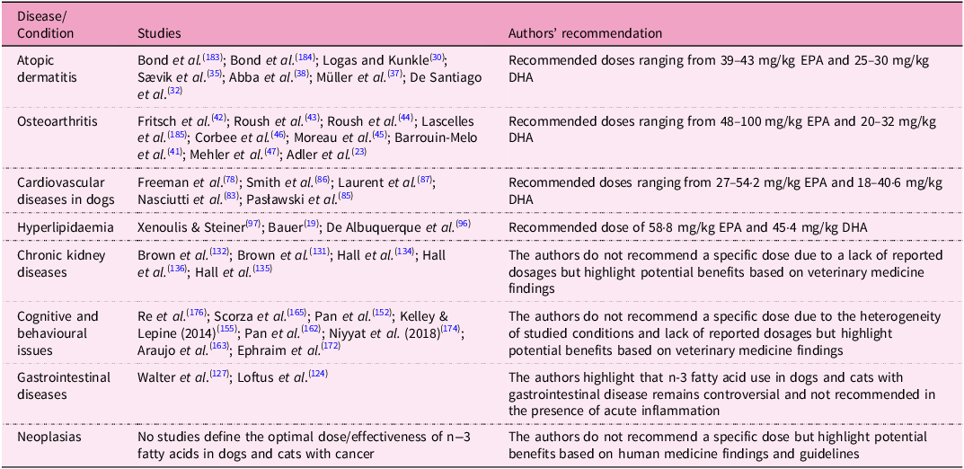
In light of all the evidence discussed, the authors’ dosage recommendations have been summarised in Table 1. These recommendations are based on key findings reported in the literature regarding the efficacy of omega-3 fatty acids in managing various diseases and conditions in dogs and cats.
Dosage recommendations for studied diseases and conditions

Risk of adverse effects
One of the primary concerns with omega-3 supplementation is its influence on platelet activity. Omega-3 fatty acids can lead to the production of thromboxane A2, which is related with the process of platelet activation. EPA and DHA are associated with the production of thromboxane A3, which is less effective in this process. This pathway was already described and mentioned in human subjects and translated to dogs and cats, although this might not be accurate. Other adverse effects include GI discomfort (mentioned earlier), especially in inflammatory conditions and with higher dosages than those usually applied. These effects raise concerns about immune function, as omega-3 can modulate the immune system, potentially altering the inflammatory pattern to the extent that it compromises immunity and overall host defences. However, the author of this work understands that omega-3 itself is not responsible for the entire scenario and this is the point where the n-6:n-3 ratio makes the difference. The balance between proinflammatory and anti-inflammatory cytokines plays an essential role in modulating immune function, and this is a matter of balance and does not cause adverse reactions in isolation.
Some researchers have already mentioned some concerns around lipid peroxidation (within cellular membranes), although the doses associated with these adverse effects are not the doses and n-6:n-3 (1·4:1) ratios typically used in clinical practice. In the light of these possible adverse effects, platelet function is the one which needs to be addressed and better understood(Reference Lenox and Bauer39). In cats, Saker et al.(Reference Saker, Eddy and Thatcher178) noted prolonged bleeding time and decreased platelet aggregation following supplementation with omega-3 fatty acids, and Bright et al.(Reference Bright, Sullivan and Melton179) did not document any measurable effects on haemostasis after supplementation with EPA and DHA. In dogs, Boudreaux et al.(Reference Boudreaux, Reinhart and Vaughn180) reported small changes in platelet reactivity but did not consider them clinically relevant. LeBlanc et al.(Reference LeBlanc, Bauer and Hosgood181) found no significant effects on platelet function after 12 weeks of supplementation with EPA and DHA. McNiel et al.(Reference McNiel, Ogilvie and Mallinckrodt182) observed no effects on platelet aggregation or platelet count in dogs with lymphoma and hemangiosarcoma compared with a control group.
Conclusions
EPA and DHA play essential physiological roles, particularly in cell membrane structure and immune modulation. Based on the available evidence, these omega-3 fatty acids effectively alleviate inflammation in atopic dermatitis, cardiovascular diseases, dyslipidaemias and chronic kidney disease in dogs. Their benefits extend to cognitive and behavioural issues, and based on human studies, they may be considered for patients with cancer. However, their use in gastrointestinal diseases remains controversial. Nonetheless, future studies should isolate the effects of EPA and DHA by ensuring nutritionally comparable diets, standardising intake based on body weight and using objective outcome measures. These efforts will strengthen evidence-based applications of omega-3 fatty acids in chronic diseases.
Supplementary material
For supplementary material accompanying this paper visit https://doi.org/10.1017/S0954422425100115
Acknowledgments
The authors thank the Sao Paulo Research Foundation – Fundação de Amparo à Pesquisa do Estado de São Paulo (FAPESP) - for the scholarship (process no. 2023/07307-6, process no. 2023/09486-5 and process no. 2023/07727-5) and grants (process no. 2022/06499-6). Balieiro, J.C.C. thanks the National Council for Scientific and Technological Development (CNPq) for the productivity grant (309189/2023-3). This study was financed in part by the Coordenação de Aperfeiçoamento de Pessoal de Nível Superior (CAPES) – Brasil – Finance Code 001. We would like to thank Grandfood Industry and Commerce LTD. (PremieR Pet) for the maintenance of the Pet Nutrology Research Center (CEPEN Pet) and the School of Veterinary Medicine and Animal Sciences for their support.
Authorship
T.H.A.V. and M.A.B. were responsible for conceptualisation and supervision; T.H.A.V., P.H.M., R.F.G.O., V.P., A.R.A., M.S.M., L.A.P., C.L., R.V.A.Z. and M.P.P. were responsible for writing the original draft; T.H.A.V., P.H.M., A.R.A., L.O.C.L. and J.C.C.B. were responsible for reviewing and editing. All authors have read and agreed to the published version of the manuscript.
Financial Support
This research received no specific grant from any funding agency, commercial or not-for-profit sectors.
Competing Interests
The authors declare none.