INTRODUCTION
Studies on surface modifications of various materials (including bio-materials) by different types of lasers are almost as old as the laser itself, and the field is constantly growing (Di Bernardo et al., Reference Di Bernardo, Batani, Desai, Courtois, Cros and Matthieussent2003; Bussoli et al., Reference Bussoli, Batani, Desai, Milani, Milan, Gakovic and Krousky2007; Alti & Khare, Reference Alti and Khare2006; Bashir et al., Reference Bashir, Rafique and Ul-Haq2007; Beilis, Reference Beilis2007; Batani et al., Reference Batani, Stabile, Ravsio, Lucchini, Desai, Ullschmied, Krousky, Juha, Skala, Kralikova, Pfeifer, Kadlec, Mocek, Präg, Nishimura and Ochi2003, Reference Batani, Dezulian, Redaelli, Benocci, Stabile, Canova, Desai, Lucchini, Krousky, Masek, Pfeifer, Skala, Dudzak, Rus, Ullschmied, Malka, Faure, Koenig, Limpouch, Nazarov, Pepler, Nagai, Norimatsu and Nishimura2007; Desai et al., Reference Desai, Batani, Bussoli, Villa, Dezulian and Krousky2008, Reference Desai, Dezulian and Batani2007, Reference Desai, Batni, Rossetti and Lucchini2005; Fernandez et al., Reference Fernández, Hegelich, Cobble, Flippo, Letzring, Johnson, Gautier, Shimada, Kyrala, Wang, Wetteland and Schreiber2005; Kasperczuk et al., Reference Kasperczuk, Pisarczyk, Kalal, Martinkova, Ullschmied, Krousky, Masek, Pfeifer, Rohlena, Skala and Pisarczyk2008; Schade et al., Reference Schade, Bohling, Hohmann and Scheel2006; Thareja & Sharma, Reference Thareja and Sharma2006; Trusso et al., Reference Trusso, Barletta, Barreca, Fazio and Neri2005; Trtica et al., Reference Trtica, Gakovic, Batani, Desai, Panjan and Radak2006a, Reference Trtica, Gakovic, Maravic, Batani, Desai and Redaelli2006b; Veiko et al., Reference Veiko, Shakhno, Smirnov, Miaskoski and Nikishin2006; Wieger et al., Reference Wieger, Strassl and Wintner2006; Schreiber, Reference Schreiber2005).
Various lasers and bio-materials have been used up to now for such studies. For fundamental and application interest, laser surface modification studies of bio-material including human teeth are very importance.
The tooth structure is known to be inhomogeneous and birefringent (Carlson & Krause, Reference Carlson and Krause1985; Zip & Bosch, Reference Zip and Ten Bosch1993; Bhaskar, Reference Bhaskar1991), the coronal portion of the tooth can be regarded as a bio-mechanical complex of two major tissues: enamel and dentine. Enamel is the outermost layer restricted to the coronal portion of the tooth. It is composed of enamel rods, which are not in direct contact with each other, but are cemented together by inter-prismatic substance. Dentine is the supporting structure and consists of specialized cells and bulky intercellular substance. The latter consists of two components: (1) collagenous fibers and (2) cementing substance mainly composed of polysaccharides (Bhaskar, Reference Bhaskar1991; Takuma et al., Reference Takuma, Tohda, Watanabe, Ogiwara and Yanagisawa1982).
The specialized cells are the odontoblasts. Each cell consists of two parts, the body of the cell that lies in the pulpal side of dentine and the odontoblastic process that extends through the full thickness of the dentine (the so-called, Tom's fiber) in the dentinal tubules. Each odontoblastic process in the dentine is found in thin walled tubes. The wall (the so-called Newman's sheath) is not formed by a different membrane, but originates from the differences in the nature of the matrix at the edge of the tubules, or from differences in the orientation of the fibers.
The interest in the studies of laser beam interaction with human teeth has risen, especially in the last two decades; Nd:YAG (Serafetinides et al., Reference Serafetindes, Khabbaz, Makropouloub and Kar1999), Ti:Sapphire (Rode et al., Reference Rode, Gamaly, Luther-Davies, Taylor, Graessel, Dawes, Chan, Lowe and Hannaford2003), TEA CO2 (Makropoulou et al., Reference Makropoulou, Serafetinides and Khabbaz1996), and different excimer (Neev et al., Reference Neev, Stabholz, Liaw, Torabinejad, Fujishige, Ho and Berns1993; Frankline et al., Reference Frankline, Chauhan, Mitra and Thareja2005) laser systems have so far been employed for these purposes.
Generally, during laser interaction with tooth surfaces, the laser energy is partially absorbed, and partially reflected. Due to the high intensity laser interaction, the initially absorbed part induces multiphoton absorption (MPA) and multiphoton ionization (MPI). Free electrons generated by means of multiphoton absorption will absorb the energy from the laser light, initiating ionization avalanche, and producing an exponential growth of free electron. Consequently, laser absorption increases exponentially as well. Due to the high laser intensity and the key role of MPA, even transparent materials become strongly absorbent, independently of the linear absorption characteristics (Neev et al., Reference Neev, Da Silva, Feet, Perry, Rubenchik and Stuart1996).
Again, in general, interaction of a short duration laser pulse with the target minimizes thermal and hydrodynamic effects (melting, shock waves propagation, etc.). However, interaction of human teeth with Nd:YAG laser beam in the picosecond time domain (Serafetinides et al., Reference Serafetindes, Khabbaz, Makropouloub and Kar1999) has not been sufficiently described in the literature in comparison to the nanosecond domain (Frankline et al., Reference Frankline, Chauhan, Mitra and Thareja2005). In this paper, our emphasis is on the study of the effects of a picosecond laser emitting in the visible region (532 nm) on human teeth. Special attention was paid to morphological surface modifications of human teeth at given laser energy fluence.
MATERIALS AND METHODS
Tooth Sample
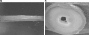
A total of 10 extracted human teeth belonging to 14–20 years old patients from Sweden were used during the experiment, non-caries premolars were transversally sectioned by a slow speed diamond disk (Model 150, MTI Corporation, Richmond, CA), which are parallel to the occlusal surface. Water is used as a coolant during the slicing of the tooth to prevent damage, due to friction. The image of the sample, prior to laser irradiation, is shown in Figure 1. The thickness of each sample was about 1 mm. The preparation process resulted in a relatively rough surface.
Transverse section of the prepared tooth sample. The thickness of each sample was about 1 mm.

Teeth were stored in distilled water until the time of the experiment, so that the structure of the tooth was not disturbed (Strawn et al., Reference Stawn, White, Marshll, Gee, Goodis and Marshall1996). Slices of the teeth were also stored in water until the start of the experiment.
The Laser System
Human teeth were irradiated by focusing the laser beam using a glass lens of 10 cm focal length. The angle of incidence of the beam with respect to the sample surface was near 0°. The irradiation was carried out in air, at atmospheric pressure of 1013 mbar, and standard relative humidity.
The laser employed is an active passive mode-locked Nd:YAG system (Model SYL P2, Quanta System Srl. Solbiate, Italy), which was characterized by Faeov et al. (Reference Faeov, Pikuz, Magunov, Batani, Lucchini, Canova and Piselli2004), and used for ablation studies (Gakovic et al., Reference Gakovic, Trtica, Batani, Desai, Panjan and Vasiljevic-Radoyic2007; Trtica et al., Reference Trtica, Gakovic, Maravic, Batani, Desai and Redaelli2007a, Reference Trtica, Gakovic, Radak, Batani, Desai and Bussoli2007b). It includes an oscillator, an amplifier, and a non-linear crystal (KD*P) to convert laser emission to second harmonic (532 nm). Pulse duration of about 40 ps is obtained by using a saturable absorber dye and an acousto-optic modulator. The laser was operated with a typical repetition rate of 1 Hz at 532 nm wavelength. Thanks to the use of an intra-cavity pin-hole, the emitted mode was quasi-TEM00.
Diagnostics Techniques
Various analytical techniques were used for the characterization of the human teeth before and after laser irradiation. The surface characteristics were monitored by optical (OM) and by scanning electron microscope (SEM). Teeth, as non-conductive samples, must be prepared before the SEM analysis. In our case, first the samples were dehydrated in graded solutions of ethanol and later a 20 nm gold coating was deposited onto the surface by using conventional sputter coater. This conductive layer was needed to minimize charging and beam drift during imaging scanning. The SEM was typically operated at 20 kV and the beam current was varying in the interval of 15–40 nA under a pressure of 10−6–10−8 mbar.
The SEM was also coupled to an energy dispersive analyzer (EDAX) for determining the surface compositions of the sample. Optical confocal microscopy (OCM) was employed in the characterization process in the reflection mode, primarily for specifying the geometry of the ablated area and measuring the depth of the crater after coating with gold. In addition, the diameter of each crater depth was measured using the OCM. In this paper, we concentrate on SEM results only.
RESULTS AND DISCUSSION
Morphological changes induced by the action of lasers on human teeth targets were studied versus laser beam characteristics: energy fluence and number of accumulated successive pulses.
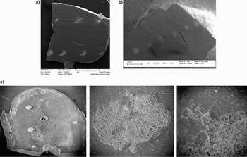
Morphological changes of the human teeth for one and three accumulated laser pulses at 532 nm are presented in Figures 2 to 4, respectively. The laser energy fluence (Φ) during the experiment was kept constant at ≅ 11 J/cm2; such fluence induced significant surface modifications on human teeth. The results of the induced modifications can be presented as follow.
View of human teeth after irradiation with picosecond Nd:YAG laser at 532 nm with fluence F = 11 J/cm2; SEM analysis. (a, b) View of enamel-dentine after irradiation with one and three pulses, respectively. (c) Only dentine after one pulse (in each position). Interaction of picosecond laser pulses at the given fluence creates the damage at enamel as well as at the dentine surface.

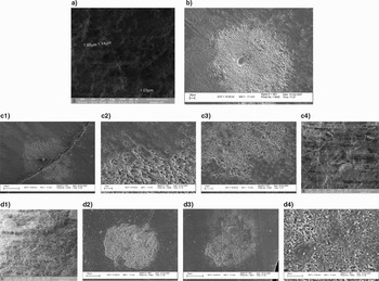
Picosecond Nd:YAG laser induced changes on human tooth. SEM analysis; F = 11 J/cm2; 532 nm. (a) view of enamel before laser irradiation, (D1) view of dentine before laser irradiation, (b) enamel after three pulses, (c) enamel-dentine junction after three pulses, (C1) enamel-dentine border, (C2, C3, and C4) view of enamel, dentine, and channel, (D2) dentine after irradiation with one pulse, (D3) dentine after irradiation with three pulses, (D4), same as D3 but with larger magnification.

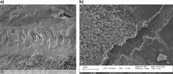
View of enamel-dentine junction before (a) and after laser irradiation with three pulses per position (b). SEM analysis; F = 11 J/cm2, λ = 532 nm. The non-irradiated junction (Fig. 4A) is filled up with collagen fibers and the junction channel can not be seen.

Interaction of picosecond laser pulses at the given fluence creates damage on enamel as well as dentine surfaces (Fig. 2). Normally, the accumulation of numerous pulses resulted in an enhancement of the modification (primarily diameter and depth of the ablation area were increased). When the laser was focused on the tooth surface, bright plasma was clearly visible, pointing toward the laser source, and a typical sparking noise was heard. In this context, the finding of Serafetinides et al. (Reference Serafetindes, Khabbaz, Makropouloub and Kar1999) is in good agreement with results that show morphology changes in the fundamental and the second harmonic Nd:YAG laser wavelengths, but the most prominent modification was recorded for 532 nm (Serafetinides et al., Reference Serafetindes, Khabbaz, Makropouloub and Kar1999).
Changes in Enamel
Detailed morphology changes on enamel are shown in Figures 3 and 4. On enamel (Fig. 3B), surface features can be characterized as: (1) appearance of a crater in the central zone of interaction and (2) existence of a diffuse periphery characterized by a cauliflower-like morphology. The crater has a conical form with a depth of 4 µm for one pulse, and 17 µm depth for three successive accumulated pulses; the crater diameter is more or less 300 µm.
Changes in Dentine-Enamel Junction
The effect of laser irradiation on the dentine-enamel junction is shown in Figures 3C1–3C4 and Figure 4B. SEM analysis showed that on the enamel and dentine side, different morphology structures were produced. Features on the enamel side posses a conical-like character (Fig. 2C3). Besides some holes exist. On the dentine side (Fig. 3C3), these features are lower in dimensions (micron and sub-micron range) with typical scales of SEM, and usually there are no holes in the damage area. Also on the dentine surface, sporadic islands of melted and resolidified materials can be detected (Fig. 3C3). The periphery is relatively sharp whereas, at larger distance from the edge of the irradiated area, some pores can be clearly recognized. These represent the dentinal tubules on the non-irradiated surface. The ablation depth on enamel and dentine in the area of the junction was different and typically amounted to 13 µm and 32 µm, respectively, after accumulation of the three pulses per position.
The difference in the recorded morphology, after laser irradiation, on the dentine-enamel junction can be primarily attributed to the different composition. Enamel includes about 96% hydroxyapatite (Ca10(PO4)6(OH)2AH), 4% protein (organic material) as well as water, which makes it mechanically hard and highly resistant to wear. Dentine is the supporting structure that lies underneath enamel, primarily composed of hydroxyapatite (70%), water and organic material like proteins (30%). Less mineralized tissues provide the tooth with toughness required to resist fracture when it is subjected to masticatory stresses. The morphology of the junction between enamel and dentine, after laser irradiation, is presented in Figures 3C4 and 4B. The non-irradiated junction (Fig. 4A) is filled up with collagen fibers and the junction channel can not be seen. Examination of the interface between enamel and dentine reveals closely arranged scalloped-shaped outlines. Several collagen fibrils that are projected longitudinally from the dentine matrix seem to converge to form a coarse collagen bundle of 80–120 nm in diameter, these are the Kroff fibers.
Irradiation with a picosecond laser causes the removal of collagen fibers from the channel as well as its modification. Since the images in Figures 3C4 and 4B correspond to three accumulated shots, we can suppose that the first pulse removes the collagen fibers, whereas the other pulses induce the modification of the channel. The bottom of the channel shows transversely oriented needles whereas the surface shows some cracking-like effect.
Changes in Dentine
Surface modifications of dentine, away from the enamel-dentine junction, are shown in Figures 3D2–3D4. Even one laser pulse was enough to induce significant ablation (Fig. 3D2) of the dentine. The region of the actual crater ablation extends to about 400 µm in diameter, and the diameter of the visible surface modification in dentine is 450 µm (i.e., it overlaps the diameter of the laser beam). The depth of ablation was 17 µm for one pulse per position. Generally, changes on dentine can be summarized as: (1) efficient ablation, (2) sharp periphery, (3) presence of melted and resolidified regions in the central damage zone, and (4) the tissue modification at the bottom of the crater shows no crack on the floor, with the presence of spherical shape particles of a few micrometers in diameter, which are formed due to hydrodynamic sputtering.
In general, the covered and exposed dentinal tubules, which are present within the area of the study, show no sign of damage after laser irradiation.
CONCLUSIONS
The interaction of 532 nm Nd:YAG laser radiation, at fluence of about 11 J/cm2, with human teeth is very efficient. Various surface morphologies on enamel and dentine were recorded. Features on enamel (away from the dentine-enamel junction) include a crater in central part (conical form) and cauliflower morphology at periphery. Instead at dentine (away from the dentine-enamel junction), the crater shows a different shape, and a relatively sharp periphery. Analyses of the dentine-enamel junction area and of regions far from the junction showed that the morphology of enamel alone or dentine alone (i.e., in locations more distant from the dentine-enamel junction area) was different. The enamel and dentine junction areas showed features like conical shape craters with sporadic melted resolidified islands. Finally, the junction channel, after irradiation, showed a removal of collagen fibers, and a needle-like bottom structure.
Generally, this investigation showed that picosecond Nd:YAG laser radiation can induce ablation on teeth surfaces almost instantaneously, meaning that by adjusting the focal spot of the laser beam, a precise cutting of the tooth can be achieved. Roughness of the inner surface and the wall of the craters, and bleached borders are beneficial to provide enough retention for the restorative (filling) material.
The appearance of plasma, in front of the teeth, indicates relatively high temperatures created above the surface. This may offer sterilizing effects, facilitating contaminant-free conditions during dental treatment.
ACKNOWLEDGMENTS
We would like to express our deep thanks to the Landau network, Centro Volta (LNCV) in particular to Prof. Riccardo Redaelli and Dr. Andrea Plebani for offering the opportunity to accomplish the research work at the University of Milano Bicooca. We acknowledged the kind advice and rich discussion with Milan Trtica (Vinca Institute of Nuclear Sciences, Belgrad, Serbia). In the same regards, we would like to thank the staff at the Istituto di Istologia, Embriologia and Neurocitologia for preparing and cooperation during scanning SEM images. Sincere thanks go to Dr. Noberto Chiodini for his help in preparing the teeth slices.