INTRODUCTION
Parasites that are successful colonizers tend to be generalists with a wide host range, or they infect a host that has a wide geographical and ecological range (Kennedy, 1994). One of the most important platyhelminths to have successfully invaded new host populations, with disastrous economic implications, is the monogenean Gyrodactylus salaris infecting Atlantic salmon (Salmo salar). This ectoparasite has devastated wild Atlantic salmon populations in Norway since its introduction from the Baltic (see Hansen et al. 2003). With its high fecundity and transmission potential, G. salaris can rapidly colonize an entire river system (Bakke et al. 1992; Johnsen et al. 1999) and even re-colonize waterways after treatment with the biocide rotenone (Johnsen et al. 1999). Experimental studies indicate that G. salaris is efficient at dispersing and infecting a wide range of hosts (Bakke et al. 2002). However, given simultaneously a choice of hosts, both G. salaris (optimal host Atlantic salmon) and G. derjavini (optimal hosts brown trout and rainbow trout) exhibit distinct preferences (Buchmann et al. 2004).
Gyrodactylids have no specific transmission stage; the viviparous worms give birth to fully grown adults, which during the birth process attach to the same host as the parent and only subsequently may transfer to a new host. Bakke et al. (1992) proposed 4 routes by which G. salaris could transfer to new hosts: (i) via contact with live hosts, (ii) via dead hosts, (iii) by detached parasites drifting in the water column, and (iv) by parasites attached to the substrate. Gyrodactylids on salmonids probably disperse using a variety of such mechanisms (Mo, 1992, 1997; Soleng and Bakke, 1997), all being temperature-dependent (Soleng et al. 1999). The most common is traditionally assumed to be through contact between living hosts, but the relative importance of the 4 different transmission routes has not been quantified. Soleng et al. (1999) found that transmission of G. salaris from the substrate or direct contact from infected live and/or dead fish was comparatively more important than transmission by drifting detached parasites. Furthermore, Bakke et al. (1991) showed experimentally that live salmon become more heavily infected when exposed to dead infected hosts than when exposed to living hosts. As gyrodactylids are dependent on the host for food, the window for transmission is restricted if most G. salaris individuals leave a host within a few hours of its death. Such behaviour has been reported by Malmberg (1970) for G. rarus and G. cryptarum, by Harris (1980) for G. gasterostei and by Cable et al. (2002a) for G. turnbulli, but is only assumed for G. salaris (see Jensen and Johnsen, 1992). Determination of all possible mechanisms that might facilitate transmission of G. salaris is essential for development of accurate risk assessments.
In the current study, we utilize a multidisciplinary approach (survival experiments, infection experiments and electron microscopy) to highlight the importance of dead hosts as a significant reservoir of infection for G. salaris.
MATERIALS AND METHODS
Host-parasite origin and maintenance
G. salaris from the River Lierelva in S.E. Norway were maintained on hatchery-reared salmon from the River Drammenselva for a minimum of 1 month prior to experimentation. Stock fish were maintained in dechlorinated water at 7·5 °C. This temperature and temperatures cited for the in vitro and in vivo experiments below varied within ±1 °C. Fish used in experiments and for maintaining parasites ranged in length from 52 to 106 mm (mean 85 mm).
For infection monitoring or removal of parasites, hosts were anaesthetized in 0·04% chlorbutanol and examined using a stereo-microscope with fibre-optic illumination. Individual parasites were removed from their host by carefully inducing them to attach to the tip of an insect pin. As gyrodactylids are sensitive to host serum (Buchmann, 1998; Harris et al. 1998) extreme care was taken to prevent contamination of parasites with host body fluids.
In vitro survival and behaviour
Single parasites were carefully removed from recently killed hosts and individually transferred to the bottom of a 96-well microtitre plate well containing 200 μl of dechlorinated water. Any parasites that showed signs of damage, or which died within 1 h of transfer, were discarded and excluded from the data set. Microtitre plates were subsequently stored at 3, 12 or 18 °C (n=75, 88, and 65 parasites, respectively). Worms were examined regularly to assess births, behaviour, location (i.e. attached/detached at the bottom of the well, in the water column or water film) and survival. Classification of behaviour and estimating time of death followed the procedures described by Cable et al. (2002b).
Behaviour and survival on dead hosts
Salmon with known infection intensities (75–320 worms per fish) were killed by a single blow to the head. Observations focused on those parasites that were attached to one side of the caudal and anal fins to avoid manipulation of the host or parasites during the course of the experiment. Selection of these fins was based on their naturally high parasite loads and the fact that the body mass of the corpse prevented their direct contact with the substrate. Worms on the reverse side of the fins and the nearby surrounding host tissue, were removed with watchmaker's forceps prior to the experiment, thus reducing migration. The fish were placed in 50 ml of water in shallow, black plastic dishes at 3, 12 or 18 °C; 10 fish per temperature. The water was changed twice daily with handling time minimized to reduce the effects of changing macro- and microenvironments. Parasites were monitored regularly to record general condition and location on the host. At 3 °C, worms were monitored twice a day during the first week and then once a day thereafter. At 12 °C, monitoring was performed twice daily throughout the experiment, whereas at 18 °C the intervals were shortened, and monitoring was carried out 4 times a day.
Re-infection potential
Naïve salmon were individually infected with 1 gravid worm that had previously been maintained at 10 °C either: (i) in vitro for 24 h (14 fish infected), or (ii) on a dead host for 72 h (14 fish infected). These 2 pre-experiment treatment times were chosen based on parasite population half-life calculated from the survival experiments described above. A control group of 14 fish was infected with worms removed directly from live hosts. Initially, fish were kept for 24 h in one of three small (20×30×10 cm) grey plastic boxes according to pre-treatment of the parasites. A possible implication of this treatment could be transfer of parasites among fish. However, only single gravid worms or, on one occasion, a mother (non-gravid worm) and daughter (newborn parasite) were observed on individual hosts, ruling out the likelihood of parasite transfer between hosts (unless parasites had transferred back and forth during this time). All fish still carrying an infection after 24 h were individually isolated in floating plastic boxes with a wire-mesh bottom to ensure free flow of fresh water into the box. Infection intensity was monitored weekly for 5 weeks.
Life-stage distribution
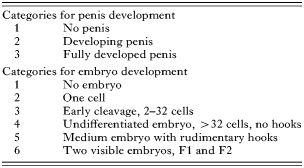
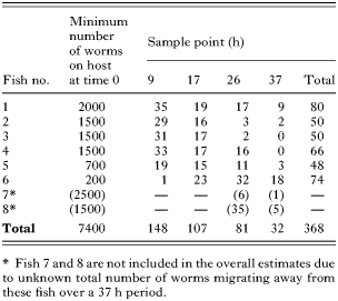
In order to determine life-stage distributions, worms were transferred to a microscope slide after which they were categorized under a stereo-microscope according to development of the F1 embryo and penis (see Table 1). Subsequent analyses revealed no distinct trends regarding these categories. Specimens were therefore re-categorized as either pre-1st birth (penis categories 1 or 2 in combination with a developing embryo, categories 5 or 6), or post-1st birth worms (all other combinations of embryo and penis development categories). Equality of distributions was tested using a Chi-square test, with Yates' correction in PAST vs. 1·32 (Hammer et al. 2001). Parasite population structure was determined from (i) an established infection, at least 1 month old. This served as a baseline for comparison with the stage structure in experimental groups (ii and iii below). (ii) Worms leaving dead hosts. Recently killed hosts with known infection intensities (see Table 2) were placed in 50 ml of water in black plastic dishes. After 9, 17, 26, and 37 h, the water in the dishes was changed and live worms attached to the bottom of the dish were staged. (iii) Individuals transferred between live hosts. Naïve fish were confined with hosts harbouring known parasite intensities in floating boxes (see above); one naïve fish (identified by clipping the adipose fin) and 1 infected fish per box. After specific intervals (<24 h), the recently infected fish were anaesthetized and the worms were removed and staged.


Ultrastructural evidence for feeding
To determine whether G. salaris can feed on a dead host, parasites that had previously been maintained for 18 h either (i) in vitro, (ii) on a dead host, or (iii) in situ on live hosts (control) were processed for transmission electron microscopy (TEM). Briefly, living parasites were fixed for 2–3 h in 2·5% glutaraldehyde buffered to pH 7·4 with 0·1 M sodium cacodylate containing 3% sucrose and 0·01 M calcium chloride, washed in several changes of cold buffer and then post-fixed for 30 min in 1% osmium tetroxide in the same buffer. Following dehydration in ethanol, the specimens were cleared in propylene oxide and embedded in Araldite resin overnight. Ultrathin sections mounted on uncoated 200 mesh copper grids or coated single whole grids, were stained in a saturated solution of uranyl acetate in 50% ethanol for 30 min followed by lead citrate for 10 min. Sections were examined using either a Philips or Joel EM microscope operating at 80 kV.
RESULTS
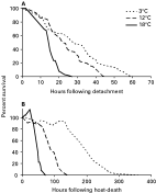
As predicted, survival of G. salaris was temperature dependent. In the absence of a host, the worms had a maximum survival of 60, 45 and 27 h, at 3 °C (n=75), 12 °C (n=88) and 18 °C (n=65 worms), respectively. Time elapsed before half the initial number of worms had died (population half-life) was 28, 22 and 15 h in the 3 groups (Fig. 1A). The same pattern of temperature-related survival was seen in worms on dead hosts, with maximum survival being 365, 142 and 72 h at 3 °C (n=422), 12 °C (n=315) and 18 °C (n=204 worms), respectively, and population half-lives of 186, 84 and 46 h at the three increasing temperatures (Fig. 1B). Maximum survival was thus 2·7–6·1 times higher among worms remaining on a dead host compared to those maintained in vitro, whereas population half-life was 3·1–6·6 times higher.

Fig. 1. Survival of Gyrodactylus salaris individuals (A) in vitro, artificially detached from Atlantic salmon parr and (B) on dead Atlantic salmon parr. Percentage of remaining individuals plotted against time post- detachment from host at 3 different temperatures. Note the different X-axis scales. At 18 °C in B, >100% indicates immigration of parasites from the body and other fins, and/or reproduction.
In vitro, the majority of parasites were attached to the bottom of the microtitre wells, occasionally moving by alternately attaching and detaching the anterior glands and haptor in a caterpillar-like locomotion. As time progressed, individual worms detached and lay wriggling, then twitching and were eventually moribund (=motionless unless gently touched with the edge of a pin in which case they contracted) at the bottom of the wells before dying. Only two worms were recorded in the water film at any time-point during the experiment (i.e. 0·88% of all parasites monitored). Parasites monitored on dead hosts were particularly active, moving short distances on the fins, during the first few hours to first day after host death. The length of this active period apparently was temperature dependent, with prolonged periods of activity at colder temperatures. Thereafter, the majority of worms were inactive with only occasional ‘steps’ in any direction. However, throughout the whole observation period parasites were observed in feeding-like activity, in which the anterior portion of the body was pressed in close contact with the host while pulse-like waves were detected passing along the length of the body. In addition, we observed 2 other distinctive behaviours: (i) stationary transmission mode (STM) in which the parasites were motionless with their slightly extended bodies held at right angles to the host; and (ii) search mode (SM) in which the parasites' body was highly extended (at least 4× its normal length) making constant circular movements and contractions pivoting around the motionless haptor. Worms exhibiting SM behaviour were frequently positioned on the very edge of a fin.
A high proportion of parasites remained with their dead hosts. This trend persisted with time even after the host had started to decay. For example, after 3 days at 12 °C, the fish carcasses were considerably degraded but even though wrapped in mucus, many living parasites were still attached to the host. The proportion of live worms actively leaving dead hosts over 37 h (Table 2) was estimated to be <5% (368 worms found alive out of a total starting population of >7400 worms on 6 hosts).
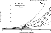
Regarding re-infection capability, 50% of the attempted transfers from live fish (7 fish out of 14) and from fish that had been dead for 72 h (7 out of 14) were successful in infecting new, naïve hosts. In contrast, only 1 of 14 parasites previously kept for 24 h in vitro was able to re-infect a new host. Subsequent monitoring of these infections indicated that population growth was reduced among the parasites from a dead host compared to the controls, but differences were not statistically significant (Student's t-test; P>0·05). Of the 7 fish that were initially infected with worms from dead hosts, 2 displayed no infection growth and both lost the infection within 1 month (see Fig. 2).

Fig. 2. Establishment and population growth of Gyrodactylus salaris on Atlantic salmon parr following 2 different pre-treatments: maintenance in vitro (n=1) or on a dead host (n=7), compared to controls (parasites transferred directly from a living host, n=7). Note the incomplete range on the Y-axis. The Y-axis is set to cross the X-axis at week 1 as infection by then is considered established.
Life-stage distribution indicates that 33% of a population of G. salaris from living hosts consists of newborn/pre-1st birth worms (Table 3). There was no significant pattern of stage-related dispersal from a dead host when compared to stage-distribution of parasites on living hosts (baseline populations) (Chi-square test, with Yates' correction; P>0·05, 1 D.F.). However, the proportion of older parasites (post-1st birth) that transmitted from live-host to live-host was significantly higher than in the baseline population (87·6% compared to 67%, Chi-square test, with Yates' correction; P<0·05, 1 D.F.).

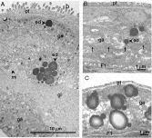
Ultrastructural examination confirmed previous observations (Cable and Harris, 2002) that the gastrodermis of G. salaris is a syncytial structure that lies on a thin basal lamina complex (Fig. 3A). Basally positioned nuclei in the gastrodermis are sparse, but are surrounded by scattered mitochondria, Golgi bodies and a variety of digestive vesicles. In recently-fed individuals (Fig. 3A), the gut lumen is greatly expanded, so that the gastrodermis often lies in close contact with the F1 embryo and the thin muscle layers underlying the surface layer of the parental tegument. Short microvilli extend into the gut lumen (Fig. 3A) or, in individuals that had expelled their gut contents, are compressed together amongst the cytoplasm of the gastrodermis. No gut contents were detected in any of the individuals that had been starved for 18 h (Fig. 3B). In all of these specimens there was a significant reduction in the number of electron-dense bodies. Rarely, these bodies were detected bound to large membrane-bound vesicles with heterogeneous contents (e.g. Fig. 3B). There was considerable variation in the appearance of the gastrodermis from worms maintained on dead fish (data not shown). In some individuals, however, digestive vesicles including electron-dense bodies were present but relatively sparse, whereas in others, the gastrodermis was packed with vesicles but cell membranes in most tissues were indistinct (Fig. 3C) indicative of degeneration.

Fig. 3. Transmission electron micrographs of Gyrodactylus salaris maintained on (A) live Atlantic salmon (controls), (B) starved in vitro for 18 h, and (C) a dead Atlantic salmon for 18 h. (A) Syncytial gastrodermis (ga) packed with numerous vesicles, including large electron-dense bodies (ed). Microvilli (mi) project into the distended gut lumen (gl) filled with host material indicating that this parasite had recently fed prior to fixation. Gastrodermis lies in close proximity to both the tegumental surface layer of the parent (pt) and embryo. Bacteria (b) are associated with the parental tegument of G. salaris. (B) Gastrodermis compressed between the tegumental surface layer of the parent (pt) and embryo (F1) and with microvilli along the mid-line of the gut. Midline of gut and compressed microvilli are shown by small unlabelled arrows. Note electron-dense body (ed) apparently releasing its contents within a larger heterogeneous vesicle. (C) Gastrodermis compressed between the tegument of the parent (pt) and F1 embryo but gastrodermis apparently actively digesting host material, packed with electron-dense bodies and heterogeneous vesicles.
DISCUSSION
In contrast to what has previously been assumed (Jensen and Johnsen, 1992), the present study shows that individuals of Gyrodactylus salaris do not necessarily leave a host after its death. Furthermore, parasites that remain with a dead host survive and maintain their infectivity for longer periods than detached worms. The main route of transmission for G. salaris is assumed to be (i) through contact between live hosts. Despite this assumption, the relative importance of direct transfer has not been directly compared with the other 3 potential transmission routes highlighted by Bakke et al. (1992), namely (ii) via dead hosts, (iii) by parasites attached to the substrate, and (iv) by detached parasites drifting in the water column.
Survival
As Mo (1988) suggested that G. salaris can survive for 6 days in vitro at 3 °C, it might be inferred that transmission routes (iii) and (iv) above (both involving parasites that are detached from a host) are restricted by a narrow window of opportunity. The current study indicates that G. salaris from the River Lierelva can only survive for 2·5 days in vitro at 3 °C and, importantly, viability is apparently low, even at the point of the populations' half-life post-host detachment. However, more interestingly, we found that a large proportion of G. salaris individuals will remain with their host after its death and survive substantially longer than off a host. Thus, the window for transmission between dead and live hosts might be wider than previously considered. Ultrastructural evidence suggests that this prolonged survival on dead hosts is due to the fact that at least some individual worms obtain nutrients from a dead host, whereas those parasites that detach from the host will starve. This probably explains why parasites remaining on the dead host have a higher re-infection potential than those which become dislodged or actively disperse to the substrate.
In our experiments, hosts were killed by a single blow to the head. Therefore, parasite survival on hosts that die naturally may be even greater than that observed in the current study. Harris et al. (1998) reported extreme sensitivity of G. salaris to complement in salmon blood and mucus. It is thus surprising that there was no evidence of higher initial-stage mortality, despite increased mucus production immediately after host death. Even more surprising, was the observation that worms remained on the dead host even when the host started to decay. Due to oxygen consumption and release of potential toxins from the carcass, the parasites' microenvironment must rapidly deteriorate. Such deterioration was predicted a priori to cause the parasites to leave the host, but there was no evidence of any such strategy. The 2 fins selected for the focus of our observations (the anal and caudal fins), turned out to be the areas of the fish least affected by host decay and fungal growth (based on visual inspections). Although worms in the immediate vicinity (i.e. those on the reverse side of the fins and nearby surrounding host tissue) were mechanically removed prior to observations, some worms remained on the body as removal of all worms would have resulted in excessive handling of the corpse. Some of these parasites migrated on the anal and caudal fins during the course of the experiment. However, this did not affect our primary goal of determining maximum life-span of parasites attached to a dead host.
Transmission
Although renowned for their conserved morphology, we suspect that gyrodactylids employ a variety of different life-history traits and behaviours related to the ecology of their hosts. One transmission strategy employed by individuals of G. turnbulli, is migration from recently dead hosts onto the water film (Cable et al. 2002a). This behaviour increases the chances of parasites contacting a new host since the main host species of G. turnbulli, the guppy, is a surface feeder. Unlike guppies, salmon parr are drift-feeders that position themselves on or close to the substrate waiting for food items to come into sight. A possible important route of transmission in such a system would thus be via the substrate. If so we might predict a life-stage distribution bias amongst G. salaris leaving their dead host comparable to that observed during transmission between live hosts. However, our studies did not reveal any such stage-distribution bias, which could reflect the superior advantage in staying on, as opposed to leaving, a dead host.
Scott and Anderson (1984) and Scott (1985a) conducting a series of experiments with G. turnbulli (originally described as G. bullatarudis, but re-described as G. turnbulli by Harris, 1986), identified transmission as a critical stage in the parasite's life; only 35–39% of parasites that attempted transfer, succeeded (Scott and Anderson, 1984). The guppy host of G. turnbulli is a shoaling fish that occupies a variety of habitats from small enclosed pools to rivers. For G. salaris, which parasitizes solitary hosts in relatively fast flowing water, the likelihood of parasites being washed away from dead as well as live hosts must be high. Scott and Anderson (1984) found that transmission of G. turnbulli from infected to uninfected hosts during host contact occurred at a greater rate from dead to live fish than between live fish, and attributed this to the tendency for guppies to cannibalize moribund fish. There is no evidence published that salmon display post-mortem cannibalism in the wild. It is, however, well established that during territorial behaviour, salmon nip the fins of conspecifics (e.g. Keenleyside and Yamamoto, 1962). Mo (1992) for example, mentioned this as a potential transmission strategy in G. salaris. In experimental and aquaculture tanks, this behaviour is also displayed towards dead conspecifics (personal observation). Our observations of G. salaris individuals remaining with a dead host therefore might be attributed to a combination of the high risk related to transmission and the increased likelihood of contacting a new host due to nipping, cannibalism or scavenging by a susceptible host.
To counteract the above-mentioned risks of transmission, gyrodactylids may have adopted a compensatory survival strategy. Our findings that worms post-1st birth are more likely to transfer (between live hosts) than those that have not given birth, are consistent with the observations of Harris et al. (1994) although in an experiment not designed for this purpose. Considering the high risk of failing to transfer to a new host (see above), leaving at least 1 offspring on a host seems the optimal strategy for continuation of a particular parasite strain. If the parental worm is lost, then assuming that at least the 1st born asexually-derived daughter remains with the host, a genetically identical clone, pre-adapted to the host, will survive.
In most other gyrodactylid – host interactions, continuous transmission is predicted to occur due to the fact that a host immune response will eventually eliminate the infection (see for example Scott, 1985b). East Atlantic salmon, in contrast to the Baltic salmon, are generally less able to mount an effective immunological response against gyrodactylids (see review by Bakke et al. 2002). In the G. salaris – Norwegian Atlantic salmon interaction, host death is a frequent end-point of the infection. As discussed above, transmission from a dead host via post-mortem cannibalism is likely to be a successful strategy among these parasites. So, if continuous transmission is a strategy to avoid the host response, such a strategy might be superfluous in G. salaris parasitizing Atlantic salmon. This poses the question: is live-host-to-live-host transmission in G. salaris based solely on inbreeding avoidance, or is it a residual strategy reflecting an ancient interaction with Baltic salmon? A final factor to be considered is that Atlantic salmon from the Atlantic Ocean and Baltic Sea, despite not being semelparous, both have high post-spawning mortality. Thus, cross-cohort host transmission may occur frequently in G. salaris between dead post-spawning adults and fry/pre-smolts. Future studies should assess the direct effect of the host immune response on parasite behaviour. For instance, do parasites die on the host, or do they leave at a stage sufficiently early to have the potential to re-infect a new host? Bakke et al. (2002) found that G. salaris surviving on innate resistant trout for 6 days were able to re-infect salmon, exemplifying the potential for survival under obviously extreme conditions.
G. salaris is a significant threat to populations of Atlantic salmon. In Norway, action is taken to control the epidemics. In the UK, on the other hand, where the parasite has never been observed, considerations and precautionary measures are made on a ‘what if’ basis (e.g. Peeler et al. 2004). The present study highlights the importance of dead hosts as a significant reservoir of infection; a fact which should be incorporated into future management polices and risk assessments.
This work was funded by a Norwegian National Centre for Biosystematics grant (Project no. 146515/420), co-funded by the NRC and the NHM, University of Oslo, to K.O., a Natural Environment Research Council Advanced Research Fellowship (NER/J/S/2002/00706) to J.C. and a NRC ‘Wild Salmon Program’ grant (Project no. 145861/720) to T.A.B. We thank two anonymous referees for their valuable comments on this manuscript.