Introduction
Inflammatory bowel diseases (IBDs), namely Crohn's disease and ulcerative colitis, are groups of heterogeneous disorders of uncertain origin that cover a broad spectrum varying from chronic indolent episodes to acute fulminant disease as a result of uncontrolled inflammation of the gastrointestinal tract (Hanauer, Reference Hanauer2006; Xavier & Podolsky, Reference Xavier and Podolsky2007). Unlike Crohn's disease, ulcerative colitis is typically restricted to the mucosal surface extending from the rectum to the entire colon. Symptoms can vary from mild disease to severe diarrhoea and rectal bleeding with mucus passage, in association with fever and gross weight loss (Garud & Peppercorn, Reference Garud and Peppercorn2009). In predisposed patients, the absence of enteric parasites, combined with various heterogeneous environmental factors associated with the lifestyle of wealthy developed countries, may trigger these intestinal disorders or affect their outcome (Danese et al., Reference Danese, Sans and Fiochi2004).
Despite the dramatic increase in the prevalence of these disorders in industrialized Western countries (Molodecky et al., Reference Molodecky, Soon, Rabi, Ghali, Ferris, Chernoff, Benchimol, Panaccione, Ghosh, Barkema and Kaplan2012; Ponder & Long, Reference Ponder and Long2013), as yet no completely successful treatment is available. Conventional attempts at treatment have included the use of drugs such as aminosalicylates, corticosteroids, azathioprine/6-mercaptopurine, ciclosporin and antibiotics, combined with surgical resection where necessary, but success is not guaranteed. Ongoing research into new alternative therapies is based on biological approaches that make use of probiotics, nicotine and fish oil, as well as new drugs such as immunosuppressants like tacrolimus, thiazolidinedione-class drugs like rosiglitazone, or monoclonal antibody treatments, including anti-tumour necrosis factor-α (infliximab) and anti-CD3 (visilizumab) (Garud & Peppercorn, Reference Garud and Peppercorn2009; Theede et al., Reference Theede, Dahlerup, Fallingborg, Hvas, Kjeldsen, Munck and Nordgaard-Lassen2013).
Recent clinical studies have provided evidence that treating patients who have Crohn's disease or ulcerative colitis with the pig whipworm Trichuris suis can alleviate the severity of these diseases (Summers et al., Reference Summers, Elliot, Urban and Thomson2005; Reddy & Fried, Reference Reddy and Fried2007). The rationale behind this therapeutic approach is supported by the ‘hygiene hypothesis’, which holds that environmental factors, such as the helminth infections common in underdeveloped countries, may protect from inflammatory bowel diseases provided that such enteric infections counteract IBD-based immunological disarrangements (Weinstock et al., Reference Weinstock, Summers and Elliot2005).
In the present study a murine model for chemically triggered ulcerative colitis was implemented to assess the efficacy of infection by Trichuris muris on the course of subsequent induced inflammatory disease.
Materials and methods
Parasitological procedures
The Edinburgh strain of T. muris was used. It was kindly provided by Professor Wakelin from the University of Nottingham in 1989 and since then has been maintained by periodic passages in outbred Swiss CD-1 mice (Charles River Laboratories, L'Arbresle Cedex, France). Maintenance of T. muris and the methods used for infection are described elsewhere (Wakelin, Reference Wakelin1976). Briefly, male Swiss CD-1 mice were orally infected with 400 embryonated eggs of T. muris. Fifty days after infection the mice were sacrificed and their caeca removed, opened lengthwise and incubated in phosphate-buffered saline (PBS) for 30 min before the worms were picked out carefully with fine forceps. The worms were then washed several times in PBS with penicillin, streptomycin, gentamycin and amphotericin B and then placed in minimum essential medium (MEM) for egg shedding by females. The medium was changed every 24 h for 4–5 days and the eggs were collected by filtration and washed several times by sedimentation in deionized water to remove the medium. They were then allowed to embryonate for 28 days at 28°C. Finally, they were suspended in distilled water in order to adjust the corresponding dose for mouse infection. Intestinal worm counting following infection was performed as described by Else et al. (Reference Else, Wakelin, Wasson and Hauda1990). Once the mice were sacrificed, their caeca were removed, opened lengthwise and incubated in PBS at 37°C for 4–5 h for worm collection. To collect the L4 larvae at day 20 post-infection (20 p.i.) the mucosa were gently scraped to allow the larvae to be released and transferred to a Petri dish. Adult worms at day 30 p.i. and onwards were carefully picked out with fine forceps. Worm counting was always carried out under a stereomicroscope.
Experimental infections of mice with Trichuris muris
Ten- to 12-week-old females from the CBA/J (H-2k) inbred strain were purchased from Charles River Laboratories and kept at the Animal House of the Complutense University under controlled conditions of light, temperature, water and food supply.
The mice were divided into groups of six and in all of the experiments each mouse was infected with 50 embryonated eggs of T. muris administered by oral gavage. A small inoculum was used because previous attempts using higher doses, ranging from 100 to 400 eggs, had caused severe disease and high mortality in groups undergoing simultaneous infection and dextran sodium sulphate (DSS) administration.
Induction of ulcerative colitis
Acute colitis was induced following the protocol described by Okayasu (Okayasu et al., Reference Okayasu, Hatakeyama, Yamada, Ohkusa, Inagaki and Nakaya1990) and modified according to our experimental requirements. Four per cent DSS (molecular weight (Mw) 100,000) purified from Leuconostoc sp. (Fluka Biochemica, Spain) was administered to each mouse in distilled drinking water for nine consecutive days. The schedule for administration was chosen following previous toxicity assays carried out with different percentage doses of DSS. After that, tap water was administered until the end of the experiment. This model was used as it was considered to be a fair representation of acute ulcerative colitis in humans (Okayasu et al., Reference Okayasu, Hatakeyama, Yamada, Ohkusa, Inagaki and Nakaya1990).
Ulcerative colitis was induced at different times after infection with T. muris. In the first experiment DSS was supplied from day 0 to day 9 p.i. Five groups of mice were involved: uninfected–DSS administration, infected–no DSS administration, infected–DSS administration, infected–DSS administration followed by worm suppression with administration of albendazole (ABZ) at 400 mg/kg/mouse (day 10 p.i.), and a control group of uninfected–no DSS administration. Clinical activity and histopathology were assessed on days 10 and 20 p.i. The second experiment involved late induction of ulcerative colitis by administering DSS from days 54 to 62 p.i. to one infected and one uninfected group, including an additional infected–no DSS administration group. Evaluations were performed on days 63 and 70 p.i. In a third experiment the DSS administration was moved up to days 27–35 p.i. with evaluations carried out on days 36 and 43 p.i.
Clinical activity and histopathology
The clinical activity of the colitis was assessed daily from the presence of blood clots around the anus, stool consistency and the general appearance of mice. Body weight was recorded at the beginning of the procedure and again just before sacrifice. Histopathological findings were in accordance with standard procedures and were applied as follows. The animals were sacrificed and underwent a midline laparotomy. The colon was resected from the ileo-caecal junction up to the rectum and its length was measured. The last 5 cm of the distant colon was then cut open and flushed with 10% buffered formalin using a 1-ml syringe with a 20G needle for about 10 min. Once fixed, the selected sample was opened lengthwise and the resulting long sheet was ‘Swiss rolled’ on to a small plastic cylinder for storage in 10% buffered formalin at 4°C, pending processing for histological examination. The presence of worms in the caecum was estimated using the methods described above.
For histological analysis, the samples were stained with hematoxylin and eosin and the mucosal morphological alterations were assessed in a blind manner by considering the score of various parameters graded on a scale of 0 to 3, as described by Gaudio et al. (Reference Gaudio, Taddei, Vetuschi, Sferra, Frieri, Ricciardi and Caprilli1999). The following parameters were estimated: destruction of the epithelium and glands; dilatation of glandular crypts; depletion and loss of goblet cells; inflammatory cell infiltration; oedema; vascular congestion; crypt abscesses; atrophy. Each parameter was graded as follows: 0 = normal aspect, 1 = focal lesion, 2 = zonal lesion, 3 = diffuse lesion. The colitis score for each individual mouse represented the sum of the subscores for the different parameters and the data were summarized as means ± standard errors of all subscores from range 0 to 24.
Data analysis
Statistical significance for body weight and colon shortening was estimated using one-way ANOVA. Significance was defined as P< 0.05. For histological studies, statistical significance of the scores for different groups was determined by the Kruskal–Wallis test. Where significant, the Wilcoxon Rank Sum test was used (SPSS v. 20 IBM software; SPSS Inc., Chicago, Illinois, USA).
Results
Induction of acute ulcerative colitis concomitant to infection with T. muris
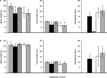
DSS-induced severe colitis manifested itself from the very start of the experiment and continued right to the end of the observations at day 20 p.i. (fig. 1). Significant decreases (P< 0.05) in body weight and colon shortening were observed by day 10 p.i. in animals administered DSS (fig. 1A). No weight reduction was evident in the infected–no DSS administration group compared to the naïve control group and no worms were recovered from the intestinal caeca. However, by day 10 p.i., 9.05 ± 4.30 and 6.24 ± 3.12 adult worms were remaining in the infected groups subjected to DSS administration, with or without additional suppressive anthelmintic treatment. Histological analysis showed ulcerative colitis lesions in all groups that had received DSS. Scores ranged from 15.2 ± 3.75 to 20.42 ± 5.03, with a marked difference between the lowest score and the highest, in which the anthelmintic treatment was applied. With infection alone, no histopathological changes were visible (score = 0.90 ± 0.43).
Clinical and histopathological changes in CBA/J mice infected with T. muris and administered DSS on days 0–9 p.i., with analysis undertaken on (A) day 10 and (B) day 20 p.i. Naïve control (left-hand bars, diagonally shaded), DSS administration (black bars), T. muris (grey bars), T. muris + DSS administration (white bars); T. muris + DSS administration + albendazole (right-hand bars, diagonally shaded); a, b, significantly different from infected and no DSS administration group (P< 0.05); c, significantly different from uninfected and DSS administration group (P< 0.05); * not determined.

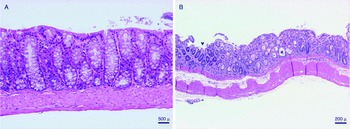
A clear improvement in clinical features was observed by day 20 post-infection in all groups that had received DSS, especially in the infected–DSS administration groups, where mean body weight rose to equal that recorded in healthy controls (fig. 1B). A similar picture could be seen with colon length restoration, although to a lesser extent since the differences still reached statistical significance (P< 0.05). However, despite clear improvement, histopathological analysis still showed signs of moderate to severe ulcerative colitis, with the presence of necrosis of surface epithelium and crypt enlargement associated with microabscesses and destruction, submucosal oedema and mixed inflammatory cell infiltration plus atrophy, all of them more marked in the distal large intestine (fig. 2B). It is important to note that 11.8 ± 6.5 adult worms were recovered from those animals that had been administered DSS, whereas they were effectively suppressed in those undergoing anthelmintic treatment which, at the same time, exhibited a histological score of 18.5 ± 4.43, comparable to that recorded on day 10 p.i. (fig. 1A). Taken as a whole, these results indicate that concomitant colitis clearly favours the persistence of infection by T. muris.
Colonic sections of (A) control mice (scale bar = 500 μm) and (B) DSS administration mice (scale bar = 200 μm) on day 20 post-infection, to show moderate to severe colitis with the presence of necrosis of surface epithelia (asterisk), crypt enlargement (black arrowhead) and mixed inflammatory cell infiltration in (B).

Later induction of acute ulcerative colitis following T. muris infection
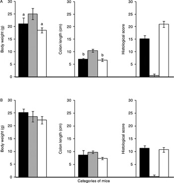
As shown in the previous experiment, administration of DSS caused severe colitis, with a significant reduction by day 63 p.i. in both body weight and terminal colon length in all animals compared to those that had only been infected. With regard to histology, infection alone did not induce any significant change (score 0.60 ± 0.55), whereas for those administered DSS the histological scores varied from 15.2 ± 1.30 in uninfected–DSS administration mice to 21.0 ± 1.22 in the infected–DSS administration group (fig. 3A), with the appearance of surface epithelial damage, crypt destruction and loss of goblet cells, together with severe submucosal oedema and mixed inflammatory cell infiltration. On day 70 p.i. no clinical improvement in body weight and terminal colon length was evident with respect to day 63, although histopathological analysis showed an early recovery, with histological scores falling to 10.4 ± 0.89 and 10.75 ± 0.96 for uninfected–DSS administration and infected–DSS administration mice, respectively (fig. 3B). The observed recovery was based on the initiation of a regenerative surface epithelium process, together with an increase in crypts and goblet cells and a decrease in inflammatory infiltration, although residual lesions with interstitial fibrosis and atrophy still remained. According to the histological scores, the overall improvement from induced ulcerative colitis was markedly higher in animals previously infected with T. muris. As expected, no worms were recorded from the caecum of any infected animal.
Clinical and histopathological changes in CBA/J mice infected with T. muris and administered DSS on days 54–62 p.i. with analysis undertaken on (A) day 63 and (B) day 70 p.i.; DSS administration (black bars), T. muris (grey bars), T. muris + DSS administration (white bars); a, b, significantly different from infected and no-DSS administration group (P< 0.05).

Earlier induction of acute ulcerative colitis following T. muris infection
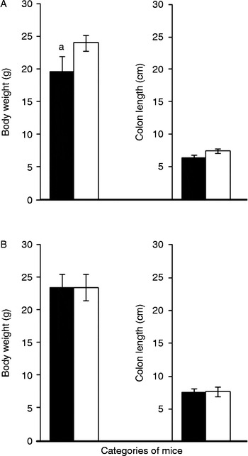
This experiment was designed after seeing the results of inducing colitis at a later stage after infection. Since histological analysis clearly showed early signs of recovery from colitis in infected animals, despite the apparent absence of infection, an additional similar experiment was designed, but with the gap between infection and the induction of colitis narrowed in order to search for effects of a supposedly early recovery that could be attributable to the recent presence of the parasite. Both groups showed clear signs of colitis after receiving DSS, although this was less intense in the previously infected group. Significant differences among both groups were recorded for body weight on days 36 but not on day 43 p.i. Moreover, by day 36 the infected group already showed an average increase in terminal colon length of nearly 17% compared to the non-infected one (fig. 4A). Interestingly, 2.6 ± 0.36 adult worms were recovered from the caeca of infected mice by day 36 p.i.
Clinical and histopathological changes in CBA/J mice infected with T. muris and administered DSS on days 27–35 p.i. with analysis undertaken on (A) day 36 and (B) day 43 p.i.; DSS administration (black bars), T. muris + DSS administration (white bars); a, significantly different from infected and DSS administration group (P< 0.05).

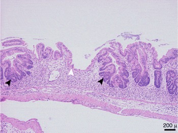
In the histopathological follow-up, the criterion set out by Gaudio et al. (1999) was not applicable here since negative scores resulting from regeneration events mainly in infected animals would give an erroneous estimation. Based on microscopic observations both groups showed evidence of active colitis by day 36 post-infection, with loss of glands, oedema and destruction of the intestinal epithelium, although these lesions were more extensive and severe in the uninfected-DSS administration group. However, in infected-DSS administration animals patches with clear signs of initial epithelial regeneration (superficial epithelization and glandular regeneration) were frequently found (fig. 5). The second determination on day 43 p.i. showed that both groups had limited areas of ulcerative colitis, but epithelization was also present in the non-infected group, although mucosal glandular regeneration still appeared to be minimal compared to that of the infected group (data not shown).
Colonic sections on day 36 post-infection of mice infected with T. muris but with DSS administered earlier on days 27–35, to show initial epithelial regeneration with superficial re-epithelization (white arrowhead) and glandular hyperplasia (black arrowheads) (scale bar = 200 μm).

Discussion
In recent years, there has been extensive application of experimental models based on the use of helminths to ameliorate chemically induced ulcerative colitis. Although results have been somewhat contradictory in cases where the Hymenolepis diminuta tapeworm was used (Reardon et al., Reference Reardon, Sanchez, Hogaboam and Mckay2001; Wang et al., Reference Wang, Fernando, Leung, Phan, Smyth and Mckay2010), most of the studies carried out with nematodes such as Trichinella spiralis or Heligmosomoides polygyrus clearly demonstrated beneficial effects (Khan et al., Reference Khan, Blennerhasset, Varghese, Chowdhury, Omsted, Deng and Collins2002; Motomura et al., Reference Motomura, Wang, Deng, El-Sharkawy, Verdu and Khan2008, Sutton et al., Reference Sutton, Zhao, Madden, Elfrey, Tuft, Sullivan, Urban and Shea-Donohue2008; Ashour et al., Reference Ashour, Othman, Shareef, Gaballah and Mayah2014). The basis for the therapeutic ability of these helminths is supported by their proven ability to modulate the immune response towards predominance of the Th2 type through triggering T and B regulatory cells, resulting in down-regulation of Th1-type responses typically associated with this kind of inflammatory disease. Despite the use of T. suis in the clinical field to alleviate these immunologically based disorders, so far only one experimental study has been conducted using the related murine species T. muris to treat experimentally induced ulcerative colitis in resistant wild-type C57BL/6 and transgenic mice (Taylor et al., Reference Taylor, Zaph, Troy, Du, Guild, Comeau and Artis2009). The approach used here was to select a mouse strain resistant to infection by T. muris, in which we induced acute ulcerative colitis by oral administration of DSS.
Selecting the appropriate mouse strain is critical for evaluating the interaction of helminth infection and ulcerative colitis, two inflammatory events that are apparently different in nature but develop in the same biological niche: the large intestine. A recent paper reported the presence of common quantitative trait loci (QTLs) and novel gene clusters for colonic inflammation induced by T. muris in susceptible AKR and resistant BALB/c mouse strains, considered as colitis ‘hotspots’ for susceptibility, with the regulation of some of them being relevant in the context of human Crohn's disease (Levison et al., Reference Levison, McLaughlin, Zeef, Fisher, Grencis and Pennock2010, Reference Levison, Fisher, Hankinson, Zeef, Eyre, Ollier, McLaughlin, Brass, Grencis and Pennock2013). In our opinion, CBA/J (H-2k) fulfils the minimum requirements for a suitable model as it allows very light, transient infections to be established which are able to prime regulatory Th2-type anti-inflammatory responses, as in the case of the pig whipworm T. suis in humans. The main aim of our work was therefore to evaluate the influence of parameters such as infection and treatment doses, together with the relative time-course for both events, as a first step towards further studies focused on the pathophysiological and immunological bases for these events and their interplay.
Following previous assays, we first established 4% DSS (100,000 Mw) given in distilled drinking water for nine consecutive days as the appropriate dose for inducing acute colitis while, at the same time, being compatible with concurrent infection by T. muris. Infection with more than 50 eggs led to severe colitis with serious lesions and a high mortality rate. This low dose, however, did not seem to produce a patent infection, since no worms were collected from the caecum by day 10 post-infection, although this did not mean that the infection was not present. At this time, worms are at the beginning of the second larval stage (L2) and it is likely that the stress caused by DSS acts both ways in treated mice, either arresting the larvae in the mucosa, thus preventing them from coming out during incubation in saline, or accelerating their early expulsion from the intestine. Contact between the parasite and the intestinal mucosa at any time appears to lower the severity of the DSS-induced colitis, as shown by the amelioration of clinical and histopathological alterations in infected animals. At the same time, it allows the worms to remain and infection to proceed.
It can therefore be concluded from this protocol assay that the simultaneous induction of T. muris infection and acute ulcerative colitis by DSS has mutual beneficial consequences, in that the acquired host's tolerance of the parasite results in an amelioration of the ulcerative disorder. Colitis induction following a previously established infection produced negligible beneficial effects on the inflammatory events. This was probably due to the loss of worms before DSS was applied (days 63 to 70 post-infection). However, 8 days after DSS was administered the histopathological scoring dropped nearly 50% with respect to day 1 in animals previously infected. Reducing the induction time for ulcerative colitis with regard to the course of the T. muris infection led to early improvement in the evolution of the disease, as shown by histopathological findings observed as early as 1 day after DSS administration was completed.
In line with these findings, a recent paper also showed that amelioration of acetic acid-induced ulcerative colitis in mice was more pronounced when infection by the related nematode T. spiralis preceded colitis induction (Ashour et al., Reference Ashour, Othman, Shareef, Gaballah and Mayah2014). This raises a question about the presence of adult worms by day 36 post-infection in earlier induction of colitis. In a previous study aimed at defining the CBA/J phenotype concerning susceptibility to T. muris infection, we found that by using standard inocula (around 200–400 embryonated eggs) worm expulsion was completed by day 23 post-infection (data not shown). Consequently, we considered CBA/J to be a resistant strain. However, previous researchers (Bancroft et al., Reference Bancroft, Else and Grencis1994) have found that the dynamics of expulsion of T. muris in the resistant BALB/K mouse strain varies in a dose-dependent manner, in such a way that higher doses (from 200 to 400 embryonated eggs) elicit stronger responses, thus promoting the rapid expulsion of worms, whereas low doses (50 or fewer embryonated eggs) allow them to persist for longer, turning the mouse phenotype from resistant into susceptible. Irrespective of this, the results of our work corroborate the beneficial role of the presence of Trichuris in alleviating ulcerative colitis in humans, and stress the critical influence of the dose and duration of the host–parasite contact with regard to the onset of the acute inflammatory event.
It is well documented that, owing to the particular location of T. muris in the large intestine, threaded along the string of enterocytes surrounding the intestinal microvilli, expulsion is based on accelerated renewal of epithelial cells from the base of the gland to the luminal face. Expulsion is triggered by a strong Th2-type response accompanied by the secretion of active factors such as thymic stromal lymphopoietin (TSLP) from the intestinal epithelial cells and resistin-like molecule β (RELMβ) from goblet cells (Herbert et al., Reference Herbert, Yang, Hogan, Groschwitz, Khodoun, Munitz, Orekov, Perkins, Wang, Brombacher, Urban, Rothenberg and Finkelman2009; Patel et al., Reference Patel, Kreider, Urban and Gause2009). TSLP is related to epithelial regeneration and crypt hyperplasia (Cliffe et al., Reference Cliffe, Humphreys, Lane, Potten, Booth and Grencis2005) while RELMβ would inhibit the chemotactic functions of the parasite (Artis, Reference Artis2006). Both factors are found to be deficient during inflammatory bowel diseases (Munitz et al., Reference Munitz, Waddell, Seidu, Cole, Ahrens, Hogan and Rothenberg2008; Sepúlveda et al., Reference Sepúlveda, Beltrán, Peralta, Rivas, Rojas, Figueroa, Quera and Hermoso2008) and it has been shown that neutralization of TSLP results in susceptibility to T. muris infection, while exacerbating inflammation in mice resistant to DSS administration (Levison et al., Reference Levison, McLaughlin, Zeef, Fisher, Grencis and Pennock2010).
To sum up, based on these premises, DSS ulcerative colitis appears be a good model for studies on cell proliferation and epithelial regeneration (Williams et al., Reference Williams, Fuller and Dieleman2001) and T. muris would be a suitable candidate for studying the intrinsic interaction between host and parasite during a process that produces mutually beneficial effects. Moreover, overall analysis of the results from the three experiments in this study clearly supports the view that histological score analysis is a highly sensitive tool for evaluating early steps in the recovery from inflammatory intestinal disorders. Ongoing immunopathological studies at the intestinal level will doubtless shed more light on the close interactions between these two types of intestinal inflammatory disorders and their potential mutually beneficial consequences. Thus, chemical ulcerative colitis would favour the permanence of T. muris as it progresses from the acute to the chronic stage of infection in the large intestine and, reciprocally, this would result in down-regulation of the immunopathological effects associated with these two inflammatory diseases of the intestine.
Financial support
This work was supported by project PR41/06-14 941 from Grupo Santander/Complutense University of Madrid, Spain.
Conflict of interest
None.
Ethical standards
Animal management was carried out in accordance with Complutense University regulations and Spanish legislation for the use of laboratory animals. All the procedures involved in this study complied with the ethical standards of the relevant national and institutional committees on the care and use of laboratory animals.