Introduction
Schwannomas are benign peripheral nerve sheath tumours arising from Schwann cells and account for approximately 5 per cent of all benign soft tissue tumours. In the head and neck region, extracranial schwannomas represent a minority of cases, with the parapharyngeal space being involved in less than 1 per cent of head and neck tumours.Reference Pang, Goh and Tan1, Reference Hamza, Fagan, Weissman and Myers2 Although relatively uncommon, vagal schwannomas account for a substantial proportion of neurogenic tumours arising in the post-styloid parapharyngeal space, along with sympathetic chain and lower cranial nerve schwannomas.Reference Sato, Imanishi, Tomita, Ozawa, Sakamoto and Fujii3
Parapharyngeal vagal schwannomas typically present as slow-growing, well-encapsulated masses, often detected incidentally or as painless lateral cervical swellings.Reference Kamath, Dosemane, Sreedharan, Majeed and Shenoy4 Up to 10–25 per cent of patients may be asymptomatic at diagnosis, contributing to delayed detection.Reference Loperfido, Celebrini, Fionda, Bellocchi and Cristalli5 Pre-operative diagnosis relies heavily on imaging. Magnetic resonance imaging (MRI) is considered the ‘gold standard’ for parapharyngeal space schwannomas, allowing accurate assessment of tumour extent, compartment of origin and relationships with the carotid artery and jugular vein.Reference Sato, Imanishi, Tomita, Ozawa, Sakamoto and Fujii3, Reference Suthiphosuwan, Bai, Yu and Bharatha6 Computed tomography (CT) is a valuable adjunct for evaluating vascular displacement patterns and excluding paragangliomas, which remain a critical differential diagnosis in the post-styloid parapharyngeal space.Reference Fermi, Botti, Chiari, Abeshi, Presutti and Miglio7 More recently, ultrasonography has been proposed as a useful tool to identify the nerve of origin in carotid space schwannomas, particularly in differentiating vagal schwannomas from sympathetic chain tumours.Reference Hamza, Fagan, Weissman and Myers2 Fine needle aspiration cytology or biopsy may be performed in selected cases; however, diagnostic accuracy remains variable, with reported diagnostic yields ranging from 40 per cent to 60 per cent, limiting its routine use.Reference Sato, Imanishi, Tomita, Ozawa, Sakamoto and Fujii3, Reference Loperfido, Celebrini, Fionda, Bellocchi and Cristalli5
Despite their benign nature, the management of parapharyngeal space vagal schwannomas is predominantly surgical and remains one of the most debated aspects of treatment. However, not all patients require immediate surgical intervention. Given the typically slow-growing and benign nature of these tumours, a watch-and-wait strategy may be appropriate in selected asymptomatic patients or in cases where surgical morbidity is expected to be significant. Therefore, the indication for a surgical procedure should be individualised and carefully balanced against potential functional sequelae. Surgical excision is generally recommended for symptomatic tumours, enlarging lesions or cases with diagnostic uncertainty.Reference Sato, Imanishi, Tomita, Ozawa, Sakamoto and Fujii3, Reference Loperfido, Celebrini, Fionda, Bellocchi and Cristalli5
The transcervical approach has traditionally been considered the standard surgical access for parapharyngeal space vagal schwannomas, providing wide exposure and optimal vascular control.Reference Pang, Goh and Tan1, Reference Sato, Imanishi, Tomita, Ozawa, Sakamoto and Fujii3, Reference Aghazadeh, Mohammadtaghi, Rikhtegar, Sharifi, Hashemiaghdam and Hashem Zade8 However, over the last two decades, transoral approaches, including endoscopic and robotic-assisted techniques, have gained increasing attention for selected tumours with medial extension, offering improved cosmetic outcomes and reduced surgical morbidity.Reference Chen, Chen, Yu, Zhou, Bao and Shang9–Reference Chiari, Filippini, Serafini, Fermi, Presutti and Maniaci13 Reported series suggest that transoral approaches may be safely adopted in carefully selected cases, although concerns remain regarding limited exposure and vascular control.Reference Chen, Chen, Yu, Zhou, Bao and Shang9, Reference Li, London, Gao, Carrau and Chen14 In more complex scenarios, particularly for large tumours with both medial and cervical components, combined approaches have been advocated to maximise exposure while maintaining surgical safety.Reference Loperfido, Celebrini, Fionda, Bellocchi and Cristalli5, Reference Chen, Chen, Yu, Zhou, Bao and Shang9
Importantly, no universally accepted guidelines or size-based thresholds exist to guide the selection of the optimal surgical approach. Based on the available literature, reported surgical decision-making appears to rely mainly on surgeon experience and qualitative tumour characteristics, such as medial extension, vascular relationships and compartmental involvement, rather than on absolute tumour size.Reference Filippini, Carosi, Querzoli, Sabbioni, Fermi and Chiari15 Given the rarity of parapharyngeal space vagal schwannomas and the heterogeneity of published data, largely limited to case reports and small retrospective series, a systematic synthesis of the available evidence focusing on surgical management is still lacking. In particular, factors influencing the choice of surgical approach and their impact on peri-operative outcomes remain insufficiently defined. Therefore, the aim of this study was to analyse the available literature on parapharyngeal space vagal schwannomas, focusing on contemporary patterns of surgical management, evolving surgical practice and the clinical factors associated with surgical approach selection.
Material and methods
Search strategy and information sources
Following the Preferred Reporting Items for Systematic Review and Meta-Analyses (‘PRISMA’) 2020 guidelines for systematic reviews and meta-analyses, a comprehensive literature review was conducted using the PICOTS framework to define the search strategy and inclusion criteria.Reference Page, McKenzie, Bossuyt, Boutron, Hoffmann and Mulrow16, Reference Shard, Adams, Owens, Keitz and Fontelo17 A computerised search was performed across the Embase, PubMed, Scopus and Cochrane Library databases to identify articles published between 1 January 1997 and 31 March 2026. The limit was set to studies published from 1997 onwards to reflect the era of modern cross-sectional imaging and contemporary surgical techniques. Earlier reports frequently lack standardised radiological assessment, detailed operative descriptions and consistent outcome reporting, which would have limited meaningful comparison with current clinical practice. The search string used for each database was ((‘Parapharyngeal Space’[Mesh] OR parapharyngeal[tiab] OR parapharyngeal space[tiab] OR parapharyngeal tumor*[tiab] OR parapharyngeal mass*[tiab] OR prestyloid[tiab] OR poststyloid[tiab] OR retrostyloid[tiab]) AND (‘Vagus Nerve’[Mesh] OR vagus[tiab] OR vagal[tiab] OR cranial nerve X[tiab] OR tenth cranial nerve[tiab] OR lower cranial nerve*[tiab] OR neurogenic[tiab]) AND (schwannoma*[tiab] OR neurinoma*[tiab] OR neurilemmoma*[tiab] OR ‘nerve sheath tumor*’[tiab] OR ‘peripheral nerve sheath tumor*’[tiab])).
The systematic review protocol has been registered in the PROSPERO database (registration number CRD420261302000).
Study selection and data extraction
After executing the search strategy, the titles and abstracts retrieved were independently screened by two authors (FC and PG). Discrepancies regarding citation inclusion were resolved through discussion and consensus. The eligibility of studies was assessed based on the PICOTS criteria, which included participants (patients of any age with vagal schwannomas arising in the parapharyngeal space, confirmed by imaging, intra-operative findings or histopathology), intervention (surgical approaches, such as the transcervical approach, transoral approaches and combined approaches), comparator (not applicable), outcomes (clinical outcomes, diagnostic outcomes and therapeutic outcomes), time (1997–2026) and study design (case report, case series, retrospective studies and observational studies).
Inclusion criteria comprised articles written in English that reported clinical, diagnostic and therapeutic data of patients of any age affected by vagal schwannomas of parapharyngeal space. Studies were excluded if they lacked an abstract or full text, were not in English or did not simultaneously report clinical, diagnostic and therapeutic data or were not exclusive of the parapharyngeal space, therefore articles providing only one or two of these domains were not eligible. A manual search of the reference lists of included studies was also performed to identify any additional relevant publications. Data from each study were extracted using a standardised data collection form. The study selection process is illustrated in the PRISMA flow diagram (Figure 1). Neurological deficits related to the nerve of origin were not classified as post-operative complications because they represent an expected consequence of tumour resection rather than approach-related morbidity.
Flow chart of the study. PPS = parapharyngeal space.

Figure 1 Long description
The flowchart details the identification, screening and inclusion process for studies via databases and registers. In the identification phase, records were identified from PubMed and/or Embase (97), Scopus (17) and Cochrane (4), totaling 118 records. Before screening, 14 duplicate records were removed. During screening, 108 abstracts were reviewed, with exclusions for unavailability of abstracts (4), non-English language (6) and topics not related to vagal PPS schwannomas (32). Full texts were screened (66), with exclusions for unavailability of patients’ data (25), unavailability of full texts (1) and features not related to PPS schwannomas (15). Full texts assessed for eligibility numbered 25, with no exclusions for high risk of bias. Finally, 25 papers were included in the review.
Quality assessment
The same two authors (FC and PG) independently assessed the quality of the included studies using a standardised tool for evaluating case series and case reports.Reference Murad, Sultan, Haffar and Bazerbachi18 This tool evaluates four domains, selection, ascertainment, causality and reporting, through eight specific questions that contribute to an overall quality score. Based on this assessment, studies were categorised as having a low, moderate or high risk of bias (Supplementary Table 1). Articles identified as having a high risk of bias were excluded from the review. To further validate the inclusion of studies with low or moderate risk of bias, the Joanna Briggs Institute Critical Appraisal Tools were applied,Reference Munn, Barker, Moola, Tufanaru, Stern and McArthur19 ensuring the methodological robustness and reliability of the selected studies.
Data analysis
A patient-level analysis was conducted by extracting individual data from the included studies. Statistical analysis was performed using BM SPSS Statistics for Windows, Version 27.0 (IBM Corp., Armonk, NY, USA). Quantitative variables were reported as median and interquartile range or as mean and standard deviation, depending on data distribution. The normality of continuous variables was assessed using the Shapiro–Wilk test. For categorical variables, comparisons were made using the chi-square test or Fisher’s exact test, as appropriate. Statistical significance was defined as p < 0.05, with a 95 per cent confidence interval.
Data analysis was performed exclusively using the available data for each individual patient. However, missing data at the patient level were not included in the analysis. Consequently, the percentages reported for specific variables are calculated as the ratio between the number of observed events and the total number of evaluable cases. Given heterogeneous and incompletely reported follow up, time-to-event analyses were not feasible.
Results
A total of 97, 17 and 4 manuscripts were initially identified through the search strategies applied to the Embase and/or PubMed, Scopus and Cochrane databases, respectively. After the removal of duplicates, 108 unique records were retained for further screening. During the title and abstracts process 42 records were excluded for the following reasons: unavailability of an abstract (n = 4), non-English language (n = 6) and different topic than parapharyngeal space vagal schwannomas (n = 32). Among the 66 full-text articles assessed for eligibility, 25 studies met the preliminary inclusion criteria and then were subjected to risk of bias assessment. A total of 41 studies were excluded at this stage for the following reasons: absence of individual patient-level data (n = 25), unavailability of full text (n = 1) or features not related to parapharyngeal space schwannomas (n = 15). Following quality appraisal, no study was excluded because of a high risk of bias, as summarised in Supplementary Table 1. In total, 25 studies were included in the final qualitative and quantitative analysis.Reference Pang, Goh and Tan1–Reference Loperfido, Celebrini, Fionda, Bellocchi and Cristalli5, Reference Aghazadeh, Mohammadtaghi, Rikhtegar, Sharifi, Hashemiaghdam and Hashem Zade8–Reference Meng, Zhong, Fang, Ma, Wang and Wei10, Reference Li, London, Gao, Carrau and Chen14, Reference Oyama, Kito, Maki, Hattori, Noda and Wada20–Reference Zhu, Chen, Yu and Xu34 Table 1 presents the pooled characteristics of the overall cohort.
Patients’ features and functional outcomes

Table 1 Long description
Published case series and case reports summarize patient demographics, suspected nerve of origin, diagnostic workup, presenting symptoms, surgical approach, complications, recurrence, and follow-up time. Sample sizes range from single cases to 38 patients, with mean ages mostly in the 30s to 50s and one series reporting follow-up beyond 80 months. When specified, the vagus nerve and cervical sympathetic chain are the most frequent nerves involved, with occasional involvement of other lower cranial nerves and plexus structures. Cervical mass is the most common presenting symptom, followed by bulging in the oropharynx and swallowing or voice complaints; several cohorts include asymptomatic patients. Imaging with computed tomography and magnetic resonance imaging is widely used, often alongside ultrasound and needle aspiration or biopsy. The transcervical approach is the predominant surgical route, while transoral and combined approaches are used in selected reports. Reported complications vary by series, but Horner syndrome, vocal fold palsy, first bite syndrome, dysphagia, and tongue changes recur across studies; rare severe events include stroke and heart failure. Recurrence is uncommon where reported, but many entries list recurrence and follow-up as not available, limiting cross-study comparisons.
NA = not available; FNAC = fine needle aspiration cytology; TC = transcervical; TO = transoral; BH = Bernard Horner; SC = cervical sympathy chain; US = ultrasound sonography; CT = computed tomography; MRI = magnetic resonance imaging; FNAB = fine needle aspiration biopsy; FBS = first bite syndrome; EATA = endoscopic-assisted transoral approach; VF = vocal fold; CBP = cervical/brachial plexus; HL = hearing loss; IX = Glossopharyngeal nerve; XII = Hypoglossal nerve; V = Trigeminal nerve; TP = transparotid; ITA = infratemporal approach; TM = transmandibular
Cohort’s features
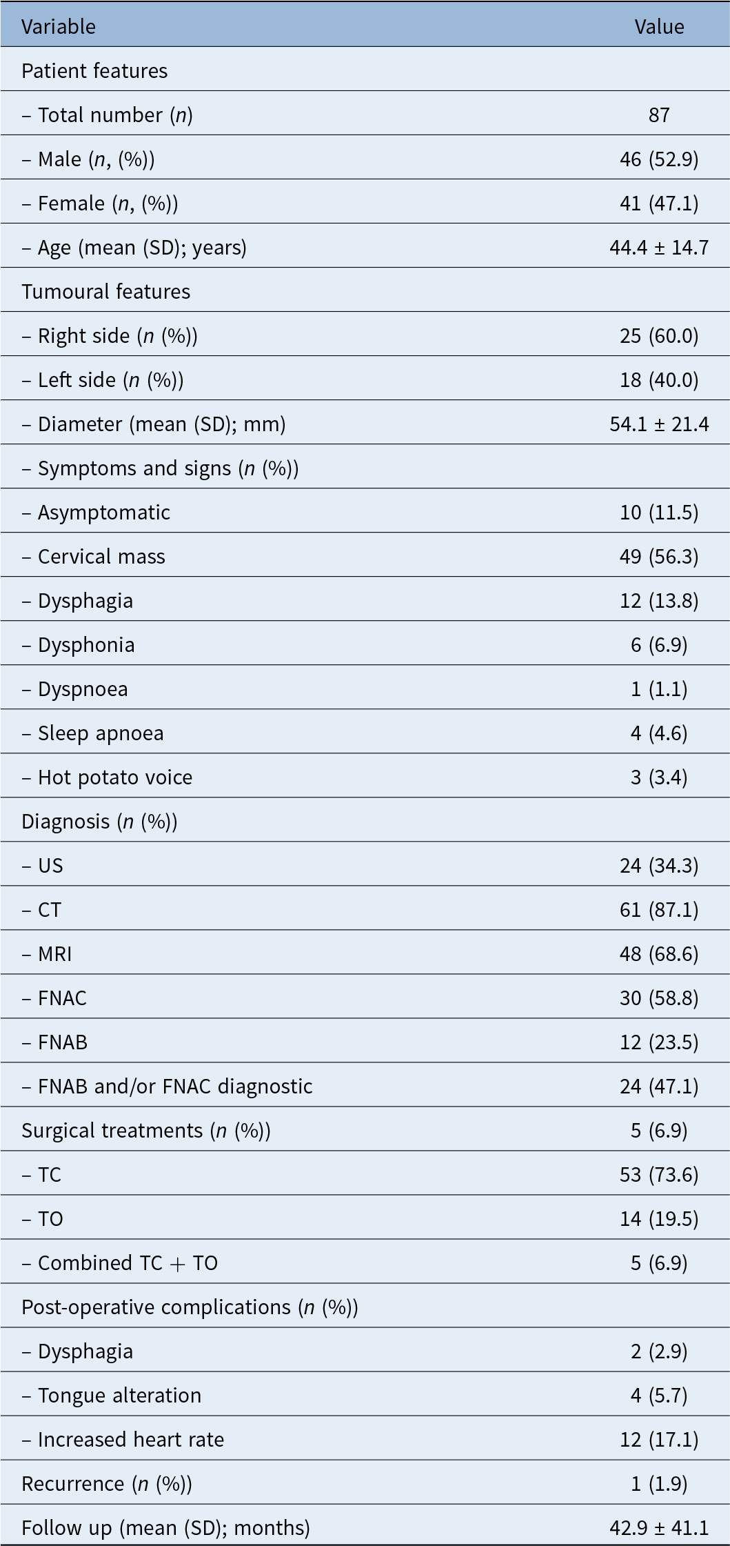
Overall, 87 patients with parapharyngeal space vagal schwannomas were included (Table 2). The cohort comprised 46 males (52.9 per cent) and 41 females (47.1 per cent), with a mean age of 44.4 ± 14.7 years. Tumour laterality was available in 43 patients, with lesions more frequently right-sided (25 of 43, 58.1 per cent) than left-sided (18 of 43, 41.9 per cent). Maximum tumour diameter was reported in 87 patients, with a mean value of 54.1 ± 21.4 mm.
Cohort’s characteristics

Table 2 Long description
Cohort characteristics are summarized for 87 patients, including demographics, tumor features, diagnostic workup, surgical approach, complications, recurrence, and follow-up. Sex distribution was 46 male (52.9 percent) and 41 female (47.1 percent), with mean age 44.4 years and standard deviation 14.7. Tumors were more often right-sided than left-sided (25 versus 18), with mean diameter 54.1 and standard deviation 21.4. Most patients were symptomatic; cervical mass was most common (49, 56.3 percent), while 10 (11.5 percent) were asymptomatic and other symptoms were less frequent, including dysphagia (13.8 percent) and dysphonia (6.9 percent). Imaging was commonly used, especially CT (61, 87.1 percent) and MRI (48, 68.6 percent), while ultrasound was reported in 24 (34.3 percent). Needle sampling included FNAC in 30 (58.8 percent) and FNAB in 12 (23.5 percent), with 24 (47.1 percent) reported as diagnostic by FNAB and/or FNAC. Surgery was mainly transcervical (53, 73.6 percent), with transoral (14, 19.5 percent) and combined approaches (5, 6.9 percent). Post-operative complications were uncommon overall, though increased heart rate was noted in 12 (17.1 percent); recurrence was rare (1, 1.9 percent). Percentages vary by variable because they are based on the number of evaluable patients for each measure.
Percentages were calculated on the number of evaluable patients for each variable. SD = standard deviation; US = ultrasound sonography; CT = computed tomography; MRI = magnetic resonance imaging; FNAC = fine needle aspiration cytology; FNAB = fine needle aspiration biopsy; TC = transcervical approach; TO = transoral approach
At presentation, 10 of 87 patients (11.5 per cent) were asymptomatic. The most common clinical finding was a cervical mass (49 of 87, 56.3 per cent), followed by dysphagia (12 of 87, 13.8 per cent) and dysphonia (6 of 87, 6.9 per cent); less frequent symptoms included sleep apnoea (4 of 87, 4.6 per cent), hot potato voice (3 of 87, 3.4 per cent) and dyspnoea (1 of 87, 1.1 per cent). Diagnostic workup was variably reported: ultrasonography was performed in 24 of 70 evaluable patients (34.3 per cent), CT in 61 of 70 (87.1 per cent) and MRI in 48 of 70 (68.6 per cent). Fine needle aspiration cytology was carried out in 30 of 51 evaluable cases (58.8 per cent), whereas fine needle aspiration biopsy was performed in 12 of 51 (23.5 per cent); a definitive pre-operative diagnosis was obtained in 24 of 51 patients (47.1 per cent) undergoing cytological or biopsy assessment.
Surgical approach was available in 72 patients: a transcervical approach was adopted in 53 of 72 (73.6 per cent), a transoral approaches in 14 of 72 (19.5 per cent) and a combined transcervical approach-transoral approaches in 5 of 72 (6.9 per cent). Post-operative complications were reported in 70 patients, with dysphagia occurring in 2 of 70 (2.9 per cent), tongue alteration in 4 of 70 (5.7 per cent) and heart rate increase in 12 of 70 (17.1 per cent). These complications refer to events related to the surgical approach and peri-operative management. Neurological deficits resulting from sacrifice or dysfunction of the nerve of origin were not classified as post-operative complications. During a mean follow up of 42.9 ± 41.1 months (available in 87 patients), one recurrence (1 of 53, 1.9 per cent) was observed. The dataset predominantly reflects surgically treated patients because conservatively managed cases are rarely reported in the literature.
Patient and tumour characteristics stratified by surgical approach
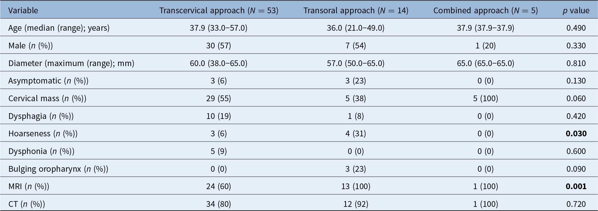
Seventy-two patients surgically treated for parapharyngeal space vagal schwannomas with available surgical data were included in the analysis and stratified according to surgical approach into transcervical approach (n = 53), transoral approaches (n = 14) and combined transcervical approach + transoral approaches (n = 5) groups. Table 3 details patient and tumour characteristics stratified according to the adopted surgical approach. No significant differences were observed among the three groups in terms of age, sex distribution or maximum tumour diameter. Tumour size was not associated with the choice of surgical approach (p = 0.81).
Patient and tumour characteristics stratified by surgical approach

Table 3 Long description
Patient and tumor characteristics are compared across three surgical approaches: transcervical (53 patients), transoral (14), and combined (5), with p values for between-group differences. Median age was similar across groups, about 36 to 38 years, and maximum tumor diameter was also similar, about 57 to 65 mm. Male proportion was comparable in transcervical and transoral groups (about mid-50 percent) and lower in the combined group (20 percent). Presenting symptoms varied: cervical mass was common in transcervical (55 percent) and universal in combined (100 percent), while bulging of the oropharynx appeared only in transoral (23 percent). Hoarseness was more frequent in transoral (31 percent) than transcervical (6 percent) and combined (0 percent), and this difference was statistically significant. Imaging differed mainly for MRI, used in 60 percent of transcervical cases versus 100 percent in transoral and combined, also statistically significant; CT use was high in all groups (80 to 100 percent) without a significant difference. Interpretation should be cautious because the combined group is very small, which can make percentages unstable and reduce power for detecting differences.
MRI = magnetic resonance imaging; CT = computed tomography
Clinical presentation differed across groups. Asymptomatic tumours were more frequently observed in the transoral approaches group compared with the transcervical approach and combined groups, although this difference did not reach statistical significance (p = 0.13). A clinically evident cervical mass was present in all patients undergoing a combined approach and in more than half of those treated via a transcervical approach, whereas it was less common in the transoral approaches group, showing a trend towards statistical significance (p = 0.06). Among symptom-related variables, hoarseness was more frequently observed in patients treated with transoral approaches, being reported in 30.8 per cent of those patients compared with 5.7 per cent of transcervical approach patients and none in the combined group (p = 0.03). Dysphagia and dysphonia were not significantly associated with surgical approach (p = 0.42 and p = 0.60, respectively). Bulging of the oropharyngeal wall, indicative of medial tumour extension, was observed exclusively in the transoral approaches group, although this difference did not reach statistical significance (p = 0.09).
Pre-operative imaging workup differed among approaches. Magnetic resonance imaging was performed in all patients treated with transoral approaches or combined approaches, compared with 60.0 per cent of those undergoing a transcervical approach (p = 0.001), whereas the use of CT did not differ significantly among groups (p = 0.72).
Discussion
In this patient-level systematic review, the transcervical approach was adopted in 73.6 per cent of cases, whereas transoral approaches and combined transcervical + transoral approaches accounted for 19.5 per cent and 6.9 per cent, respectively. This distribution closely reflects that reported in most surgical series on parapharyngeal space schwannomas, where the transcervical approach remains the predominant strategy, with reported rates ranging from 60 per cent to 85 per cent.Reference Pang, Goh and Tan1, Reference Sato, Imanishi, Tomita, Ozawa, Sakamoto and Fujii3, Reference Aghazadeh, Mohammadtaghi, Rikhtegar, Sharifi, Hashemiaghdam and Hashem Zade8 Similar surgical distributions have also been reported for schwannomas arising from lower cranial nerves in the post-styloid compartment, including the vagus nerve.Reference Oyama, Kito, Maki, Hattori, Noda and Wada20 The lower proportion of transoral procedures in earlier reports likely reflects historical concerns regarding surgical exposure and vascular control in the post-styloid compartment.Reference Pang, Goh and Tan1, Reference Hamza, Fagan, Weissman and Myers2
A key and clinically relevant finding of the present analysis is that tumour size was not associated with the choice of surgical approach (p = 0.81). Importantly, the present analysis is descriptive and does not allow conclusions regarding the appropriateness or superiority of specific surgical approaches because treatment selection was not standardised and is inherently subject to selection bias. Accordingly, the present findings should be interpreted as descriptive associations reflecting real-world surgical decision-making patterns rather than evidence of comparative superiority between approaches. Mean tumour diameter exceeded 5 cm across all groups, including patients treated exclusively via transoral approaches. This observation challenges the traditional surgical paradigm whereby large parapharyngeal space schwannomas were considered unsuitable for transoral resection.Reference Loperfido, Celebrini, Fionda, Bellocchi and Cristalli5, Reference Ahn, Kwak, Lee and Sohn21 In contrast, several contemporary series have demonstrated that tumours larger than 5–6 cm can be safely resected transorally when favourable anatomical conditions are present.Reference Sato, Imanishi, Tomita, Ozawa, Sakamoto and Fujii3, Reference Chen, Chen, Yu, Zhou, Bao and Shang9, Reference Li, London, Gao, Carrau and Chen14, Reference Ansarin, Tagliabue, Chu, Zorzi, Proh and Preda22
From a practical standpoint, these data suggest that tumour size should be regarded as a secondary parameter, useful for anticipating technical complexity but insufficient to exclude transoral approaches. Instead, surgical planning should prioritise tumour orientation, medial extension and neurovascular relationships. From a clinical perspective, the indication for surgery itself should be carefully considered. Given the benign and often slow-growing nature of parapharyngeal space vagal schwannomas, a watch-and-wait strategy may be appropriate in selected asymptomatic patients or in cases where the risk of functional deficits is substantial. Therefore, the present findings primarily reflect patterns observed in surgically treated cases rather than criteria for surgical indication.
Among clinical variables, hoarseness emerged as the most relevant practical indicator associated with transoral approaches selection. Hoarseness was present in 30.8 per cent of patients treated with transoral approaches, compared with 5.7 per cent in the transcervical approach group and 0 per cent in the combined group (p = 0.03). This finding has immediate surgical implications because pre-operative vocal symptoms likely reflect medial displacement or functional involvement of the vagus nerve, a configuration frequently associated with tumours protruding toward the oropharynx. Similar associations between pre-operative voice alterations and medially located vagal schwannomas have been described in previous case reports and small series.Reference Kamath, Dosemane, Sreedharan, Majeed and Shenoy4, Reference Carroll, Jagatiya, Kamel and Siddiqi23 Rare presentations related to upper airway compromise, including sleep apnoea secondary to parapharyngeal mass effect, have also been described.Reference Walshe, Smith, Coakeley, Dunne and Timon24 In clinical practice, systematic pre-operative voice assessment should therefore be considered an integral component of surgical decision-making because it may provide indirect yet valuable information regarding tumour anatomy and suitability for transoral approaches.
Conversely, the presence of a clinically evident cervical mass was not strongly associated with transoral approaches. In the current cohort, a cervical mass was observed in 100 per cent of patients undergoing a combined approach and in 54.7 per cent of those treated via the transcervical approach, compared with 38.5 per cent in the transoral approaches group (p = 0.06). These findings are consistent with previous surgical series in which tumours with a dominant cervical component were preferentially managed through the transcervical approach to ensure adequate exposure and vascular control.Reference Pang, Goh and Tan1, Reference Aghazadeh, Mohammadtaghi, Rikhtegar, Sharifi, Hashemiaghdam and Hashem Zade8 Extensive cervical vagal schwannomas with significant longitudinal growth have been reported, often requiring a wide transcervical approach to achieve safe resection.Reference Chiun, Tang, Prepageran and Jayalakshmi25 In addition, vagal schwannomas may occasionally masquerade as parotid gland tumours, complicating pre-operative assessment and reinforcing the importance of surgical exploration.Reference Shetty, Avinash and Auluck26
When both medial and cervical extensions coexist, several authors have advocated planned combined approaches to avoid incomplete exposure or uncontrolled bleeding.Reference Loperfido, Celebrini, Fionda, Bellocchi and Cristalli5, Reference Chen, Chen, Yu, Zhou, Bao and Shang9 Practically, these data support a stepwise surgical strategy: isolated transoral approaches for predominantly medial tumours without a significant cervical component, the transcervical approach for laterally or inferiorly extending lesions, and combined approaches for anatomically complex tumours spanning both compartments.
The presence of oropharyngeal bulging, used as a simple clinical surrogate marker of medial tumour extension, was observed exclusively in the transoral approaches group (23.1 per cent), although this difference did not reach statistical significance (p = 0.09). Despite the limited sample size, this finding is highly relevant from a surgical perspective. Several authors have emphasised visible medial displacement of the pharyngeal wall as a prerequisite for safe transoral resection, particularly when the endoscopic-assisted transoral approach or robotic-assisted techniques are employed.Reference Chen, Chen, Yu, Zhou, Bao and Shang9, Reference Meng, Zhong, Fang, Ma, Wang and Wei10, Reference Li, London, Gao, Carrau and Chen14 Meng et al.Reference Meng, Zhong, Fang, Ma, Wang and Wei10 specifically reported consistent oropharyngeal bulging as a key selection criterion in their early experience with the endoscopic-assisted transoral approach for parapharyngeal space tumours. Taken together, the combination of oropharyngeal bulging, pre-operative hoarseness and favourable imaging findings should prompt consideration of a transoral approach, whereas their absence should raise caution. These clinical findings should be interpreted as indirect indicators of tumour anatomy. In particular, medial extension toward the oropharynx, relationship with major vascular structures and compartment involvement represent the key factors guiding surgical approach selection.
Although vagal schwannomas are benign lesions, rare but severe complications have been described and should be acknowledged when planning surgical treatment. Peri-operative ischaemic stroke related to vascular manipulation during parapharyngeal space schwannoma resection has been reported and represents a potentially devastating event.Reference Shaikh, Pradhan, Routray, Das, Adhikari and Parida27 In addition, exceptionally rare cases of malignant transformation, including angiosarcoma arising within a pre-existing vagal schwannoma, have been described, underscoring the importance of careful surgical planning and long-term follow up.Reference Shaikh, Khyani, Diwan, Naseem and Abbas28 Their benign nerve sheath origin is confirmed histologically by the presence of Antoni A and B areas and strong S-100 protein positivity.Reference Budu, Bulescu, Popp, Mocanu and Mogoantă29
Although recurrence after complete excision of parapharyngeal space vagal schwannomas is uncommon,Reference Carinci, Carls, Grasso, Pelucchi and Pastore30 recurrent or plexiform lesions involving deep parapharyngeal space or skull base regions have been reported, particularly when radical resection is challenging.Reference Ijichi, Muto, Masaki and Murakami31, Reference Luzzi, Giotta Lucifero, Del Maestro, Marfia, Navone and Baldoncini32 In tumours extending toward the skull base or jugular foramen, alternative open surgical corridors, including anterolateral or skull base-oriented approaches, may be required to ensure adequate exposure and neurovascular control.Reference Shinohara, Matsumoto, Kiga, Tojyo and Fujita33 While surgical excision remains the standard of care, minimally invasive techniques such as ultrasound-guided percutaneous microwave ablation have been preliminarily explored for cervical vagal schwannomas, although their role in parapharyngeal space lesions remains investigational.Reference Zhu, Chen, Yu and Xu34
Based on the present findings, several descriptive considerations may be proposed: tumour size alone should not contraindicate a transoral approach, pre-operative hoarseness represents a useful clinical marker of medial tumour extension and transoral approaches feasibility, a dominant cervical component favours a transcervical approach or combined access, combined approaches should be planned rather than improvised, and surgical decision-making should be anatomy-driven and individualised rather than based on rigid criteria.
• Vagal schwannomas of the parapharyngeal space are rare benign tumours for which no standardised criteria currently guide surgical approach selection
• This systematic review included 87 surgically treated patients from 25 studies and found that the transcervical approach remained the most frequently adopted surgical strategy, followed by transoral and combined approaches
• Tumour size was not associated with surgical approach selection
• Pre-operative hoarseness and oropharyngeal bulging were more frequent in transoral cases, suggesting a role as practical markers of medial tumour extension
• Cervical mass was more commonly associated with transcervical and combined approaches
• These findings support an anatomy-driven and individualised strategy for surgical approach selection
Limitations and future perspectives
The current study has several limitations that should be acknowledged. Firstly, despite the systematic methodology and patient-level data extraction, the analysis is inherently limited by the retrospective nature of the included studies, which predominantly consist of case reports and small case series. This inevitably introduces selection bias and limits the generalisability of the findings. Secondly, heterogeneity in reporting represents a major limitation. Clinical presentation, radiological characteristics, intra-operative findings and post-operative outcomes were not uniformly described across studies. In particular, functional outcomes such as vocal fold mobility, swallowing function and quality-of-life measures were inconsistently reported, preventing a more detailed comparative analysis of functional morbidity across surgical approaches.
Thirdly, the definition and classification of surgical approaches varied among studies. Transoral approaches encompassed a wide spectrum of procedures, including conventional transoral approaches, the endoscopic-assisted transoral approach and robotic-assisted techniques, which could not be reliably analysed as separate subgroups because of limited sample sizes and incomplete technical descriptions. Fourthly, although a patient-level analysis was performed, missing data at the individual level limited the statistical power of some comparisons and precluded multivariable modelling. As a consequence, the associations identified in this study should be interpreted as exploratory rather than predictive. Finally, follow-up duration was heterogeneous and often incompletely reported, limiting the ability to assess long-term functional outcomes and recurrence rates. Time-to-event analyses could not be performed and late complications may therefore be underestimated.
Conclusion
Parapharyngeal space vagal schwannomas represent a rare and surgically challenging entity for which surgical management and approach selection remain largely experience-based and influenced by anatomical tumour characteristics. The transcervical approach remains the most commonly adopted strategy and is particularly suited for tumours with a predominant cervical component, while transoral approaches represent a valid and safe option in carefully selected cases, especially in the presence of medial extension and pre-operative vocal symptoms. Combined approaches should be reserved for complex lesions requiring both cervical control and medial access. Overall, surgical decision-making should be anatomy-driven and individualised, integrating clinical presentation and tumour extension rather than relying on rigid size-based criteria. The findings of this review support a tailored and anatomy-driven surgical strategy and provide descriptive insights into contemporary approach selection in the management of parapharyngeal space vagal schwannomas. Further prospective, multicentre studies with standardised reporting are warranted to validate these conclusions and refine evidence-based surgical algorithms.
Supplementary material
The supplementary material for this article can be found at https://doi.org/10.1017/S0022215126105325
Acknowledgements
The authors thank all colleagues who contributed to the discussion and critical revision of the manuscript.
Competing interests
None declared