Kawasaki disease is a febrile disease of childhood and the leading cause of acquired heart disease in children of developed countries.Reference Newburger, Takahashi and Gerber 1 Coronary artery dilatations occur in up to 30% of patients during the acute phase of the disease and are most often transient.Reference Printz, Sleeper and Newburger 2 , Reference McCrindle, Li and Minich 3 The finding of coronary artery dilatation in febrile patients can be an important clue to Kawasaki disease diagnosis, especially in patients with incomplete clinical criteria.Reference Newburger, Takahashi and Gerber 1 Although coronary artery dilatation can be attributed to coronary vasculitis, the effect of myocardial inflammation in Kawasaki disease on coronary artery dilatation is probably underestimated. Coronary artery dilatations in Kawasaki disease are best determined by elevated coronary artery Z-scores (⩾2.5), normalised to patients’ body habitus. Coronary artery Z-score formulas, however, do not take into account coronary artery dominance. The aim of the present study was to describe the impact of coronary artery dominance on dilatation in Kawasaki disease.
Methodology
Population
We carried out a retrospective study including Kawasaki disease patients with coronary artery dilatation who had undergone cardiac catheterisation at the CHU Sainte-Justine (Montreal, Canada) between 2002 and 2014. Patients with coronary artery aneurysms, normal coronary arteries (maximum coronary artery Z-score<2.5), and insufficient echocardiographic data were excluded. Patients with co-dominance were excluded from the analysis, as it was not possible to assess the impact of dominance on coronary artery Z-scores. The main outcome was coronary artery Z-score variation in time in the first 6 months following Kawasaki disease diagnosis in accordance with coronary artery dominance. Secondary outcomes compared clinical and laboratory values between patients with dilatations ipsilateral and contralateral to the dominance. This retrospective study was approved by the local ethics review boards (CHU Sainte-Justine).
Data collection and definitions
We retrospectively reviewed hospital medical records for clinical course of the illness, echocardiograms, and angiograms. Coronary artery angiograms were assessed by two radiologists (J. D., C. L.) to determine coronary artery dominance. Coronary arteries were measured on echocardiograms during follow-up, and Z-scores were calculated for analysis. Early coronary artery dilatation was defined as a Z-score⩾2.5 in the 1st month after diagnosis.Reference Dallaire and Dahdah 4 Sustained coronary artery dilatation was defined as a Z-score⩾2.5 persisting for more than 3 months after Kawasaki disease diagnosis. Left coronary artery or right coronary artery dominance was defined when the left coronary artery or the right coronary artery supplied both the diaphragmatic left ventricular wall and the posterior interventricular septum. Co-dominance was defined when the right coronary artery supplied the posterior interventricular septum and when the left coronary artery supplied the diaphragmatic left ventricular wall. Patients with dilatation ipsilateral to the dominance were classified as ipsilateral dilatation. Patients with dilatation contralateral to the dominance were classified as contralateral dilatation, as well as patients with dilatation of both the right and the left coronary arteries.
Statistical analysis
Quantitative variables are summarised as mean±SD or median [range] and categorical variables as frequencies or percentages. The Shapiro–Wilk’s test was used to test for normal distribution. Comparison of clinical and laboratory data between patients with ipsilateral and contralateral dilatation was performed using Student’s t-test for continuous variables with normal distribution or the Mann–Whitney U-test for continuous variables with non-normal distribution. The Fisher’s exact or the χ2 tests were used for categorical variables. A mixed analysis of variance was performed to compare the mean difference in coronary artery Z-scores between patients with ipsilateral and contralateral dilatation over time. All analyses were performed with SPSS Statistics version 23 (IBM, Chicago, Illinois, United States of America). A two-tailed p value of <0.05 was deemed significant.
Results
Patient characteristics
During the study period, 28 patients met inclusion criteria, of whom 15 had right coronary artery dominance, and 13 had left coronary artery dominance. All 28 patients had early coronary artery dilatation, which persisted at the 3-month follow-up in 16 patients (57%).
Baseline characteristics
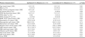
The median age at Kawasaki disease diagnosis was 2.6 [0.2–10.1] years, and 19/28 (68%) patients were boys. Patients had a median of 5 diagnostic criteria [range 2–6], 9/28 (32%) of whom presented with incomplete clinical criteria. Diagnosis was confirmed at 6.7±2.3 days of fever, and 2 g/kg intravenous immunoglobulin was administered in 23/28 (82%) patients, 7.0±2.3 days after onset of fever. Intravenous immunoglobulin treatment was given after 10 days of fever in two patients (9%), and eight (36%) had resistance to intravenous immunoglobulin defined as persistent or relapsing fever >24 hours after completion of the first intravenous immunoglobulin dose. All patients showed signs of systemic inflammation, with increased C-reactive protein (110.1±93.7 mg/ml), erythrocyte sedimentation rate (48.5±14.8 mm/hour), white blood cell counts (13.8±6.3×10⁹ cells/L), and neutrophils (0.65±0.16%) and decreased haemoglobin (105.2±12.3 g/L), haematocrit (0.31±0.04%), and albumin (27.4±6.9 g/dl). There was no significant difference between patients with ipsilateral or contralateral dilatations with respect to basic characteristics, intravenous immunoglobulin therapy, or laboratory values (Table 1).
Patient characteristics according to coronary artery (CA) dilatation.
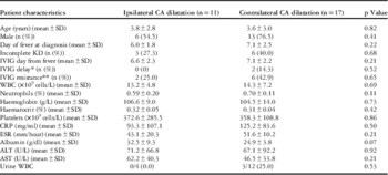
ALT=alanine aminotransferase; AST=aspartate aminotransferase; CRP=C-reactive protein; ESR=erythrocyte sedimentation rate; IVIG=intravenous immunoglobulins; KD=Kawasaki disease; WBC=white blood cell count
* IVIG received after >10 days of fever
** Defined as persistent or relapsing fever >24 hours after completion of the first IVIG dose
Coronary artery dilatation
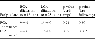
During the 1st month of follow-up, three (11%) patients had right coronary artery dilatation, 15 (54%) patients had left coronary artery dilatation, and 10 (36%) patients had dilatations of both the right and the left coronary arteries, compared with late follow-up (3 months) with two (13%), 12 (75%), and two (13%) patients, respectively. Dilatations of the left coronary artery were more frequent than dilatations of the right coronary artery (25/28 versus 13/28, p<0.001, at early follow-up, 14/28 versus 4/28, p<0.001, at late follow-up). In patients with right coronary artery dominance, dilatation involved the right coronary artery in 2/15 (13%) patients, the left coronary artery in 6/15 (40%), and both the right and the left coronary arteries in 7/15 (47%) at early follow-up, compared with 2/15 (13%), 4/15 (27%), and 2/15 (13%) patients, respectively, at late follow-up. In patients with left coronary artery dominance, dilatation involved the right coronary artery in 1/13 (8%) patients, the left coronary artery in 9/13 (69%) patients, and both the right and the left coronary arteries in 3/13 (23%) patients at early follow-up, compared with only left coronary artery dilatation (eight patients) at late follow-up. Patients with left coronary artery dominance were more likely to have left coronary artery dilatation (p=0.02 at early follow-up and p=0.002 at late follow-up), whereas there was no difference in patients with right coronary artery dominance (p=0.21, p=0.36, respectively) (Table 2).
Early and late coronary artery dilatation according to dominance.

LCA=left coronary artery; RCA=right coronary artery
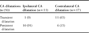
Dilatation was ipsilateral to the dominance in 11/28 (39%) patients during early follow-up, and persisted in 10/16 (63%) patients at late follow-up, whereas 17/28 (61%) patients had contralateral dilatation during the early phase, which persisted in 6/16 (38%) at late follow-up (p=0.12). Contralateral dilatations were more often transient than ipsilateral dilatations (11/12 versus 1/12, p=0.004) (Table 3). Most of the ipsilateral dilatations were present at the time of initial diagnosis (82%), as opposed to contralateral dilatations, which were present in only 6/17 (35%) patients at diagnosis, but peaked to include 16/17 (94%) patients at the 2-week follow-up (p=0.02 at diagnosis, p=0.29 at 2 weeks).
Transient and persistent coronary artery (CA) dilatations during follow-up.

p=0.004 Fisher exact test ipsilateral versus contralateral CA dilatation
Coronary artery Z-score variation
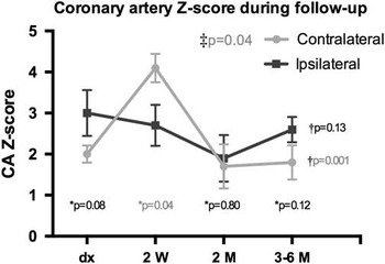
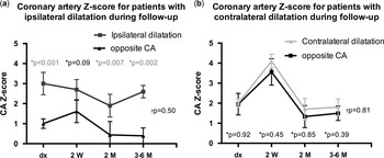
A mixed analysis of variance found a statistically significant difference between ipsilateral and contralateral dilatations with respect to coronary artery Z-score variations with time (p=0.04). At initial diagnosis, there was a trend towards higher Z-scores in patients with ipsilateral dilatation (3.0±0.9) compared with contralateral dilatation (2.0±2.2), without reaching statistical significance (p=0.08). At 1–2 weeks, coronary artery Z-scores increased in patients with contralateral dilatation (4.1±1.8), whereas it decreased slightly in patients with ipsilateral dilatation (2.7±1.1), p=0.04. At 2 months, coronary artery Z-scores of patients with contralateral dilatations regressed significantly to return to baseline values. At the 2-month and 3–6-month follow-up, coronary artery Z-scores were similar between both groups (1.9±1.3 versus 1.7±2.2, p=0.80 and 2.6±1.5 versus 1.8±1.2, p=0.12, respectively). A significant variation of coronary artery Z-scores over time was recorded in patients with contralateral dilatation (p=0.001), unlike in patients with ipsilateral dilatations (p=0.13) (Fig 1).
Coronary artery (CA) Z-scores during follow-up of patients with ipsilateral and contralateral dilatations. ‡Mixed analysis of variance (ANOVA) CA Z-score variation with time for ipsilateral versus contralateral dilatations; †repeated-measures ANOVA CA Z-score variation with time; *ipsilateral versus contralateral CA Z-scores.

Comparison of coronary artery Z-scores from the dilated coronary artery to the opposite coronary artery was performed to verify whether or not the chronological Z-score pattern was a random observation. Accordingly, the observed patterns of coronary artery Z-scores in the ipsilateral and the contralateral groups were replicated at the level of the opposite segments (p=0.50, 0.81, respectively). In addition, the dominant segments in patients with ipsilateral dilatation had higher coronary artery Z-scores than the non-dominant segments at diagnosis (p<0.001), as well as late follow-up (p=0.002). Such difference was not shown in the contralateral dilatation group, however (Fig 2).
Coronary artery (CA) Z-scores of patients with ( a ) ipsilateral or ( b ) contralateral dilatation during follow-up. †Mixed analysis of variance CA Z-score variation with time for ipsilateral/contralateral dilatation versus opposite CA; *ipsilateral/contralateral dilatation versus opposite CA Z-score.

Discussion
This study focusses on the impact of coronary artery anatomical dominance on the Z-scores of coronary arteries for determining Kawasaki disease-induced coronary artery dilatation. Coronary artery Z-scores in patients with ipsilateral dilatation is maximal at onset of the disease, which differs from patients with contralateral dilatations in whom coronary artery Z-scores peak 2 weeks after onset, with subsequent regression. These two observations suggest that the differential onset of coronary artery dilatation in acute Kawasaki disease underlines two different pathophysiologies leading to such increases in Z-scores – first, an increase in coronary artery Z-score related to vasodilatation of the coronary artery as a physiological response to Kawasaki disease carditis and, second, a phenomenon secondary to coronary artery vasculitis. The former explanation of coronary artery dilatation would be the reflection of physiological response to an increase in oxygen demand of an inflammatory myocardial state and/or to the febrile status during the acute inflammatory phase of the disease. In this perspective, although Kawasaki disease is the most common cause of coronary artery dilatation,Reference Muniz, Dummer, Gauvreau, Colan, Fulton and Newburger 5 other aetiologies have been reported, such as infectious and autoimmune disorders.Reference Muniz, Dummer, Gauvreau, Colan, Fulton and Newburger 5 – Reference Konishi, Nishimura, Ono, Satou and Ueda 9 The proposed mechanism is by coronary artery dilatation to increase coronary artery blood flow and meet the increased metabolic demands in the setting of fever.Reference Muniz, Dummer, Gauvreau, Colan, Fulton and Newburger 5 A review on the regulation of coronary blood flow during exercise supports this theory, where the increased myocardial oxygen demand during exercise is principally met by augmenting coronary blood flow, from a combination of coronary vasodilatation and decrease in coronary vascular resistance.Reference Duncker and Bache 10 A study on coronary artery diameter in febrile children without Kawasaki disease found greater coronary artery dimensions in febrile children compared with normative values for all coronary arteries (p<0.001 for the left main coronary artery, p=0.03 for the left descending artery, and p=0.03 for right coronary artery). In this series, however, only two patients had elevated coronary artery Z-scores that met the definition for coronary artery dilatation.Reference Muniz, Dummer, Gauvreau, Colan, Fulton and Newburger 5 A higher percentage of coronary artery dilatation (21%) was found in patients with acute myocarditis.Reference Rached-d’Astous, Boukas, Fournier, Raboisson and Dahdah 11 It is shown that myocarditis is almost universal in the acute stage of the disease, independent of the presence or absence of coronary artery dilatation, based on classic histological reports.Reference Yutani, Go and Kamiya 12 Moreover, N-terminal B-type Natriuretic peptide (NT-proBNP), which is secreted by the myocardium in response to shear stress, is elevated in the acute phase of Kawasaki disease,Reference McNeal-Davidson, Fournier and Spigelblatt 13 and linearly correlated with a decrease in myocardial contractibility,Reference Adjagba, Desjardins, Fournier, Spigelblatt, Montigny and Dahdah 14 further supporting myocardial inflammation in Kawasaki disease. Our study supports this finding with higher coronary artery Z-scores at the time of diagnosis in the dominant segment, which could represent the physiological adaptation to the higher metabolic demand in the context of febrile disease and myocarditis.
The American Heart Association guidelines recommend echocardiographic evaluation of the coronary arteries at the time of diagnosis, at 2 weeks, and at 6–8 weeks after onset of the disease for uncomplicated cases.Reference Newburger, Takahashi and Gerber 1 As aneurysms, the undeniable Kawasaki disease vasculitis, rarely form before 10 days of fever, initial echocardiogram can be normal with subsequent development of aneurysms, which is the reason why repeat echocardiograms are necessary to identify all patients with coronary artery complications.Reference Newburger, Takahashi and Gerber 1 , Reference Scott, Ettedgui and Neches 15 In our series, patients with contralateral dilatations showed an increase in coronary artery Z-scores in the first 2 weeks after Kawasaki disease diagnosis, which is a similar timing compared with pathology-proven timing of coronary artery aneurysms and vasculitis.Reference Orenstein, Shulman and Fox 16 Determination of coronary artery dominance in the clinical setting is not commonly performed; however, an echocardiography subcostal view allows following the right coronary artery to the posterior interventricular branch when there is right coronary dominance. From this perspective, determining the coronary dominance by echocardiography could be eventually useful. Otherwise, and based on our angiography observation, the timing of coronary artery dilatation as determined by echocardiography follow-up may help stratify patients between dominance-related coronary dilatation and vasculitis-induced dilatation. In essence, patients with maximal dilatation at the time of diagnosis followed by early coronary artery diameter normalisation could be considered at lower risk, compared with patients who develop coronary artery dilatation at the 2-week follow-up or later.
In this series, both the dominant and the non-dominant segments were similarly affected in patients with contralateral dilatation, as opposed to patients with ipsilateral dilatation where only the dominant segment is affected. This difference suggests that diffuse vasculitis is the cause for coronary artery Z-score increase at 2 weeks from onset of fever in patients with contralateral dilatation, as opposed to ipsilateral dilatations, where myocarditis and physiological increase in coronary artery blood flow during the febrile period would explain the Z-score variation in time. Thus, a proportion of patients with ipsilateral dilatation are falsely diagnosed with coronary artery complications, due to increase in coronary artery Z-scores above the cut-off values of the dominant coronary artery during the febrile period. We believe that distinguishing between early-onset dilatation and peaked dilatation 2 weeks from onset could play an important role in the understanding and future expectations of coronary artery function following Kawasaki disease.
From an anatomical perspective, coronary artery dominance plays an important role as reported in a study evaluating coronary artery blood flow in 1322 vessels, from 496 patients.Reference Sakamoto, Takahashi and Coskun 17 Accordingly, there is a higher coronary artery blood flow in the left circumflex artery in cases with left-dominant or balanced circulation, compared with patients with right coronary artery dominance (113±43 versus 72±37 ml/minute, p<0.001), and lower blood flow in the right coronary artery in patients with left-dominant or balanced circulation, compared with patients with right coronary artery dominance (56±40 versus 113±49 ml/minute, p=0.003). Another study evaluated the relationship between coronary artery dominance and coronary artery diameter in 220 adults.Reference Kaimkhani, Ali and Farugi 18 Patients with left coronary artery dominance had smaller mean diameter of the right coronary artery, compared with patients with right- or co-dominance (p<0.001). Similarly, patients with right coronary artery dominance had smaller circumflex diameter (p<0.001). From this perspective, echocardiography-based coronary artery Z-score equations do not take into account such differences,Reference Dallaire and Dahdah 4 as dominance can only be determined on angiography. On the basis of a large series of coronary angiography studies, 82% had right coronary artery dominance, 12% left coronary artery dominance, and 6% co-dominance.Reference Altin, Kanyilmaz and Koc 19 From this perspective, right coronary artery dominance is under-represented in our series (54 versus 82%), whereas left coronary artery dominance is over-represented (45 versus 12%). Expectedly, normative echocardiography coronary artery Z-score equations do not take into account coronary artery dominance, and are probabilistically based on children with right coronary artery dominance in the most part, which may explain the higher prevalence of left coronary artery dilatation, especially in patients with left coronary artery dominance in a febrile state.
There are limitations to this study related to the small sample size and selection bias. First, most patients with coronary artery dilatation without aneurysms do not typically require cardiac catheterisation, which inherently limits the number of patients included in this study. Second, patients without coronary artery aneurysms who had clinically indicated coronary catheterisation often had persistent coronary artery dilatation unless cardiac symptoms were the indication for diagnostic catheterisation. Above all, the selection bias in this series could be portrayed by the excessive presence of the left coronary artery dominance anatomy comparatively with the expected ratio of such dominance. Nevertheless, the unexpected over-representation of the left coronary artery dominance anatomy further supports our study hypothesis, and may not be taken as a limitation per se.
Conclusion
To our knowledge, this is the first report portraying the impact of coronary artery dominance on coronary artery dilatation in Kawasaki disease. Accordingly, a peak coronary artery dilatation 2 weeks from onset of fever may be the actual reflection of true dilatation secondary to vasculitis, as opposed to onset coronary artery dilatation followed by regression of coronary artery Z-scores, which we attribute to vasodilatation as a physiological response to carditis. In this view, further validation with vasculitic markers and myocardial inflammatory markers may bring valuable precisions for future considerations of coronary artery function later in life.
Acknowledgements
None.
Financial Support
This research received no specific grant from any funding agency or from commercial or not-for-profit sectors.
Conflicts of Interest
None.
Ethical Standards
The authors assert that all procedures contributing to this work comply with the ethical standards of the relevant national guidelines on human experimentation in Canada and with the Helsinki Declaration of 1975, as revised in 2008, and has been approved by the institutional committees of CHU Sainte-Justine.