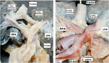
Crossing of the right and left pulmonary arteries at their origin from the pulmonary trunk is a rare congenital malformation. Initially described by Jue et al,Reference Jue, Lockman and Edwards 1 the essence of the lesion is the origin of the left pulmonary artery in superior and rightward position relative to the right pulmonary artery, with the right and left pulmonary arteries then crossing extrapericardially as they proceed to their respective lungs. The index case was a newborn with trisomy 18, also having a sinus venosus defect (Fig 1a). Subsequent to this account, Becker and EdwardsReference Becker, Becker and Edwards 2 described three examples of crossed pulmonary arteries in the setting of common arterial trunk (Fig 1b).
The image shows the arrangement in the index case of crossed pulmonary arteries, first published by Jue, Lockman, and Edwards in 1966. The left pulmonary artery (LPA) takes its origin from the pulmonary trunk above and to the right relative to the origin of the right pulmonary artery (RPA), with the pulmonary arteries then crossing each other as they proceed to their respective lungs. In panel B, we show the situation in one of the three cases of crossed pulmonary arteries coexisting with common arterial trunk, as initially reported by Becker and Edwards in 1970. The crossing of the pulmonary arteries is directly comparable to that shown in panel A. The specimens are drawn from the Minnesota Jessie Edwards Cardiovascular Registry, and have been rephotographed for this presentation. DAo=descending aorta; P trunk=pulmonary trunk.

In their report, Becker and EdwardsReference Becker, Becker and Edwards 2 noted that, in one of their specimens, the left pulmonary artery arose superiorly relative to the right pulmonary artery, but without crossing the right artery as it extended into the pulmonary hilum. They considered this to be a lesser form of the anomaly. Such crossing of the pulmonary arteries at the bifurcation of the pulmonary trunk is now known to be associated with multiple other congenital cardiac defects, and with extracardiac anomalies.Reference Chaturvedi, Mikailian and Freedom 3 22q11.2 deletion has also been reported in association with crossed pulmonary arteries when seen in isolation, and may be present in several anomalies involving the aortic arch.Reference Recto, Parness, Gelb, Lopez and Lai 4 It is noteworthy that the first examples of the lesion were all diagnosed either by angiography or postmortem examination. Potential difficulties in noting the crossing of the arteries using cross-sectional echocardiography are now ameliorated by the advent of color Doppler imaging, and by improvements in the resolution of echocardiographic machines.
Computed tomography angiography, or magnetic resonance angiography, both permitting three-dimensional reconstruction, are now recognised as the preferred diagnostic tools for confirming the presence of crossed pulmonary arteries.Reference Cuturilo, Drakulic and Krstic 5 Using computed tomographic angiography, we have now encountered such crossed pulmonary arteries in six of our patients in association with hypoplasia of the transverse aortic arch, some with additional discrete coarctation. To the best of our knowledge, this association has not previously received emphasis. As surgical repair in patients with hypoplasia of the transverse arch is very challenging, and short-term restenosis is often seen,Reference Brown, Roedefeld and Ruzmetov 6 we describe here our experience in this unusual setting.
Methods
Institutional Review Board approval was obtained for retrospective chart review and complies with the declaration of Helsinki. The institutional congenital advanced imaging database was queried for patients with crossed pulmonary arteries from 2011 to 2013. Patient information including hospital course and management were obtained from patient charts. It is the current practice of our Heart Center programme to obtain accurate reconstruction of cross-sectional images when assessing complex lesions of the aortic arch, thus better elaborating defects and revealing any associated anomalies. To achieve these goals, computed tomographic angiography was performed on a dual-source Siemens SOMATOM Definition Flash scanner using intravenous contrast enhancement according to previously reported protocols.Reference Siripornpitak, Pornkul, Khowsathit, Layangool, Promphan and Pongpanich 7 , Reference Kim and Newman 8 Paediatric Flash modality was utilised with prospective electrocardiographic triggering, following dose minimisation protocols.Reference Goske, Applegate and Boylan 9 Patients were not sedated, but were wrapped and made comfortable for the scan. Image post-processing was carried out on a Voxar 3D imaging system (Toshiba Medical Visualization Systems, Edinburgh, Europe). No additional imaging was necessary for the study. Photographs from the original specimens with crossed pulmonary arteries were taken at the Jessie Edward Cardiovascular Registry in St. Paul, Minneapolis, Minnesota.
Results
Prenatal diagnosis
Prenatal diagnosis had been made of associated intracardiac lesions in three of our six patients; however, in none of the six patients did the images interpret prenatally as showing crossing of the pulmonary arteries (Table 1). Indeed, in only one of our patients, the crossed pulmonary arteries were observed during postnatal echocardiographic interrogation, which had been interpreted as showing aortic coarctation. As our familiarity with crossed pulmonary arteries has increased, its prenatal and postnatal identification by echocardiography outside of this series has also increased.
Summary of the findings in our patients with crossed pulmonary arteries and hypoplasia of the transverse aortic arch.

ASD=atrial septal defect; BAV=bicuspid aortic valve; CS=coronary sinus; CTA=computed tomography angiography; Echo=echocardiography; HLHS=hypoplastic left heart syndrome; LSCV=left superior caval vein; LV=left ventricle; MS=mitral stenosis; TA=tricuspis atresia; TAPVC=total anomalous pulmonary venous connection; VSD=ventricular septal defect
Summary of the findings in our patients with crossed pulmonary arteries and hypoplasia of the transverse aortic arch
* Prenatal diagnosis of the associated lesions. No prenatal diagnosis of crossed pulmonary arteries and hypoplastic transverse arch was made
** Echo diagnosed HLHS and coarctation of the aorta. No transverse arch hypoplasia or crossed pulmonary arteries were suspected
Imaging
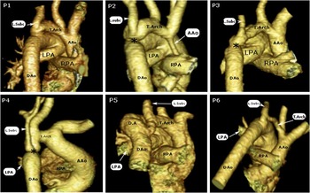
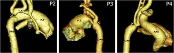
In all of the patients, important elements of the cases were revealed using computed tomographic angiography, the images showing, to great effect, both the crossing of the pulmonary arteries at their origin from the pulmonary trunk, and the degree of hypoplasia of the transverse component of the aortic arch (Fig 2). In three of the patients, the tomographic images also showed the presence of a discrete waist lesion at the insertion of the aortic isthmus into the major pathway from the arterial duct to the descending aorta (Fig 3). In our fourth patient (P4), the left subclavian artery was hypoplastic and its takeoff was at the discrete waist where the arterial duct inserts into the descending aorta (Fig 3). The remaining patients had a normal left subclavian artery diameter with a proximal insertion relative to the ductal arch. In our second case, the images also revealed the gross hypoplasia of the ascending aorta expected in the setting of hypoplastic left-heart syndrome (Fig 2, P2).
Computed tomography angiography of our six patients. The left pulmonary artery (LPA) takes its origin from the pulmonary trunk above and to the right relative to the origin of the right pulmonary artery (RPA), with the pulmonary arteries then crossing each other as they proceed to their respective lungs. The transverse aortic arch (T Arch) is hypoplastic. In our second patient, there is gross hypoplasia of the ascending aorta in the setting of hypoplastic left heart syndrome. Patients 2, 3, and 4 have a discrete waist (*) at the insertion of the aortic isthmus into the major pathway from the arterial duct to the descending aorta. Patient 4 also has a significant hypoplasia of the left subclavian artery (L.Subc.), inserted at the ductal arch-descending aorta junction. AAo=ascending aorta; DAo=descending aorta; P=patient).

Computed tomography angiography of the three patients with additional discrete coarctation of the aorta (P2, P3, P4). Images are shown from a different angle emphasising the discrete waist lesion at the insertion of the aortic isthmus into the major pathway from the arterial duct to the descending aorta (arrows). In patient 4 (P4), the left subclavian artery is hypoplastic and it is inserted adjacent to the connection of the ductal arch with the descending aorta. AAo=ascending aorta; DAo=descending aorta; P trunk=pulmonary trunk; T Arch=transverse arch.

Surgical and hospital course
In five of our patients, the diagnosis had been made such that the surgery to correct the obstructive lesions in the aortic pathway could be performed before the age of 13 days. One patient with mild hypoplasia of the transverse arch was initially monitored, and ultimately underwent surgical repair at 3 months of age. The surgery, in all instances, was devoted to repair of the aortic arch and the associated intracardiac anomalies. No specific surgical procedure had been deemed necessary to correct the crossed nature of the pulmonary arteries. In two patients, we banded the pulmonary trunk at the time of the repair of the arch, thus attenuating the extent of flow to the lungs in the setting of a large ventricular septal defect. Both required revision of the band owing to impingement on the abnormal origin of the right pulmonary artery. In another two of the patients, restenosis within the transverse arch occurred subsequent to the surgical repair. This was initially treated in both instances by balloon dilation. In our final case (P6), it was also necessary to place a patch surgically at the age of 3 months so as to augment the aortic arch (Table 1). This patient also underwent additional operations for the associated lesions, including the totally anomalous pulmonary venous connections, at 6 months of age.
The remaining patients have not developed restenosis of the aortic arch nor other problems. We were able to obtain genetic analysis in four of our six patients, but only the patient with tricuspid atresia was found to have 22q11 deletion, although the genetic testing was inconclusive in another patient. Owing to the relatively short follow-up time, we have yet to analyse their ongoing progress.
Associated lesions
Bicuspid aortic valve was found in four of our six patients, and septal defects in four patients, two with atrial septal defects and two with ventricular septal defects. Left ventricular hypoplasia was identified in two patients and tricuspid atresia with totally anomalous pulmonary venous connection in one case. Half of the patients also exhibited discrete aortic coarctation in addition to the hypoplasia of the transverse arch (Table 1).
Discussion
Crossing of the pulmonary arteriesReference Jue, Lockman and Edwards 1 is found when the left pulmonary artery originates superiorly from the pulmonary trunk relative to the origin of the right pulmonary artery. In most instances, as seen in all of our patients, the pulmonary arteries then cross as they extend to their respective lungs.Reference Jue, Lockman and Edwards 1 – Reference Chaturvedi, Mikailian and Freedom 3 The crossing of the pulmonary arteries does not, in itself, produce tracheal compression. This is in contrast to the pulmonary arterial sling where the left pulmonary artery arises extrapericardially from the right pulmonary artery, and then courses between the trachea and the oesophagus to reach the left lung.
In the setting of crossed pulmonary arteries, the crossing occurs anterior to the trachea. As yet, there is no convincing explanation for the developmental basis of the anomalous origins of the pulmonary arteries, either when crossed, or when forming a sling. The right and left pulmonary arteries, nonetheless, are known to canalise within the pharyngeal mesenchyme, taking their origin from the undersurfaces of the arteries coursing through the sixth pharyngeal arches.Reference Anderson, Chaudhry and Mohun 10 The outflow tract itself is known to rotate as it extends into the heart at the arterial pole, taking its origin from the so-called second heart field.Reference Bajolle, Zaffran and Kelly 11 Therefore, it is possible that excessive rotation during development could explain the abnormal origin of the pulmonary arteries from the pulmonary trunk at the margins of the pericardial cavity.
As shown by our experience, the crossing of the pulmonary arteries can be found with various intracardiac malformations. Indeed, the lesion can also be found in isolation. The unexpected finding in all of our patients, however, was hypoplasia of the transverse aortic arch. In this respect, the anatomic criteria for defining such hypoplasia remain controversial. One accepted definition is to find the proximal transverse arch at a ratio of <60% the diameter of the ascending, or the distal transverse arch at <50% of the diameter.Reference Brown, Roedefeld and Ruzmetov 6 All of our patients met these criteria, which were confirmed visually by the surgeon at time of operation. In addition, three of the six patients showed the more typical discrete waist lesion at the insertion of the isthmus into the ductal arch. Previous histologic studies have shown that the hypoplastic segment of the arch has a significant increase in elastin lamella and collagen, and decrease in α-actin positive cells, features which could contribute to its decreased ability to distend.Reference Lacour-Gayet, Bruniaux and Serraf 12
As already indicated, although not previously reported in association with hypoplasia of the transverse aortic arch, crossed pulmonary arteries have been found in cases with multiple other lesions, such as interruption of the aortic arch, common arterial trunk,Reference Becker, Becker and Edwards 2 tetralogy of Fallot,Reference Chaturvedi, Mikailian and Freedom 3 atrial septal defects, persistent left superior caval vein,Reference Cuturilo, Drakulic and Krstic 5 ventricular septal defects, and double outlet right ventricle.Reference Zimmerman, Berdusis, Wright and Alboliras 13 Within our small series (Table 1), in addition to the hypoplasia of the transverse arch and discrete coarctation, we found bicuspid aortic valve, hypoplastic or borderline left ventricles, ventricular and atrial septal defects, and mitral stenosis. Most of these are well known to be associated with reduced aortic flow during foetal development, and therefore are not necessarily unexpected. It remains to be seen whether abnormal flow is also associated with the crossing of the pulmonary arteries.
The recognition of the presence of crossed pulmonary arteries when relying on echocardiography for diagnosis can pose a major challenge. Of course, the so-called two-dimensional echocardiography is itself a cross-sectional technique. Our experience emphasises how it is the ability to reconstruct cross-sectional images using techniques such as computed tomography, or magnetic resonance imaging, that has revolutionised the recognition of the lesion. In our cases, the tomographic technique also served to reveal the full extent of the hypoplasia of the transverse aortic arch. It is noteworthy that Xiong et alReference Xiong, Gan, Liu, Tao, Wang and Wu 14 were able to diagnose an isolated instance of crossed pulmonary arteries ultrasonographically in a foetus at 23 weeks of gestation. However, in only one of our patients, the last in our series, the crossing of the arteries was noted during postnatal transthoracic echocardiography. As with other cases described in the literature, the majority of our patients were initially diagnosed as having coarctation of the aorta. Half of our patients exhibited the typical waist lesion at the insertion of the aortic isthmus into the ductal arch (Fig 3).
As we have emphasised, it was the ability to reconstruct the cross-sectional images obtained using computed tomography that delineated the exact anatomy of the extrapericardial aortic pathways, revealing the hypoplastic transverse segments of the arch. Many non-interventional catheter-based angiograms are now being replaced in our programme by computed tomographic or magnetic resonance angiography. Computed tomographic angiography can now be obtained with significantly lower doses of radiation, acquired in rapid manner without sedation, and offers significantly more possibilities for post-processing. Such computed tomographic angiograms have been classically used sparingly because of the dose of radiation required for their production, and hence concerns for the potential risk of cancer.Reference Brand, Sommer and Achenbach 15 , Reference Miglioretti, Johnson and Williams 16 Advances in the hardware, software, and protocols used in computed tomographic imaging have markedly reduced the radiation doses required for paediatric cardiac imaging.Reference Ou, Celermajer and Marini 17 , Reference Achenbach, Goroll and Seltmann 18 , Reference Goo, Seo and Yun 19 Imaging can now be achieved with estimated dose rates of 0.2–0.5 milliSievert. Estimates of the effective dose of radiation were calculated by multiplying the system-reported phantom appropriate dose length product by the conversion factor appropriate for age, as reported by Thomas and Wang.Reference Thomas and Wang 20 For reference, the estimated annual background radiation dose while living in the United States of America is 3 milliSievert, and an adult chest x-ray, when obtained in 2 planes, requires 0.05–0.1 milliSievert. 21 , Reference Verdun, Bochud, Gunddinchet, Aroua, Schnyder and Meuli 22 Although the dose of radiation during computed tomographic angiography has been significantly reduced, radiation, and its potential risk, is not completely eliminated, and must be weighed against the benefit of theoretical decreases in cardiopulmonary bypass time and reduced intraoperative surprises. Magnetic resonance angiography is another potential modality for replacing invasive angiography, but often still requires sedation in the young child. It is now well recognised that the high spatial resolution provided by computed tomographic angiography allows smaller vessels and stenosis to be more accurately depicted.Reference Langley, Sunstrom, Reed, Rekito and Gerrah 23 In our series, computed tomographic angiography was performed at the request of the surgeons; however, we appreciate the fact that magnetic resonance angiography also has the capacity to demonstrate these lesions without the radiation exposure to the child, and may be the preferable method to obtain this information.
In most instances, the presence of the crossed pulmonary arteries has few haemodynamic implications. In two of our patients with large ventricular septal defects, it was proved necessary to band the pulmonary trunk at the time of the initial repair of the aortic arch because of the small size of the patient. This subsequently caused significant impingement of the band on the right pulmonary artery, with the need for reintervention. This is almost certainly the consequence of the more caudal origin of the right pulmonary artery. This raises the question as to whether complete early repair should preferentially be attempted in this setting, rather than initially undertaking palliative procedures.Reference Shepard, Kochilas and Rosengart 24 If pulmonary arterial banding is inevitable, it may be preferable to utilise the technique of intraluminal pulmonary arterial banding, as described by Piliuko et al,Reference Piluiko, Poynter and Nemeh 25 rather than externally banding the pulmonary trunk.
In four of our patients, the surgeon approached the hypoplastic transverse arch via a lateral thoracotomy. In two of these patients, there was restenosis requiring balloon dilation, and patch augmentation in one of them. In both the patients approached through a median sternotomy, in contrast, there was no need for reintervention. Although our numbers are small, we believe this difference may be significant. It is established that, in the setting of coarctation, the ductal tissue extends beyond the point of insertion of the aortic arch into the descending aorta.Reference Ho and Anderson 26 , Reference Russell, Berry, Watterson, Dhasmana and Wisheart 27 Many surgeons have advocated an anterior approach when dealing with this problem, thus permitting extensive distal resection, and hence preventing restenosis.Reference Brown, Roedefeld and Ruzmetov 6 , Reference Rakhra, Lee and Iyengar 28 With our initial experience of restenosis after a lateral approach, our practice has changed to use an anterior approach.
Although we searched for 22q11.2 microdeletion in four of our patients, a conclusive result was obtained only in the patient with totally anomalous pulmonary venous connection and tricuspid atresia. The presence of 22q11 deletion was inconclusive in a second patient with an isolated bicuspid aortic valve. To the best of our knowledge, only three cases with crossed pulmonary arteries have been associated with 22q11.2 microdeletion, all of them with additional ventricular septal defects.Reference Cuturilo, Drakulic and Krstic 5 , Reference Siwik, Everman and Morrison 29
In conclusion, we find it surprising that, since its first description, no cases of crossed pulmonary arteries have been described in association with coarctation, and in particular, with hypoplasia of the transverse aortic arch, the more so as we have now encountered this combination in six patients in less than 2 years. We believe that this probably represents the previous difficulties in recognising the crossing of the pulmonary arteries. Our experience suggests that standard banding of the pulmonary trunk may give sub-optimal results, and either complete repair or intraluminal banding should be considered as alternatives. Although crossed pulmonary arteries can be observed in isolation, identification of this lesion should inspire a thorough investigation for additional intracardiac or extracardiac anomalies. Further longitudinal data, with longer follow-up and larger sample size, are needed in this population.
Acknowledgements
The authors owe special thanks to Dr James Moller and the staff of the Jesse Edwards Cardiovascular Registry (Saint Paul, Minnesota) for finding and preparing the original specimens for photography.
Financial Support
This research received no specific grant from any funding agency, commercial, or not-for-profit sectors.
Conflict of Interest
None.