INTRODUCTION
The life history of an organism comprises its reproductive strategies and peculiarities, which are influenced by the organism's body plan, lifestyle and physiological conditions, and by environmental factors, including food supply, predators and other aspects of the environment (Stearns, Reference Stearns1989; Flatt & Heyland, Reference Flatt and Heyland2011). Consequently, these relationships influence the individual's life chronology (e.g. reproductive age, offspring size, lifespan), reflected in its adaptation to the environment (Flatt & Heyland, Reference Flatt and Heyland2011). For that reason, it is particularly interesting to study the life history of non-native species.
The success or failure of a bioinvasion event depends on environmental traits, but also on the life history of the organism. Usually, non-indigenous species have opportunistic life history strategies, presenting short life cycles, constant reproduction with large broods, high competition and the ability to thrive in very different environmental conditions (Sakai et al., Reference Sakai, Allendorf, Holt, Lodge, Molofsky, With, Baughman, Cabin, Cohen, Ellstrand, McCauley, O'Neil, Parker, Thompson and Weller2001; Berasategui et al., Reference Berasategui, Hoffmeyer, Biancalana, Fernandez Severini and Menendez2009; Jänes et al., Reference Jänes, Kotta and Herkül2015). The calcareous sponge Sycettusa hastifera (Row, 1909) (Calcarea, Calcaronea, Heteropiidae) was primarily described in the Red Sea (Sudan); later on, it was reported in the Indian Ocean (Dendy, Reference Dendy1913), and more recently in the south-western Atlantic (Brazil) (Muricy et al., Reference Muricy, Hajdu, Custodio, Klautau, Russo, Peixinho and Magoon1991; Azevedo & Klautau, Reference Azevedo and Klautau2007; Cavalcanti et al., Reference Cavalcanti, Skinner and Klautau2013). On the Brazilian coast, this species has been observed only in Rio de Janeiro State, where it is considered cryptogenic (Cavalcanti et al., Reference Cavalcanti, Skinner and Klautau2013). Sycettusa hastifera was first detected in the port of Arraial do Cabo in 1991, when the population was restricted to a few individuals (Muricy et al., Reference Muricy, Hajdu, Custodio, Klautau, Russo, Peixinho and Magoon1991) and it was also found in Angra dos Reis (Azevedo & Klautau, Reference Azevedo and Klautau2007). However, in the last decade, this species has become one of the most conspicuous sponges on the rocky shores of this locality. Therefore, investigating aspects of the life history and the reproduction of S. hastifera serves two purposes: it is important not only to understand and predict its invasive dynamics in Arraial do Cabo and putatively in other regions, but also allows a proactive approach to understand how reproductive biology is related to species invasiveness (Sakai et al., Reference Sakai, Allendorf, Holt, Lodge, Molofsky, With, Baughman, Cabin, Cohen, Ellstrand, McCauley, O'Neil, Parker, Thompson and Weller2001; Rilov & Crooks, Reference Rilov and Crooks2009; Cavalcanti et al., Reference Cavalcanti, Skinner and Klautau2013).
The few studies on the reproductive cycle of Calcarea have shown that, in this class, the species seem to reproduce seasonally in temperate waters, but there are also species that reproduce continuously throughout the year. Only the life cycle of another cryptogenic calcareous sponge, Paraleucilla magna Klautau, Monteiro & Borojevic, 2004, was studied in tropical waters, and it was observed that it can be either seasonal or continuous (Lanna et al., Reference Lanna, Monteiro, Klautau, Custódio, Lôbo-Hajdu, Hajdu and Muricy2007, Reference Lanna, Paranhos, Paiva and Klautau2015). Paraleucilla magna shows a high reproductive effort (when compared with demosponges), which raises the question whether this characteristic is a phylogenetic trait of Calcarea or a prerequisite of invasive species (Lanna et al., Reference Lanna, Paranhos, Paiva and Klautau2015). Thus, in the present work, we describe some aspects of the life history of S. hastifera from Arraial do Cabo, Rio de Janeiro, Brazil. We aimed to answer whether S. hastifera has reproductive traits that are characteristic of an invasive species, expecting to find that it has a set of traits associated with an opportunistic (r-selected) life history strategy, i.e. the constant presence of propagules, early reproduction, short generation time and high fecundity (Sakai et al., Reference Sakai, Allendorf, Holt, Lodge, Molofsky, With, Baughman, Cabin, Cohen, Ellstrand, McCauley, O'Neil, Parker, Thompson and Weller2001). We focused on the length of the reproductive activity and on their fecundity. We also tested whether: (i) the reproductive effort varies depending on the season; (ii) the reproductive effort is different depending on the sponge body part (top vs base); (iii) the density of reproductive elements in the choanosome is related to the wet weight of the individuals; and (iv) the dynamics of the reproductive elements are related to seawater temperature.
MATERIALS AND METHODS
Sampling site
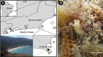
This study was carried out at the south-western rocky shore of the Forno Beach, Arraial do Cabo, Rio de Janeiro State, Brazil (23°52′S 42°01′W, Figure 1A). This region is characterized by an upwelling system that usually occurs during the austral spring and summer, decreasing the water temperature from 23 to ~14°C (Fernandes et al., Reference Fernandes, Quintanilha, Monteiro-Ribas, Gonzalez-Rodriguez and Coutinho2012). Forno Beach is localized next to an international port, with intense traffic of commercial ships, and is a touristic destination especially during the summer, resulting in the threatening of marine communities of the non-protected areas. For instance, in the last decade, formerly abundant species disappeared or became rare in the region (e.g. Clathrina aurea Solé-Cava, Klautau, Boury-Esnault, Borojevic & Thorpe, 1991) while others appeared or thrived and are now very abundant (e.g. Sycettusa hastifera, Paraleucilla magna and Darwinella sp.).
Localization of the sampling site ‘Forno Beach’ (black circle) in Arraial do Cabo (Rio de Janeiro, Brazil) (A), where the specimens of Sycettusa hastifera were collected (B).

Fixation and processing of samples
Ten specimens of S. hastifera were collected monthly randomly by snorkelling from 1 to ~4 m depth from September 2008 to December 2009 (16 months). During each sampling, the superficial seawater temperature (from here termed ‘temperature’) (±0.5°C) was measured with a calibrated thermometer. The specimens were collected in bowls filled with seawater and transferred to tubes containing Bouin's solution fixative. After 24 h, the specimens were transferred to 70% ethanol and then decalcified with 5% EDTA (ethylenediaminetetraacetic acid) solution. After confirmation of spicule elimination and removal of associated organisms, the excess liquid was wiped from the specimens and their masses were measured in a precision balance. We chose to estimate the ‘size’ of the sponges by their wet weight because most of the specimens were too small to measure volume by liquid displacement. The specimens were then split into two parts (top and base), dehydrated through an ethanol series (50, 70, 80, 90, 100%), cleared in xylene, and embedded in paraffin wax. Thin sections (5 µm) were made and mounted on glass slides, stained using Harris's haematoxylin and eosin or Heidenhain's iron haematoxylin (Bancroft & Stevens, Reference Bancroft and Stevens1996; Lanna et al., Reference Lanna, Paranhos, Paiva and Klautau2015).
Quantification and measurement of reproductive elements
To describe and count the number of reproductive elements (oocytes, embryos (i.e. from the first cleavage to the stomoblastula stage) and larvae) in the choanosome of S. hastifera, we took five random photographs (final magnification = 100×) of the base and five random photographs of the top of each individual. We preferentially photographed different regions of non-serial sections, in order to avoid repeat counts of the reproductive elements. The five pictures covered a total area of 3.5 mm2 of each fragment, totalling 7 mm2 for each sponge. Then, the reproductive elements were counted using the ImageJ software (http://rsb.info.nih.gov/ij/index.html). The density of reproductive elements was calculated at three levels: (a) – fragment (average of the five pictures), (b) – individual (average of the 10 pictures taken of each fragment of the specimen), and (c) – population (average of the averages of the 10 specimens collected that month). We estimated the monthly fecundity of the population considering both potential (eggs) and realized (embryos and larvae) fecundities (Ramirez-Llodra, Reference Ramirez-Llodra2002). Therefore, fecundity was calculated by summing the average densities of oocytes, embryos and larvae of each specimen and, after that, calculating the average of this sum for each month. To estimate if oocytes present in different months of the study were a single yield that grew continuously throughout the months or if they were repeatedly produced, we estimated the diameter of oocytes by measuring their largest diameter. Only oocytes with a recognizable nucleus were measured and, whenever oocytes were present, we measured at least 15 eggs per sponge fragment. The oocytes, and the other reproductive elements, were measured using the software AxioVs40 V 4.6.3.0 (Carl Zeiss Image Solution).
Statistical analyses
All data sets were tested for normality and homoscedasticity prior to all analysis. When the data were not normal, we chose to use non-parametric tests, instead of data-transformation.
SPATIAL DISTRIBUTION IN THE SPONGE BODY
To test whether the density of the reproductive elements and the fecundity was different depending on the sponge body part (top vs base), we performed the non-parametric Wilcoxon matched-pairs test (W) (Sokal & Rohlf, Reference Sokal and Rohlf1995). To check whether the ‘size of oocytes’ was different depending on the sponge body part (top vs base), we ran the non-parametric Mann–Whitney (U) unpaired test. For these tests, we used the specimens from September 2008 to November 2009, as no specimens were found in December 2009. Individuals without the reproductive element being tested were excluded from the analysis.
SEASONALITY
To evaluate whether the dynamics of the sponge wet weight, oocyte size and the density of the reproductive elements of S. hastifera were different depending on the season, we applied a nested analysis of variance (Sokal & Rohlf, Reference Sokal and Rohlf1995). We defined ‘season’ (spring 2008; summer, autumn, winter and spring 2009) as groups and ‘months’ (spring = October, November and December; summer = January, February and March; autumn = April, May and June; and winter = July, August and September) as subgroups nested within seasons (Sokal & Rohlf, Reference Sokal and Rohlf1995).
LIFE HISTORY PARAMETERS' PREDICTORS
To check whether the wet weight of the individuals, the oocyte diameters and the dynamics of the reproductive elements could be predicted by the temperature fluctuation at the region, we fitted the month average of the life history parameters on the temperature data through a linear model. The same analysis was applied to check whether the wet weight of the individuals could predict the reproductive effort of the organism (Sokal & Rohlf, Reference Sokal and Rohlf1995). Except for temperature, quantitative data presented along the text are averages with standard deviation (±). All tests were performed with a two-tailed P value, and a confidence interval of 95% (P < 0.05). All statistical analyses were performed using the R environment (R Development Core Team, 2015).
RESULTS
Some aspects of the life history of S. hastifera
In the study area, Sycettusa hastifera is one of the most conspicuous calcareous sponges. It was usually observed in vertical walls growing preferentially among the calcareous algae Amphiroa sp. and Jania sp. (Figure 1B). Several small organisms were found associated with S. hastifera – e.g. diatoms, polychaetes, nematodes and crustaceans. These organisms were usually epibionts observed living between or hanging on the diactines that protrude through the sponge body.
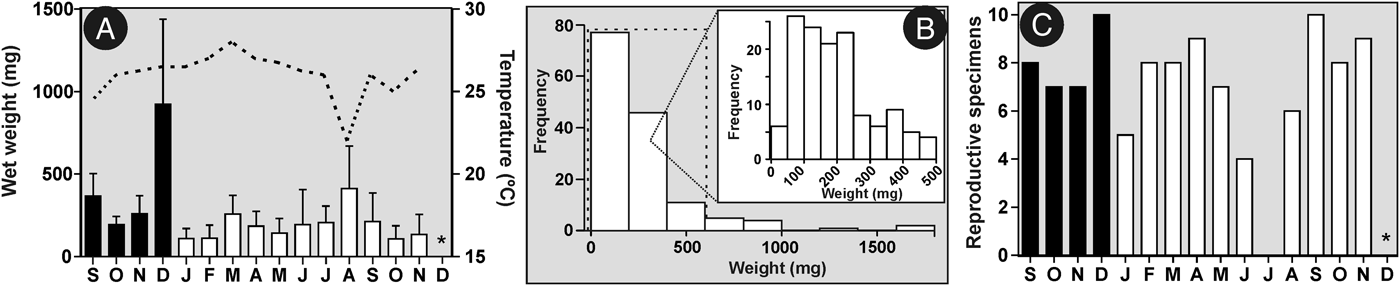
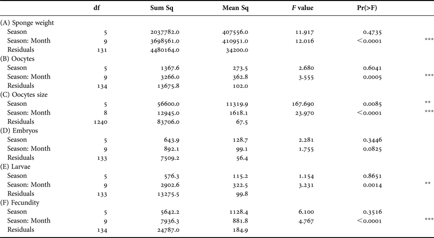
Individuals of S. hastifera were found in all sampling months except December 2009. The wet weight of the collected individuals varied from 25 mg (October 2009) to 1770 mg (December 2008) (Figure 2A), with an average of 262 (±265) mg during the whole study period (Figure 2B). On average, sponges were heavier in December 2008 (924 ± 514 mg), followed by August 2009 (412.6 ± 259 mg) and September 2008 (368 ± 135 mg), and no seasonality on the sponge wet weight was observed during the study period, although high variance was observed inside the seasons (Figure 2A, Table 1). Lighter individuals were more common: of the 146 analysed individuals, 132 (90.4%) had less than 500 mg (Figure 2B). The wet weight of the specimens of S. hastifera could not be predicted by water temperature (linear model, R 2 = 0.001; P = 0.87; df = 14).
Some aspects of the life history of Sycettusa hastifera at Arraial do Cabo (Brazil) from September 2008 to December 2009. (A) Variation of the average (and standard deviation) values of the wet weight of the soft tissue of the specimens collected each month (bars) and seawater temperature at the region during the sampling period (line) (* – no specimen was found). (B) Size frequency of the randomly collected individuals (N = 146) of S. hastifera measured as soft tissue's wet weight. Insert shows the size frequency of small individuals (0–500 mg), which were more abundant in the studied population. (C) Variation in the number of reproductive specimens along the study period. A total of 10 specimens were collected each month (* – no specimen was found). (In A and C, black bars mean 2008 and white bars mean 2009.)

Analysis of the seasonal variation of different aspects of the life history of Sycettusa hastifera. This table summarizes the results of the nested ANOVA. Months are nested within seasons.

***P < 0.001, **P < 0.01.
The population of Sycettusa hastifera was reproductively active in Arraial do Cabo throughout the study period, with at least four of 10 specimens showing some stage of reproduction (oogenesis, embryogenesis or larva) in each month, except for July 2008, when no specimen was reproducing and December 2009, when no specimen was found (Figure 2C). All identified reproductive individuals were females or, at least, hermaphrodites, as we were not able to recognize spermatogenesis. This species was viviparous, brooding its embryos until the larval stage.
Dynamics of reproductive elements of S. hastifera
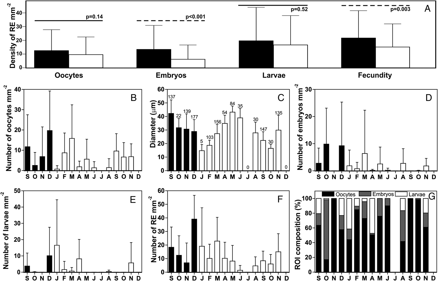
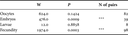
In addition to being reproductive throughout most of the study period, Sycettusa hastifera presented a strong reproductive effort. Oocytes, embryos and larvae were observed constantly and could reach densities up to 64.6 oocytes mm−2 (in an individual from December 2008), 50.1 embryos mm−2 (in an individual from March 2009), 69.9 larvae mm−2 (in an individual from January 2009) and a fecundity of 70.4 reproductive elements mm−2 (in an individual from January 2009). Oocytes and larvae did not seem to have any difference in their distribution along the osculum-base axis, but embryos and the fecundity were significantly different, with higher values observed at the base for both variables (Table 2, Figure 3A). On the other hand, the reproductive elements were distributed qualitatively evenly in the radial axis of the sponge, being found all along the choanocyte chambers, from prosopyle to apopyle.
Quantitative aspects of the reproduction of Sycettusa hastifera at Arraial do Cabo (Brazil) from September 2008 to December 2009. In December 2009, no specimen was found. (A) Comparison of the mean density (bars ± SD) of the different reproductive elements in the base (black bars) and the top (white bars) of the sponge's body. Dashed line means that there was significant difference between top and base; (B–F) Dynamics of the densities (mean ± SD) of the reproductive elements of the population of S. hastifera along the study period (black bars – 2008 and white bars – 2009); (B) Dynamics of the density of oocytes; (C) Dynamics of the diameter of the oocytes (value above the bars means the number of oocytes measured in that month); (D) Dynamics of the density of embryos; (E) Dynamics of the density of larvae; (F) Dynamics of the fecundity (density of all reproductive elements (RE) in the choanosome); (G) Percentage of the reproductive elements comprising the fecundity of each month of the study period.

Summary of the Wilcoxon paired test testing whether the reproductive elements and the fecundity are differently distributed between the base and top of the specimens of Sycettusa hastifera.

***P < 0.001.
Gametogenesis (oocytes)
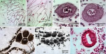
Different stages of oogenesis were observed. Overall, this gamete presented a rounded shape, with a granular cytoplasm and a large nucleolated nucleus (Figure 4A, B). Oocytes were present throughout the study period, except for July 2009, when no reproductive specimen was observed. The density of oocytes varied from 0.7 (±1.6) mm−2, in January 2009, up to 19.7 (±19.5) mm−2, in December 2008 (Figure 3B). We did not observe significant differences of oocyte density among seasons, even though it varied significantly among the months (Table 1), indicating that variation among months is greater than observed in different seasons.
Reproductive aspects of Sycettusa hastifera at Forno Beach (Arraial do Cabo, Brazil). (A) Transversal section through the choanosome of a non-reproductive specimen showing the syconoid aquiferous system with inhalant canals (ic), the elongated choanocyte chambers (cc), the apopylar region (ap), and the atrium (at) (cx – cortex). (B) Transversal section through the choanosome of a reproductive specimen showing the oocytes (oo) distributed evenly along the choanocyte chambers (cc) (at – atrium; cx – cortex). (C) More detailed view of the mature oocyte showing its nucleolated (nu) nucleus (n) (ci – inhalant canal; cc – choanocyte chamber; sc – sclerocyte). (D) The fertilization process is aided by two carrier cells (cc – choanocyte chamber, n – oocyte nucleus, nc – nurse cells). (E) Early embryo (em) after finishing the second cleavage in between the choanocyte chamber (cc) and the endopinacocytes (pi). (F) Late embryo (em) located between two choanocyte chambers (cc) showing its close contact with choanocytes (ch). (G) Amphiblastula with ciliated cells (ci) and granular cells (gc) typical of this kind of larva (cc – choanocyte chamber; ch – choanocytes).

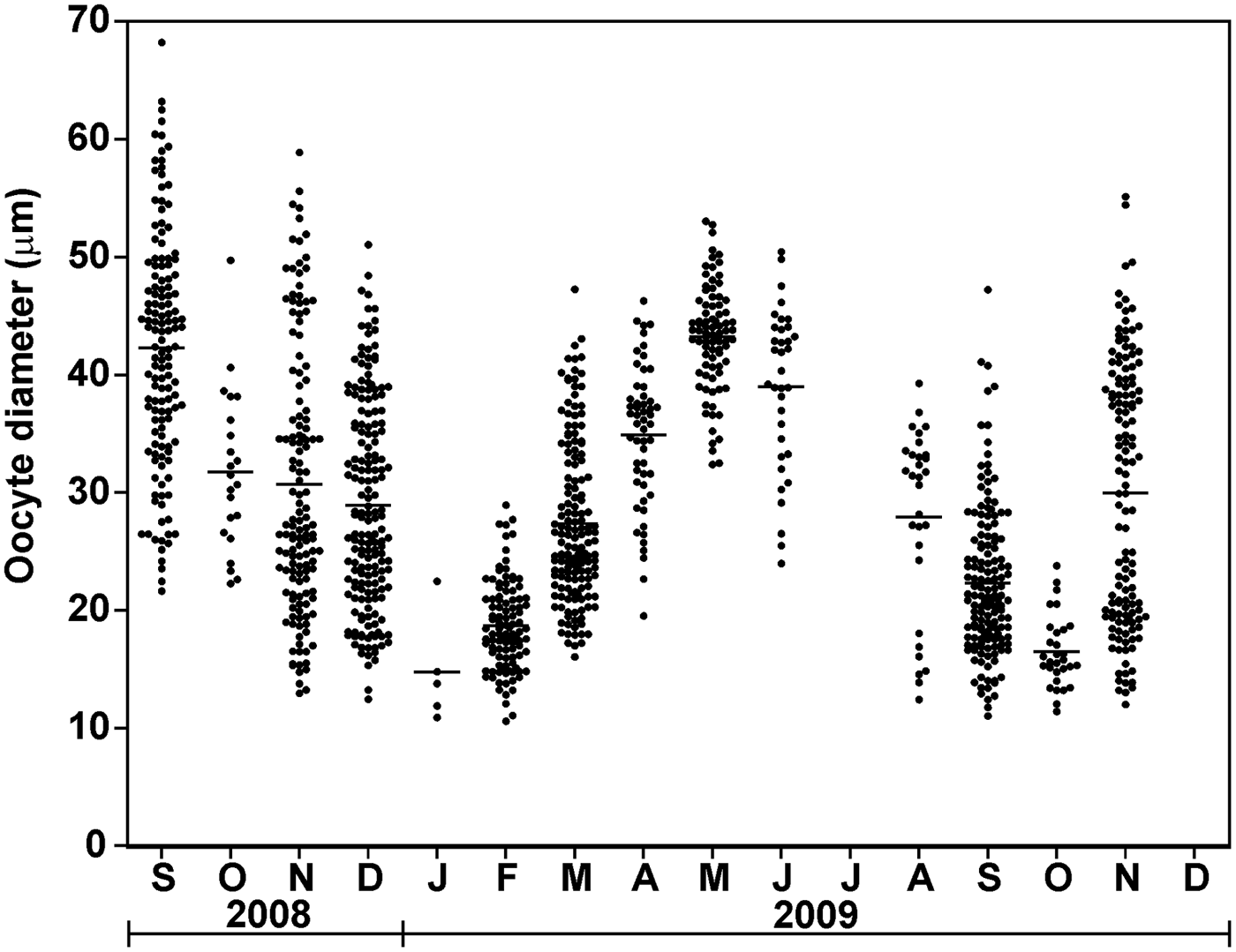
The diameter of the oocytes varied from 10.6 to 68.2 µm, with an average of 30.0 (±11.1) μm (N = 1254). Oocytes seemed to be a little larger at the base (30.7 ± 11.2 µm, N = 646) than at the top (29.3 ± 10.8, N = 608) of the sponges (Mann–Whitney–Wilcoxon test, U = 182700, P = 0.0333). At least three periods of oocytes production were observed during this study: September–December 2008 (winter–spring), January–June 2009 (summer–autumn) and August–November 2009 (winter–spring) (Figures 3C and 5). Oocytes size was significantly different amongst seasons during the study period (Table 1). These differences were probably caused by the larger diameters observed during the spring 2008 and autumn 2009 compared with the lower diameters observed in summer and winter 2009 (Figures 3C, 5). The mean diameter of oocytes did not show any relationship with the mean density of oocytes found in each month (linear model, R 2 = 0.0432; P = 0.45; df = 13).
Size distribution of oocytes found in the choanosome of S. hastifera during the study period. It was possible to observe three different cohorts: from September to December 2008, from January to June 2009, and from August to November 2009 (horizontal bars show the mean value observed for each month).

Embryogenesis
After fertilization, the egg started cleavages, forming the embryo and, later on, the larva. Cleavages occurred in the thin mesohyl of the sponge, between the choanoderm and the endopinacocytes lining the choanocyte chambers (Figure 4E, F). Different stages of embryogenesis were observed throughout the study period, but were absent in November 2008, and July and September 2009 (Figure 3D). When present, the density of embryos in the choanosome varied from 0.05 (±0.15) mm−2 in October 2009 to 9.25 (±16.1) mm−2 in December 2008 (Figure 3D). In the study period, the embryos did not present either difference among or within seasons (Table 1). Early embryos (from the first to the fifth cleavage, Figure 4E) measured from 27 to 62 µm, with an average of 42.0 (±6.2) μm (N = 150). Late embryos (during the formation of the blastocoel and the stomoblastula, Figure 4F) measured from 29 µm to 66.5 µm, with an average of 47.0 (±7.7) μm (N = 150). After differentiating into stomoblastula, embryos started the inversion process. In this species, the embryo inverts directly into the choanocyte chamber, becoming an amphiblastula larva (Figure 4G).
The larval size varied from 30.3 to 78.0 µm, with an average of 47.3 (±9.7) μm (N = 150). The amphiblastula larvae were the most irregularly observed reproductive elements in the choanosome of S. hastifera. They were not observed in November 2008, from March to July, and from September to October 2009. Nevertheless, when present, the larvae could reach overwhelming densities (as mentioned above). In a population level, larval density varied from 0.08 (±0.27) mm2 in October 2008 to 16.5 (±27.9) mm2 in January 2009 (Figure 3E). We did not find significant differences among seasons for the larval density, but, as observed for the other reproductive elements, they were significantly different among months (Table 1), indicating that variation amongst months is greater than among seasons.
Fecundity
The studied population of S. hastifera had a fecundity above 10 reproductive elements (RE) mm−2 in most of the study period. In June 2009, the population had the lowest fecundity observed in this study (1.44 ± 3.52 RE mm−2) while the largest value was observed in December 2008 (39.11 ± 17.51 mm−2) (Figure 3F). The fecundity of S. hastifera was strongly influenced by the density of oocytes, which comprised usually more than 50% of the reproductive elements observed throughout the study period (Figure 3G). The single month in which oocytes were not the most important elements was October 2008, when embryos accounted for 81% of the reproductive elements in the choanosome. We also found no evidence of seasonality on fecundity when considering all RE together. However, the fecundity was significantly different among the months (Table 1), indicating that variation amongst months was greater than in different seasons.
Life history parameters predictors
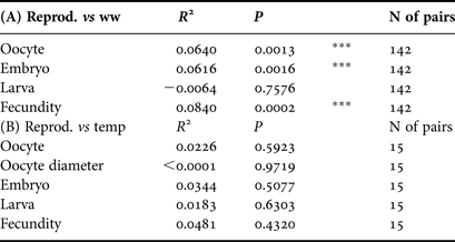
The density of oocytes, embryos and fecundity were significantly related to the wet weight of the studied individuals (but with very low R2 values), indicating that the heavier the sponge the more reproductive it became (Table 3). The temperature in the study region varied from 19°C in September 2008 to 26°C in March 2009, with an annual average of 22.5 (±1.6)°C (Figure 2A). We found that neither the density of reproductive elements/fecundity nor the size of the oocytes could be predicted by the temperature (Table 3).
Summary of the statistics related to the predictors of the density of reproductive elements of Sycettusa hastifera.

(A) Summary table of the linear model test between the reproductive elements (Reprod.) and the wet weight (ww) of the individuals. (B) Summary of the linear model between the reproductive elements and the oocyte size of S. hastifera with the seawater temperature at Arraial do Cabo (Brazil) from September 2008 to December 2009.
***P < 0.001.
DISCUSSION
Reproductive period and sexuality of S. hastifera
Sycettusa hastifera produced reproductive elements continuously during the study period. This result differs from what has been observed in most of the Calcarea studied so far. Lanna et al. (Reference Lanna, Monteiro, Klautau, Custódio, Lôbo-Hajdu, Hajdu and Muricy2007) showed that most of the calcareous sponges investigated in the temperate region of the northern hemisphere reproduce in the warmer seasons and that the same occurred with Paraleucilla magna (from 2004 to early 2006) in the tropical southern hemisphere. In a more recent study it was observed that the same population of P. magna (studied from late 2006 to 2008) reproduced continuously (Lanna et al., Reference Lanna, Paranhos, Paiva and Klautau2015), indicating that, like other marine invertebrates, calcareous sponges can reproduce all year round, at least those living in the tropics (e.g. Bauer, Reference Bauer1989; Hellström et al., Reference Hellström, Kavanagh and Benzie2010; De Paula et al., Reference de Paula, Pires and Creed2014). Orton (Reference Orton1920) postulated that tropical species should have continuous reproduction due to the absence of strong fluctuations of the environment, whilst temperate species should have a seasonal reproductive pattern. However, continuous reproduction of calcareous sponges is not restricted to the tropics. Some populations of Sycon ciliatum (Fabricius, 1780) and the Italian population of P. magna in the Mediterranean Sea are also capable of reproducing continuously throughout the year (Sarà & Orsi, Reference Sarà and Orsi1975; Franzen, Reference Franzen1988; Longo et al., Reference Longo, Pontassuglia, Corriero and Gaino2012). The extended reproductive period observed in different species of Calcaronea in England was attributed to two successive gametogenic cycles in the same individuals (Orton, Reference Orton1914, Reference Orton1920). As in the current study no repeated sampling was performed in the same individuals, so this explanation cannot be used to explain the continuous reproduction of S. hastifera (the same can be said of the Rio de Janeiro population of P. magna (Lanna et al., Reference Lanna, Paranhos, Paiva and Klautau2015)). On the other hand, with our data (Figure 5) we can infer that at the population level there were three different reproductive periods during the study period: September–December 2008, January–June 2009, and August–November 2009. The presence of reproductive specimens throughout the year has also been observed in Homoscleromorpha and Demospongiae. However, in these groups, some species present an extended period of vitellogenesis, taking almost the whole year to produce a single yield of oocytes, while in the calcareous sponges S. hastifera and P. magna the oocytes and embryos seem to be produced repeatedly during the year (Meroz-Fine et al., Reference Meroz-Fine, Shefer and Ilan2005; Riesgo et al., Reference Riesgo, Maldonado and Durfort2007; Abdo et al., Reference Abdo, Fromont and McDonald2008; Lanna et al., Reference Lanna, Paranhos, Paiva and Klautau2015).
Localization of reproductive elements
Sycettusa hastifera is a tubular syconoid sponge that presents an obvious polarity along the osculum-base axis. Although we had hypothesized that there was no difference of the reproductive elements between top and base, we found that embryos and fecundity presented a significant difference in density comparing base and top of the sponge body (Figure 3A). A similar difference was previously observed in the leuconoid species P. magna, in which also only embryos showed statistical difference between top and base, being more abundant in the latter (top = 1.37 ± 2.44 embryos mm−2; base = 2.96 ± 4.75 embryos mm−2; W = −487.0; P = 0.05; N = 57 pairs; data set from Lanna et al., Reference Lanna, Paranhos, Paiva and Klautau2015). This similarity may be just an artefact of statistical methods, as no difference was observed in previous (oocytes) or subsequent (larvae) developmental stages in either species, and there is no evidence for the migration, in the mesohyl, of the early embryo towards the base or the late embryo towards the top of the sponge. Nevertheless, in some demosponges embryos and larvae are always found only at the base of the individuals, possibly as an adaptation against predation and hydrodynamics (Reiswig, Reference Reiswig1973; Fell, Reference Fell1976; Fromont, Reference Fromont1994). The cause of the difference in the distribution of reproductive elements between top and base in calcareous sponges is still undetermined and deserves further investigation.
We expected to find a qualitative difference in the distribution of the reproductive elements in the radial axis of S. hastifera (atrium – cortex), as other studied syconoid sponges (belonging to the genus Sycon Risso, 1826) presented a strong regionalization of the reproductive elements. In those species, earlier stages of oogenesis occur next to the apopyle and later stages of development close to the prosopyle (Sarà & Orsi, Reference Sarà and Orsi1975; Gallissian, Reference Gallissian1981). Surprisingly, in S. hastifera the reproductive elements are distributed evenly in the radial axis of the sponge. It is likely that an organized distribution of the reproductive elements is not a characteristic of the sponges possessing syconoid aquiferous system, but rather a characteristic of Sycon (Sarà & Orsi, Reference Sarà and Orsi1975; Gallissian, Reference Gallissian1981). Further studies investigating the reproductive elements distribution in other species of these and other syconoid genera (e.g. Amphoriscus Haeckel, 1870, Grantessa Lendenfeld, 1885, Heteropia Carter, 1886) may elucidate whether this characteristic is related to the aquiferous system type (which is considered a homoplastic character in Calcarea, Manuel et al., Reference Manuel, Borchiellini, Alivon, Parco, Vacelet and Boury-Esnault2003, but see Voigt et al., Reference Voigt, Wülfing and Wörheide2012 for a different perspective) or is a synapomorphy of Sycon.
Gametogenesis and embryogenesis
We were not able to recognize spermatogenesis in S. hastifera. In fact, the knowledge on spermatogenesis in the class Calcarea is very scarce, restricted to few observations of the ripe spermatozoon of some species and the description of some steps of the spermatogenesis of P. magna (Lanna & Klautau, Reference Lanna and Klautau2010 and references therein). In other syconoid sponges, spermatic cysts were observed at the distal region of the choanocyte chambers (Sarà & Orsi, Reference Sarà and Orsi1975), but nothing related to the production of male gametes was observed in S. hastifera. However, we observed that oocytes were present throughout the year and their densities were not significantly different amongst seasons. This pattern was also observed in P. magna indicating that in both species, at least in the study areas, this reproductive element occurs in similar densities in all seasons (Longo et al., Reference Longo, Pontassuglia, Corriero and Gaino2012; Lanna et al., Reference Lanna, Paranhos, Paiva and Klautau2015). Such a pattern raises the question of whether tropical calcareous sponges are different from temperate sponges or if these species are adapting themselves to the conditions of a new area. The size of oocytes of S. hastifera was significantly different among seasons (Table 1), which was caused by the presence of different cohorts of oocytes depending on the period of the year (Figure 5). Repeated production of oocytes has already been observed in tropical sponges, corals, shrimps, echinoderms and other marine invertebrates (Bauer, Reference Bauer1989; Meroz & Ilan, Reference Meroz and Ilan1995; Ventura & Pires, Reference Ventura, Pires, Soares-Gomes and Pereira2009; Hellström et al., Reference Hellström, Kavanagh and Benzie2010; De Paula et al., Reference de Paula, Pires and Creed2014).
Embryos and larvae were less abundant and less frequent than oocytes in the choanosome of S. hastifera (Figure 3G), but their densities did not vary significantly among seasons. As in other sponges and marine invertebrates (e.g. Ettinger-Epstein et al., Reference Ettinger-Epstein, Whalan, Battershill and de Nys2007; Ventura & Pires, Reference Ventura, Pires, Soares-Gomes and Pereira2009; Hellström et al., Reference Hellström, Kavanagh and Benzie2010; De Paula et al., Reference de Paula, Pires and Creed2014; Lanna et al., Reference Lanna, Paranhos, Paiva and Klautau2015), the extended period of larval release (which can be inferred from the presence of late embryos or larvae in the choanosome) observed in S. hastifera indicates a lack of synchrony during the embryogenesis of this species. Along the Brazilian coast, several species of corals spawn or release their planulae in consecutive months during the whole year (Ventura & Pires, Reference Ventura, Pires, Soares-Gomes and Pereira2009; De Paula et al., Reference de Paula, Pires and Creed2014). This asynchrony in the reproduction of marine invertebrates may reflect an adaptation of the species to assure the survival of the population's offspring, escaping of a single unfavourable environmental event that could hamper the population's reproductive success (Ettinger-Epstein et al., Reference Ettinger-Epstein, Whalan, Battershill and de Nys2007; Whalan et al., Reference Whalan, Battershill and de Nys2007; Hellström et al., Reference Hellström, Kavanagh and Benzie2010). In addition, a continuous production of larvae is considered an important trait of the life history of invasive species, being a key strategy to colonize new areas (Johnston et al., Reference Johnston, Piola, Clark, Rilov and Crooks2009; Lanna et al., Reference Lanna, Paranhos, Paiva and Klautau2015).
Reproductive effort
As mentioned before (Whalan et al., Reference Whalan, Battershill and de Nys2007; Riesgo & Maldonado, Reference Riesgo and Maldonado2008; Lanna et al., Reference Lanna, Paranhos, Paiva and Klautau2015), the quantification of the reproductive elements of S. hastifera was very useful to make comparisons of the reproductive output within and with other species. Sycettusa hastifera presented a high reproductive output, which seems to be a characteristic of calcaronean sponges, although differences in the proportion of reproductive elements depend on the species (e.g. Dendy, Reference Dendy1914; Orton, Reference Orton1914; Vacelet, Reference Vacelet1964; Eerkes-Medrano & Leys, Reference Eerkes-Medrano and Leys2006; Lanna et al., Reference Lanna, Paranhos, Paiva and Klautau2015). While in S. hastifera the most abundant reproductive element is the oocyte, in P. magna embryos and larvae are the most constant elements in the choanosome. Vitellogenesis in S. hastifera is long (~3 months), involves the participation of nurse cells and results in large oocytes (Figure 5), while in P. magna vitellogenesis is faster, oocytes are nourished by choanocytes and are small (Lanna & Klautau, Reference Lanna and Klautau2010). This longer period of vitellogenesis in S. hastifera explains why oocytes are so abundant in this species. Both species also show different nursing strategies. Embryos of S. hastifera invert into the choanocyte chamber, becoming larvae and being released, while in P. magna larvae are nourished for some time in the epilarval trophocyte epithelium (ETE, Lanna & Klautau, Reference Lanna and Klautau2012; Lanna et al., Reference Lanna, Paranhos, Paiva and Klautau2015). Hence, while S. hastifera seems to spend more energy producing oocytes, P. magna seems to spend its energy nursing embryos that reached the larval stage. This difference in nursing strategies is probably related to a phylogenetic constraint of the species, but a proper investigation about this topic is still necessary. However, with these contrasting characteristics, we suggest that calcaronean sponges that do not brood their larvae in ETE, i.e. the stomoblastula inverts directly into the choanocyte chamber, will probably show lower densities of larvae in histological investigations.
Growth vs reproduction
In S. hastifera, the density of oocytes, embryos and the fecundity were related to the wet weight of the individual (Table 3). However, the appearance of reproductive elements was not related to the size of the individuals of the sampled population, and apparently there is no minimum size to start producing gametes and embryos. This result is not in accordance with the generalized idea that sexual maturity is size dependent in invertebrates. The strategy ‘grow first, reproduce later’ would allow differential allocation of energy to the growth of the young individuals, avoiding the risk of mortality associated with their small size (e.g. see Hellström et al., Reference Hellström, Kavanagh and Benzie2010 and references therein). In the present species, energy and nutrients are likely to be allocated to the high reproductive effort whilst somatic maintenance and survival are reduced. Actually, the high reproductive effort experienced by the individuals of S. hastifera may have a strong influence in their death, as occurs with semelparous invertebrates and vertebrates (Futuyma, Reference Futuyma2009). This is plausibly indicated by the low percentage of individuals that reached large wet weights (Figure 2B), suggesting that specimens of S. hastifera could die soon after their high reproductive activity. This strategy seems to be widespread in the class Calcarea (Lanna et al., Reference Lanna, Monteiro, Klautau, Custódio, Lôbo-Hajdu, Hajdu and Muricy2007, Reference Lanna, Paranhos, Paiva and Klautau2015), but is different from what has been observed in demosponges. In general, demosponges have long lifespan and usually attain large sizes (e.g. Bell, Reference Bell2008; McMurray et al., Reference McMurray, Blum and Pawlik2008). This long lifespan can be related to their relatively small reproductive effort (compared with calcareous sponges) (see Lanna et al., Reference Lanna, Paranhos, Paiva and Klautau2015). A trade-off between somatic maintenance (body growth, defence and lifespan) and reproduction has been advocated as one of the main causes of the evolution of different life histories (Stearns, Reference Stearns1989; Ramirez-Llodra, Reference Ramirez-Llodra2002) and in these two classes of Porifera, at least from a general point of view, the reproductive effort observed in their species seems to be ultimately (evolutionarily) regulated.
Temperature vs reproduction
The reproduction of S. hastifera was not statistically related to seawater temperature in Arraial do Cabo, contrasting with previous observations for many species of Porifera and other marine invertebrates (e.g. Giese & Pearse, Reference Giese, Pearse, Giese and Pearse1974; Fromont, Reference Fromont1994; Castro & Pires, Reference Castro and Pires2006; Ettinger-Epstein et al., Reference Ettinger-Epstein, Whalan, Battershill and de Nys2007; Abdo et al., Reference Abdo, Fromont and McDonald2008; Riesgo & Maldonado, Reference Riesgo and Maldonado2008; Muramatsu & Silveira, Reference Muramatsu and Silveira2008; Lanna et al., Reference Lanna, Paranhos, Paiva and Klautau2015). It may be argued that the variation in the water temperature in Arraial do Cabo (ΔT = 7°C) during this study was not sufficient to influence the reproductive activity of S. hastifera, as in other locations this variation is of greater order (e.g. ΔT = 12°C in Rio de Janeiro during the study with P. magna – Lanna et al., Reference Lanna, Paranhos, Paiva and Klautau2015). This absence of relationship, however, is not a total surprise, as a wide range of organisms, especially in tropical regions, does not show any relationship between temperature and reproduction. An alternative explanation for the onset and maintenance of the reproduction of marine invertebrates in the tropics is food availability, photoperiod and wind regimes (Hails & Yaziz, Reference Hails and Yaziz1982; Bauer, Reference Bauer1989; Muthiga et al., Reference Muthiga, Kawaka and Ndirangu2009), but these hypotheses need confirmation for the phylum Porifera.
Sycettusa hastifera has life history traits of invasive species
Sycettusa hastifera has been considered as a cryptogenic species because the evidence to determine whether it is native or introduced in Brazil (or, alternatively, in the Red Sea) is still insufficient (Cavalcanti et al., Reference Cavalcanti, Skinner and Klautau2013). It has a wide distribution (Red Sea, Indian Ocean and SW Atlantic), which is unexpected for an organism reproducing with lecitotrophic larvae that last less than 24 h in the plankton, and its location close to commercial ports, which is considered as a source of alien species, could be used as an argument in favour of an ‘alien’ status for S. hastifera. Nonetheless, the knowledge about its reproductive biology could help to understand if it is an invasive species or not.
So far, there is no consensus for the characteristics of the life history of an invasive species (reviewed in Sakai et al., Reference Sakai, Allendorf, Holt, Lodge, Molofsky, With, Baughman, Cabin, Cohen, Ellstrand, McCauley, O'Neil, Parker, Thompson and Weller2001). However, Sakai et al. (Reference Sakai, Allendorf, Holt, Lodge, Molofsky, With, Baughman, Cabin, Cohen, Ellstrand, McCauley, O'Neil, Parker, Thompson and Weller2001) highlighted that ‘characteristics common to successful colonists across taxa include r-selected life histories (use of pioneer habit, short generation time, high fecundity and high growth rates) and the ability to shift between r- and K-selected strategies’. We found here that S. hastifera presents several of these traits: (i) it reproduces throughout the year releasing a large number of larvae (high fecundity); (ii) most individuals of the population have small size, indicating that perhaps the life cycle is short; and (iii) small individuals are already sexually mature, which may suggest that reproduction starts early in the life of this sponge. In addition, Cavalcanti et al. (Reference Cavalcanti, Skinner and Klautau2013) showed that S. hastifera is one of the first species to recruit to granite plates (use of pioneer habitat) and has no clear preference for substrate type. Therefore, if S. hastifera is proven to be a native species in Brazil, at least some of its life history parameters demonstrate that it has a strong potential to colonize new areas.
ACKNOWLEDGEMENTS
We are thankful to V. Padula, R. Cavalcanti, H. Orofino, E. Resende (and the Littoral Sub crew) and the LaBiPor staff for their help with the fieldwork. We also thank the LABIM/IB/UFRJ, A.L. Rossi and M. Farina for the use of the microscope with digital camera.
FINANCIAL SUPPORT
Grants and fellowships were provided by the Coordenacão de Aperfeiçoamento de Pessoal de Nível Superior (CAPES), Fundacão Carlos Chagas de Amparo à Pesquisa do Estado do Rio de Janeiro (FAPERJ) (E-26/111.541/2008), PROPCI-PRODOC/UFBA and the Brazilian National Council of Technological and Scientific Development (CNPq) (480368/2008-2; 302442/2011-1; 477227/2013-9). This work was part of the Doctoral Thesis of E. Lanna, presented to the Post-Graduation Programme in Zoology of the Museu Nacional do Rio de Janeiro/UFRJ.