I. INTRODUCTION
The development of cost-effective, simple, sensitive, and selective biosensing platforms is essential for designing biological assays and point-of-care (POC) diagnostic tests. Generally speaking, biosensors include two elements, a recognition element that is responsible for recognizing a target of interest and a transducer that converts the recognition activity into a measurable signal. Functional nucleic acids, simplified here as FNAs, particularly DNA aptamers and DNAzymes, have been regarded as excellent recognition elements because they can be made to have excellent recognition specificity and affinity, in addition to high chemical stability and convenient availability via chemical synthesis. Reference Liu, Cao and Lu1,Reference Navani and Li2 Graphene-based materials, on the other hand, have emerged as an excellent platform to set up various effective signal transduction mechanisms, owing to their remarkable physicochemical and structural properties. Reference Chen, Feng and Li3–Reference Wang, Li, Wang, Li and Lin7 Two outstanding properties make graphene oxide, simplified as GO in this review, a favorite choice of material for the design of optical biosensors with FNAs: its varying affinity for structured and unstructured nucleic acids, and its ability to function as an effective quencher for fluorescence. In this mini-review, we will use selective examples to illustrate the usefulness of GO in the design of optical biosensors employing DNA aptamers and DNAzymes.
A. Functional DNA molecules: DNA aptamers and DNAzymes
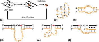
DNA is renowned for its role as the genetic information carrier of life. However, certain single-stranded DNA molecules can fold into well-defined tertiary structures to carry out intricate functions including molecular recognition and enzymatic catalysis. Reference Cech8–Reference Ponce-Salvatierra, Wawrzyniak-Turek, Steuerwald, Hobartner and Pena10 DNA molecules capable of recognizing a target of interest are called aptamers and those with ability to speed up a chemical transformation are termed DNAzymes. DNA aptamers and DNAzymes have not been found in biological systems but they can be isolated from a large pool of DNA sequences through a process called in vitro selection. Reference Tuerk and Gold11–Reference Robertson and Joyce13 A general scheme for selecting functional DNA species is outlined in Fig. 1(a). This method begins with a library that contains as many as 1016 unique single-stranded DNA sequences, some of which are presumed to adopt an intricate tertiary structure to achieve specific recognition toward a molecular target or execute catalysis for a specific chemical transformation. The pool of DNA is subjected to a selection step that enriches active sequences, followed by an amplification step where polymerase chain reaction (PCR) is applied to create more copies of these sequences. The cycle of selection and amplification is repeated until the sequences that can perform the expected function with the greatest efficiency are isolated.
Representative DNA aptamers and DNAzymes generated by in vitro selection. (a) A general in vitro selection scheme for isolating DNA aptamers and DNAzymes. (b–f) Secondary structures of thrombin-binding DNA aptamer, an ATP-binding DNA aptamer, the GR5 RNA-cleaving DNAzyme, the 8-17 RNA-cleaving DNAzyme, and Cu(II)-dependent DNA-cleaving DNAzyme.

This method has been applied in numerous studies to date, leading to the discovery of a great array of man-made DNA aptamers and DNAzymes. In theory, in vitro selection allows for the isolation of DNA aptamers and ligand-responsive DNAzymes for essentially any target of choice, including those for which antibodies are difficult to obtain, such as toxic metal ions and protein toxins. In addition, DNA is chemically stable and easy to obtain via automated solid-phase synthesis. Furthermore, additional functional groups can be conveniently grafted onto DNA aptamers and DNAzymes during or after chemical synthesis. These advantages have contributed greatly to the growing popularity of embracing DNA aptamers and DNAzymes as recognition elements for the design of biosensors. Reference Liu, Cao and Lu1,Reference Navani and Li2,Reference Mok and Li14 Here we will briefly introduce a few examples of DNA aptamers and DNAzymes frequently used in proof-of-concept biosensing studies. It is noteworthy that there are also many functional RNA molecules (ribozymes and RNA aptamers) that can also be used as molecular recognition elements. However, RNA is less stable than DNA and is more difficult to synthesize. For these reasons, they are not as popular as their DNA counterparts in biosensing applications. To date, only a few studies examined the use of RNA aptamers for the design of GO/FNA biosensors. Reference Wang, Li, Weber, Hu, Lin, Li and Lin15,Reference Ling, Jiang, Li, Tao, Qiu and Li16
One popular aptamer for biosensing studies is a DNA aptamer that binds human thrombin [Fig. 1(b)]. Reference Bock, Griffin, Latham, Vermaas and Toole17 It is relatively small, as it contains only 15 nucleotides, but binds thrombin with high affinity (K d of ∼100 nM). Structural studies have revealed that this aptamer forms an intramolecular guanine quadruplex (G-quadruplex) structure with two layers of guanine tetrads stacking on each other through π–π interactions. Reference Wang, Krawczyk, Bischofberger, Swaminathan and Bolton18–Reference Padmanabhan, Padmanabhan, Ferrara, Sadler and Tulinsky20 Another commonly used DNA aptamer is a 27-nucleotide sequence that binds adenosine 5′-triphosphates or ATP [Fig. 1(c)], which has a K d of 6 µM. Reference Huizenga and Szostak21 Structural analysis shows that this DNA aptamer actually binds two ATP molecules in its structural fold. Reference Lin and Patel22
Several DNAzymes have also been repeatedly used in biosensor studies. One such DNAzyme is known as GR5 [Fig. 1(d)], and it is the first-ever DNAzyme, reported by Joyce and Breaker in 1994. GR5 is an RNA-cleaving DNAzyme that cuts a single RNA linkage embedded in a DNA sequence in the presence of Pb2+. Reference Breaker and Joyce23 Another common DNAzyme for biosensor studies is 8–17 [Fig. 1(e)], which is also an RNA-cleaving DNAzyme. Reference Santoro and Joyce24 Sequence variants of 8–17 are capable of using a variety of divalent metal-ions as cofactors, including Pb2+, Mg2+, Ca2+, Mn2+, and Zn2+. Reference Cruz, Withers and Li25,Reference Schlosser and Li26 A DNA-cleaving DNAzyme that requires Cu2+ as a cofactor has been used in a number of biosensor studies [Fig. 1(f)]. Reference Carmi, Balkhi and Breaker27 More recent efforts have led to isolation of RNA-cleaving DNAzymes that are dependent on trivalent metal ions, Reference Huang, Lin, Cao, Vazin and Liu28 in addition to monovalent metal ions such as Na+ and Ag+. Reference Torabi, Wu, McGhee, Chen, Hwang, Zheng, Cheng and Lu29,Reference Saran and Liu30 While DNAzymes commonly used in biosensing applications often employ metal ion-dependent DNAzymes, it is worth noting that DNAzymes can also be engineered to respond to more complex biological targets. For example, our group has reported RNA-cleaving DNAzymes that can be activated by protein targets from bacterial pathogens such as Escherichia coli and Clostridium difficile. Reference Tram, Kanda and Li31–Reference Shen, Wu, Chang, Zhang, Tram, Lee, Kim, Salena and Li33
B. FNA/GO interactions
Oxygen-containing functional groups and aromatic domains within GO are functional moieties for FNA/GO interactions. The carboxyl and hydroxyl groups that decorate the basal planes and edges of GO can interact with FNAs through electrostatic forces. The hexagonal cells of GO and the ring structures of nucleobases are an excellent fit for strong π–π stacking interactions, which is the key factor behind the increased affinity for single-stranded DNA (ssDNA) by GO with higher C/O ratios. Reference Hong, An, Compton and Nguyen34 According to the density functional theory calculations, the binding energy of the nucleobases on GO varies in the order of G > A > T > C > U. Reference Gowtham, Scheicher, Ahuja, Pandey and Karna35 These calculations are consistent with experimental results obtained using isothermal titration calorimetry. Reference Varghese, Mogera, Govindaraj, Das, Maiti, Sood and Rao36
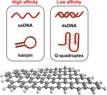
The length and the structure of FNAs can significantly affect FNA/GOs interactions (Fig. 2). In general, GO exhibits higher affinity toward ssDNA DNA species (such as unstructured DNA or DNA hairpins) than double-stranded DNA (dsDNA) or DNA molecules with rigid structures, such as G-quadruplex and i-motifs (Fig. 2). For example, hairpin structures contain both a helical region and a loop region. Because the loop region is in the single-stranded form, it is expected to have high affinity for GO. Many aptamers have hairpin structures; the well known ATP-binding DNA aptamer and PDGF-binding DNA aptamer are two excellent examples. Reference Huizenga and Szostak21,Reference Green, Jellinek, Jenison, Östman, Heldin and Janjic37 In the absence of their cognate targets, these aptamers bind GO strongly, indicating hairpin structures have excellent affinity for GO. GO usually exhibits higher affinity for longer DNA sequences than short ones, although longer sequences may create stable secondary structures that can significantly reduce their affinities to GO. This is because π–π stacking interactions between conjugated rings of GO and the nitrogenous bases of the nucleic acids are largely responsible for GO-DNA affinity, and many nucleobases of well-structured DNA molecules are engaged in intramolecular base-pairing and unavailable for stacking with GO.
Graphic illustration of varying affinities of different DNA species. Generally, more structured DNA molecules, such as duplex DNA and G-quadruplex, have lower affinity to GO than less structured DNA molecules, such as single-stranded DNA that lacks Watson-Crick base pairs or DNA hairpins with short stems.

In addition to these factors, the interaction between GO and FNAs is also strongly dependent on the local environment, such as pH, salt concentrations, and solvent. Reference Wu, Kempaiah, Huang, Maheshwari and Liu38,Reference Lei, Mi, Zhou, Chen, Hu, Guo and Zhang39 Several research and review papers have comprehensively examined or reviewed adsorption and desorption reactions between DNA molecules and graphene-based materials under various conditions, which are excellent resources on this topic. Reference Liu, Sun, Zhang and Liu40–Reference Liu, Salgado, Maheshwari and Liu43
II. SOLUTION-BASED FNA/GO OPTICAL SENSORS
There are two important properties of GO that make it a fitting nanomaterial for coupling with FNAs: variation of affinity for ligand-free and ligand-bound FNAs, and an excellent ability to quench fluorescence. The latter property reflects the fact that electrons within the extensive conjugated π system of GO can be excited by wavelengths that fall within the emission spectrum of most fluorophores. In fact, many studies have shown that GO can efficiently quench surface-bound fluorescent species such as various organic dyes, Reference Li, Wang, Cao, Chen, Tang, Wang, Zhu and Huang44 quantum dots (QDs), Reference Dong, Gao, Yan, Ji and Ju45 silver nanoclusters, Reference Liu, Wang, Aizen, Yehezkeli and Willner46 and upconversion nanoparticles. Reference Liu, Wang, Jia and Li47 Theoretical calculations have predicted that GO is a long-range dynamic quencher (up to 30 nm). Reference Swathi and Sebastian48 The quenching effect is mainly derived from fluorescence (or Förster) resonance energy transfer (FRET), or nonradiative dipole–dipole coupling between the fluorescent species and GO or reduced GO. Reference Loh, Bao, Eda and Chhowalla6,Reference Liu, Zhao, Quan, Chen and Fan49 The quenching efficiency of GO is observed to be significantly improved after reduction, and such materials are often referred to as reduced graphene oxide (rGO).
GO’s preference for binding unstructured or less structured DNA molecules compared to more structured variants offers a simple way to design optical biosensors via adsorption of fluorescently labeled FNA probes onto the GO surface and target-promoted probe release. In such sensors, the FNA acts as a recognition element, and the fluorophore/GO pair works as a transducer that converts target recognition into a signal, often as a result of the target-induced change in the structure of the FNA. The proximity-induced quenching of fluorescence by GO when the target is absent makes it a suitable matrix for immobilization of FNA biosensors to achieve sensitive detection. Thus the two properties of GO, DNA binding and fluorescence quenching, are largely intertwined in their role in FNA/GO biosensors. Both the sequence design of an FNA and optimal tuning of GO’s DNA binding affinity are important factors that influence the performance of FNA/GO biosensors. In principle, the GO surface should have adequate affinity for a free (unbound, unstructured, or less structured) FNA but should have significantly reduced affinity for the ligand-bound (structured) FNA. This approach has been applied repeatedly in the design of biosensors capable of detecting various analytes that include metal ions (e.g., Pb2+, Cu2+), Reference Zhao, Kong, Zhang, Meng, Liu, Tan, Shen and Yu50–Reference Liu, Zhao, Chen, Yu, Zhang and Quan52 small molecules (e.g., ATP, ochratoxin A), Reference Wang, Li, Hu, Lin, Li and Lin53,Reference Sheng, Ren, Miao, Wang and Wang54 proteins (e.g., thrombin, insulin, mucin 1, cellular prion protein), Reference Chang, Tang, Wang, Jiang and Li55–Reference Zhuang, Zhen, Wang and Huang58 and bacteria (e.g., Staphylococcus aureus, Listeria monocytogenes). Reference Huang, Chen, Xia, Wu, Duan, Ma and Wang59–Reference Duan, Ding, He, Wu, Wei and Wang61 In addition, this approach has been extended to multiplexed detection using different aptamer probes, each carrying a fluorophore with a distinct emission (e.g., UCNPs, QDs). Reference Wu, Duan, Ma, Xia, Wang, Wang and Zhang62,Reference Kurt, Yüce, Hussain and Budak63 Quantification of fluorescence signal can be achieved via fluorescence intensity measurements or fluorescence polarization, with the former being the more common approach. The focus of this review will be limited to studies assessing fluorescence intensity, although there are a number of interesting GO-coupled fluorescent FNA biosensors in which fluorescence polarization was used for detection. Reference Liu, Wang, Jiang, Hu, Li, Yang, Li, Yang, Tan and Huang64–Reference Yu, Liu, Zhen and Huang66
Many studies have used GO in its capacity as a quencher and an immobilization platform to engineer FNA-based biosensors. In general, there are two common approaches to coupling GO with fluorophore-labelled FNAs, both of which will be discussed below.
A. Molecular recognition prior to GO addition
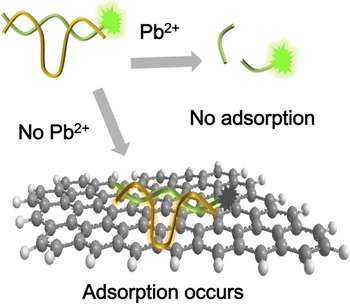
An excellent example of this approach involves the use of a well-known Pb2+-responsive DNAzyme along with a fluorescent substrate. Reference Zhao, Kong, Zhang, Meng, Liu, Tan, Shen and Yu50 The design takes advantage of the observation that uncleaved substrate (longer in size) has higher affinity for GO than the cleavage products (shorter in size), and therefore, in the presence of Pb2+, the solution exhibits a higher level of fluorescence despite the addition of GO because the cleaved products have reduced affinity toward GO (Fig. 3).
A GO sensor for lead ion detection. In the absence of Pb(II), the DNAzyme construct is adsorbed by GO leading to low-level of fluorescence. In the presence of Pb(II), the fluorescent substrate is cleaved by the DNAzyme into small fragments that do not adsorb well on GO surface.

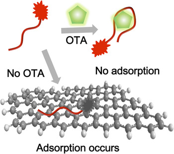
Aptamers have also been used in this approach. For example, an aptamer/GO biosensor has been reported using a fluorophore-labeled DNA aptamer that binds ochratoxin A (OTA), a mycotoxin released by fungi commonly found in grains. Reference Sheng, Ren, Miao, Wang and Wang54 The design takes advantage of the fact that ligand-free aptamers in general are less structured than ligand-bound aptamers. In the absence of OTA, the fluorescent aptamers are adsorbed by GO and their fluorescence is quenched. However, in the presence of OTA, the formation of more rigid structure of aptamer–OTA complex significantly reduces the adsorption of the aptamer by GO (Fig. 4). The same strategy has been further extended for the detection of two different mycotoxins in a single sensor using two aptamers labeled with two different fluorophores. Reference Wu, Duan, Ma, Xia, Wang, Wang and Zhang62
An aptamer/GO sensor for the detection of ochratoxin A (OTA). In the absence of OTA, the unstructured aptamer is adsorbed by GO leading to quenching of fluorescence. In the presence of OTA, the aptamer forms a tight complex that does not adsorb well on GO surface.

B. Molecular recognition in the presence of GO
Conversely, a number of interesting examples of FNA/GO coupling involve immobilization onto GO prior to the addition of target. An interesting example of this approach is a biosensor based on a Cu2+-dependent DNA-cleaving DNAzyme containing both duplex and triplex structural elements. Reference Liu, Zhao, Chen, Yu, Zhang and Quan51 These pairing elements can be utilized for fluorescence signaling through the use of intercalating fluorescent dyes. In the presence of GO, the fluorescence of the DNAzyme/fluorophore complexes is quenched. Upon Cu2+-induced cleavage, the DNA fragments come off the GO surface, resulting in the increase of fluorescence. The same approach has also been applied to DNAzymes containing covalently attached fluorophores. Reference Liu, Zhao, Chen, Yu, Zhang and Quan52 Although these metal-ion detection sensors use nucleic acid-cleaving DNAzymes as molecule recognition elements, the same approaches could also be extended to metal-ion binding aptamers. Reference Rajendran and Ellington67,Reference Wu, Zhan, Wang and Zhou68
Fittingly, this design principle is also applicable to aptamers that recognize protein biomarkers. These include DNA aptamers that bind human thrombin, Reference Chang, Tang, Wang, Jiang and Li55 insulin, Reference Pu, Zhu, Han, Liu, Liu, Liao, Zhang and Tan56 and cellular prion protein. Reference Zhuang, Zhen, Wang and Huang58 In all these cases, protein binding to the aptamer creates a tightened complex structure that exhibits lower affinity for GO. In addition to the use of traditional organic dyes as the fluorophores, newer nanomaterials could also be used to achieve fluorescence signaling. For example, silver nanoclusters have been used together with the DNA aptamer for thrombin on graphene to achieve a detection limit of 0.5 nM, which is even comparable with signal amplification approaches discussed in later sections. Reference Liu, Wang, Aizen, Yehezkeli and Willner46
Aptamer-based biosensors are powerful tools for detection of small molecules such as heavy metal ions and small molecules, or even the human proteins described above. Furthermore, aptamers for important infectious bacteria such as Staphylococcus aureus, Reference Huang, Chen, Xia, Wu, Duan, Ma and Wang59,Reference Huang, Chen, Duan, Wu, Wang, Wei and Wang60 and Listeria monocytogenes, Reference Duan, Ding, He, Wu, Wei and Wang61 have also been successfully coupled with GO.
C. Special considerations
A particularly noteworthy property of FNA/GO coupled biosensors is their nuclease resistance, due to GO’s ability to protect adsorbed nucleic acids from degradation by nucleases, thereby enabling use of FNA-based biosensors in clinical samples as well as in intracellular delivery. One example of biosensing in clinical samples is a sensor that used an aptamer for glycated albumin in serum, which may be used as a tool to monitor diabetes mellitus. Reference Apiwat, Luksirikul, Kankla, Pongprayoon, Treerattrakoon, Paiboonsukwong, Fucharoen, Dharakul and Japrung69 The protection of nucleic acids by GO has also been demonstrated by aptamer/GO sensors for intracellular biosensing. Immobilization onto graphene has enabled intracellular detection of ATP, Reference Wang, Li, Hu, Lin, Li and Lin70 as well as simultaneous visualization of ATP and GTP in live cells. Reference Wang, Li, Weber, Hu, Lin, Li and Lin15 Although we will limit our scope to intracellular delivery for the purpose of biosensing, we are cognizant of the fact that there are a tremendous array of therapeutic uses of FNAs that can be made possible by rendering them resistant to nucleases by GO.
A key challenge of FNA/GO sensors is that adsorbed FNAs can be displaced by unintended targets, thus generating false-positive results. This is particularly problematic for intracellular detection where high concentrations of proteins and other nucleic acids may compete with FNAs for GO binding. One approach for solving this problem is to use a reference probe to track for false-positive signals. Reference Tan, Chen, Xiong, Mao, Zhu, Yasun, Li, Zhu and Tan71 Another approach is to covalently immobilize an FNA on GO to eliminate the possibility of desorption by unintended targets. Reference Huang and Liu72,Reference Liu, Chen, Liu, Wu, Zhou, He, Ding and Liu73
Conversely, false negatives can also be an issue. Since the length of the binding region of an FNA is of great significance in determining its ability to generate a true positive signal, there is a need to balance the affinity of an FNA for GO. If the ligand-free FNA binds GO with great strength, the FNA may not be able to be released upon target induction. Our group has recently reported a general strategy that is useful in tuning the affinity of an existing aptamer to GO. The approach employs a blocking DNA strand to weaken the interaction of an RNA aptamer with the GO before the addition of target, thereby facilitating target-promoted release of the aptamer from the GO surface. Reference Song, Lau, Liu, Shuang, Dong and Li74 This work demonstrated that while it took approximately 15 min following addition of high concentrations of target for maximum signal enhancement to be produced, the same level of increase in signal was observed within 1 min in the presence of a blocking strand.
III. SOLID-PHASE FNA/GO OPTICAL BIOSENSORS
Solution-based assays are not necessarily the optimal approach for POC diagnostics and field applications due to repeated pipetting and measurement inconvenience. Contrastingly, solid-phase sensors such as lateral-flow or microfluidic devices can significantly simplify experimental procedures making them well suited for these applications. This is the key driving force behind efforts of making solid-phase FNA/GO sensors.
GO can be overlaid onto solid supports such as glass slides, upon which fluorescent aptamers can be adsorbed to create simple solid-phase sensors. This type of sensor has been demonstrated by Ueno and coworkers using protein-binding DNA aptamers in several recent studies. Reference Furukawa, Ueno, Tamechika and Hibino75–Reference Ueno, Furukawa, Matsuo, Inoue, Hayashi and Hibino77 For example, they have designed a solid-phase biosensor for thrombin detection using the well-known thrombin-binding DNA aptamer. Reference Furukawa, Ueno, Tamechika and Hibino75 They have also shown that these sensors can be further tuned to achieve better performance through the use of a pyrene tag to increase the binding affinity of the aptamer to GO, and incorporation of a suitable DNA spacer to optimize FRET efficiency. Reference Ueno, Furukawa, Matsuo, Inoue, Hayashi and Hibino76 They further engineered a multichannel linear-array GO/aptamer sensor for thrombin detection, and have also demonstrated the versatility of their approach by engineering similar sensors for two additional protein targets, prostate specific antigen (PSA), and hemagglutinin (HA). Reference Ueno, Furukawa, Matsuo, Inoue, Hayashi and Hibino77
To simultaneously detect multiple targets, more tunable fluorescent probes, such as quantum dots, are better options than traditional organic fluorophores. An interesting study has been published in which a unique microfluidic paper-based device was designed to detect multiple types of cancer cells using quantum dot/aptamer/GO conjugates. Reference Liang, Su, Li, Lan, Yang, Ge, Yu and Song78 More specifically, DNA aptamers for three different cancer cell lines, MCF-7, HL-60, and K562, were conjugated to quantum dots with different emission properties and adsorbed by GO. Such materials were then used to make multi-zone microfluidic paper-based analytical devices, with a designated aptamer in each detection zone for a specific cancer cell.
Another interesting approach worth noting is a silicon/paper/glass hybrid microfluidic system that has recently been developed to achieve multiplexed pathogen detection. Reference Zuo, Li, Dominguez and Ye79 The design takes advantage of aptamers as the recognition elements, chromatography paper for adsorbing aptamer-functionalized GO, and silicon on a glass surface to create multiple microchannels and inlet reservoirs. This method was applied for the detection of bacterial pathogens Lactobacillus acidophilus, Staphylococcus aureus and Salmonella enterica using specific DNA aptamers for these bacteria. The key innovation of the design is the use of the paper substrate for facilitating the integration of aptamer/GO biosensors on the microfluidic class chip, thus simplifying immobilization of aptamers in a glass-only microfluidic system.
IV. SIGNAL AMPLIFICATION STRATEGIES
Achieving high detection sensitivity is a key goal in the design of analytical tests. However, standard analytical methods without signal amplification, including optical FNA/GO sensors discussed above, may not be able to deliver the necessary detection sensitivity often needed in real-life applications. There is considerable interest in developing FNA/GO biosensors that incorporate signal amplification strategies. In this section we will discuss some common approaches that have been successful in devising FNA/GO sensors with great signal amplification capabilities.
A. Polymerase-assisted target recycling
Target recycling generally refers to analytical strategies in which the target is liberated and released from its bound FNA, thereby recycling the target by freeing it for interaction with another FNA molecule—ultimately amplifying the signal from a single target molecule. However, the approach used in enabling each individual target molecule to take part in many cycles of molecular recognition/signal production events can vary.
Aptamers are most often unmodified nucleic acid molecules which can still function as templates for DNA replication by DNA polymerases. This property has been elegantly utilized in the design of sensing mechanisms where DNA polymerases drive the recycling of the target. Reference He, Wu, Zhou, Wang, Jiang, Shen and Yu80–Reference Huang, Chen, Yang, Zhu, Zhu, Yang, Wang and Tan82 An interesting example involves a cocaine-binding DNA aptamer configured into a hairpin DNA probe, which, in the presence of cocaine, forms a complex structure that exposes an originally hidden sequence element for binding to a DNA primer. The formation of the target-induced primer-probe duplex permits DNA polymerization. Thus DNA polymerization converts the complex from a primer-bound DNA probe with a small duplex region into a full DNA duplex, thereby causing releases of the target—enabling it to bind another aptamer and restart the cycle. This strategy has been applied with end-labelled and intercalating fluorophores, with the intercalating approach possessing a lower detection limit by two-orders of magnitude, at 2 nM. Reference He, Wu, Zhou, Wang, Jiang, Shen and Yu80
Additionally, this strategy has recently been modified for the creation of an amplified GO-aptamer biosensor for the detection of interferon-gamma (IFN-γ). In this specific example, an anti-IFN-γ aptamer is labeled with a fluorophore and adsorbed on the surface of GO to achieve fluorescence quenching. The target binding event enables aptamer release, followed by primer hybridization, DNA polymerization, and target release. It was reported that this amplification method was able to detect as low as 1.5 fM. This sensitivity is about 3–5 orders of magnitude higher than that of traditional unamplified aptamer-based assays. Reference Hu, Liu, Chen, Huang, Zhao, Tian and Zhang83
More recently, a modified GO-aptamer-based strand displacement amplification method was developed for the detection of ricin B-chain (RTB). Ricin-binding aptamer (RBA) first hybridized with a short DNA blocker and then immobilized on magnetic beads (MBs). In the presence of RTB, the blocker is released due to the formation of aptamer-RTB complex. After separation, the free DNA blocker could trigger the isothermal strand-displacement polymerase reaction (ISDPR) to open numerous hairpin probes, which then cannot be effectively adsorbed onto the GO surface, resulting in high fluorescence intensity. In the absence of RTB, the hairpin probe can be adsorbed and quenched by GO. A detection limit of 0.6 μg/mL was reported, with a linear response range of 0.75–100 μg/mL. Reference Li, Xiao, Tao, Wang, Huang and Zhen84
B. Nuclease-assisted target recycling
A different target-recycling strategy involves the use of nucleases to digest aptamers. For example, DNase I has been used to enable target recycling in aptamer/GO sensors. Reference Pu, Zhu, Han, Liu, Liu, Liao, Zhang and Tan56,Reference Lu, Li, Lin, Wang, Yang, Chen and Chen85 In the absence of target, the dye-labeled aptamer probes are adsorbed on the GO surface. The adsorption brings about two functions in these cases: fluorescence quenching and protection of aptamers against DNase I digestion. Upon the addition of target, the aptamers are released from the GO, leading to the recovery of fluorescence, and more importantly, enabling the nuclease to digest the released aptamer, driving target recycling. More recently, this strategy was applied for simultaneous detection of plant hormones, such as zeatin and systemin with a detection limit of 60 nM and 35 nM, respectively. Reference Su, Liu, Chen, Chen and He86
Beyond DNase I, other enzymes capable of cleaving duplexes, including exonuclease III and RNA-cutting RNase H, have also been used in the design of target-recycling assays. Reference Guo, Yang, Zhang, Ning, Yao and Zhang87–Reference Cui, Chen, Zhu, Lin, Chen and Yang90 One such example involved the use of lysozyme-binding DNA aptamer in a design similar to polymerase-assisted target recycling described earlier, in which a hairpin is disrupted by target binding in a manner that exposes a region free to form a duplex with another oligonucleotide. The formation of a duplex with a fluorophore-labeled complement in this newly exposed region produces a region that can be cleaved by exonuclease III, thereby leading to release of free fluorophores. Reference Chen, Zhao, Jiang and Yu91 In the absence of target, the region remains occupied in the hairpin and is unable to bind the fluorophore-labeled complementary oligonucleotide, and the aptamer remains long enough to bind GO thereby quenching its fluorescence. If the fluorophore-labeled oligonucleotide is unable to hybridize to its complimentary region in the hairpin, it is not degraded by exonuclease III since its exonuclease activity is specific to duplex DNA. This amplification method was shown to be capable of detecting as low as 0.08 μg/mL, with a linear range from 0.125 μg/mL to 1 μg/mL.
Another nuclease-based target recycling strategy employs an exonuclease capable of degrading ssDNA. It involves the use of Staphylococcal enterotoxin B-binding DNA aptamer that is hybridized to two oligonucleotides separated by a quencher and a fluorophore, to which it remains bound until the presence of target induces the release of the oligonucleotides, resulting in the increase in fluorescence. Cleavage of the aptamer by exonuclease I then frees up the target so it can bind another aptamer and displace another fluorophore-quencher pair. The system is shown to have a detection limit of 0.3 pg/mL. Reference Wu, Duan, Ma, Xia, Wang and Wang92
In a very recent report, a nicking endonuclease (NEase)-mediated cyclic amplification strategy was also developed for the detection of cancer cells. The system consists of hairpin aptamer probe (HAP) that binds T-cell acute lymphoblastic leukemia cells (CCRF-CEM cells), dye-labeled linker DNA, NEase, and GO. Without the target cells, the HAP maintains a stable hairpin structure, thus the linker DNA binds to GOs leading to fluorescence quenching. In contrast, the binding of HAP toward target cells triggers its conformational change, allowing it to bind to the dye-labeled linker and attract NEase for cleavage, resulting in the release of free fluorophore-labelled fragments. Since NEase cleaves only the dye-labeled strand, the HAP becomes free to initiate another cycle. The detection limit of this design was determined to be 25 cells, which was 24 times lower than that of normal fluorescence aptasensor without amplification. Reference Xiao, Liu, Chen, Zhang and Kong93
C. Signal amplification via rolling circle amplification
The amplification strategies mediated by GO are not limited to the use of target recycling approaches, as more direct coupling between signal amplification and FNA/GO systems have also been developed. An amplified biosensor that combines the FNA/GO and rolling circle amplification (RCA) was reported to allow highly sensitive detection. The key feature of this strategy is the use of a DNA probe that contains a primer region and an aptamer region. Without the target, the adsorbed DNA probe is unavailable for RCA. However, the presence of target will dissociate the DNA probe and frees up the primer region to hybridize with its circular template to initiate RCA (Fig. 5). Reference Liu, Song, Shuang, Dong, Brennan and Li94 The resulting product can then bind multiple fluorescently labeled oligonucleotides, thereby amplifying the signal from target-aptamer recognition. This method was applied for the detection of thrombin and was shown to be capable of delivering a detection sensitivity of 10 pM, 2 orders of magnitude better than conventional unamplified methods.
Aptamer/GO sensors coupled with rolling circle amplification. The binding of a primer-aptamer sequence to GO prevents the binding of the primer domain to the circular template. However, in the presence of the target for the aptamer, the primer-aptamer construct is released from the surface of graphene and the primer sequence can bind the circular template and participate in the rolling circle amplification process.

D. Non-enzymatic amplification
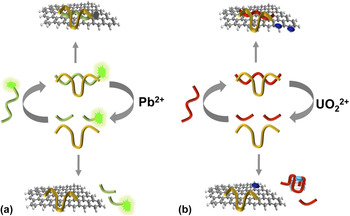
Although the above-mentioned enzyme-mediated amplification strategies can achieve high sensitivity, they may not be highly compatible with real environmental or biological samples ridden with molecules that can significantly inhibit the activity of enzymes. In this case, enzyme-free amplification is highly desirable. Several GO-DNAzyme based amplification methods have been developed with this desire in mind. For example, a GO-DNAzyme based biosensor was reported for amplified fluorescence-based detection of Pb2+. Without Pb2+, GO can bind the DNAzyme-substrate complex and quench its fluorescence. While in the presence of Pb2+, the DNAzyme is activated and cleaves the substrate into two DNA fragments, which have low binding affinity toward GO, thus resulting in the high fluorescence signal. The cleavage event also releases Pb2+ to induce another round of binding and cleavage, producing an amplified signal [Fig. 6(a)]. A limit of detection of 0.3 nM was established for this method. Reference Zhao, Kong, Zhang, Meng, Liu, Tan, Shen and Yu50 A different nonenzymatic amplification strategy has also been described for UO2 2+ detection. Instead of using fluorophore-labeled RNA-containing substrate, this study utilized N-methylmesoporphyrin IX (NMM), a dye with a highly conjugated ring as the signal generating component. Reference Li, Wang, Cao, Chen, Tang, Wang, Zhu and Huang44 Since G-quadruplexes are largely stabilized by π–π stacking interactions by guanine-tetrads, having a guanine-rich sequence within the substrate enables cleavage-induced production of a G-quadruplex for binding to NMM [Fig. 6(b)]. The benefits of this combination go beyond the obvious signal generation capability of NMM, since its ability to engage in π–π stacking interactions enables GO to act as an adsorbing agent capable of quenching the fluorescence of free NMM, thereby diminishing background signal. A limit of detection of 86 pM was reported for this method.
DNAzyme/GO sensors with non-enzymatic amplification mechanisms for the detection of (a) lead ion and (b) uranyl ion. In each case, the cleavage of the substrate by the DNAzyme results in the desorption of the cleavage fragment and the recycling of the metal ion for another round of action. Fluorophore-tagged substrate was used in (a) and N-methyl-mesoporphyrin IX (NMM), a fluorescent small molecule was used in (b).

V. CONCLUSIONS AND OUTLOOK
As illustrated with multiple examples in this short review, uniting FNAs with GO represents a unique and impactful approach toward engineering highly effective optical sensors for the detection of a wide variety of analytes, from simple metal ions to small biological cofactors, proteins, and bacterial and cancer cells. FNAs brings to this marriage the great versatility in target recognition, particularly considering the fact that DNA aptamers or DNAzymes can be derived for any target of interest from synthetic DNA pools using in vitro selection experiments. GO, on the other hand, brings to this union a simple and effective material for DNA immobilization and fluorescence quenching. We expect that research and development on biosensing with FNA/GO will continue to be a vibrant research area in the years to come.
DNA aptamers and DNAzymes not only can act as recognition probes, they can also be manipulated by many enzymes, such as nucleases and polymerases. GO can negatively influence the accessibility of the DNA species by these enzymes by shielding them through physical adsorption, and yet release them upon target binding and target-promoted catalysis. This unique interplay permits the implementation of various signal amplification mechanisms to achieve ultra-sensitive detection, as illustrated by the selective examples featured in this review. Most of the demonstrated signal amplification strategies use simple procedures that are compatible with POC applications.
DNA molecules are inherently more stable and their stability is further improved when coupled with GO, as the adsorption by graphene can prevent nucleases from degrading DNA. However, certain FNA/GO biosensors do use labile components such as protein enzymes and labile small-molecule substrates, therefore it may be necessary to incorporate protection mechanisms. One simple approach involves the use of pullulan, a water-soluble polysaccharide, which upon forming films can not only protect labile molecules from denaturation or degradation, but also dissolves readily in test solutions. Reference Jahanshahi-Anbuhi, Pennings, Leung, Liu, Carrasquilla, Kannan, Li, Pelton, Brennan and Filipe95 We have recently shown that biosensing paper sensors for DNA amplification can be engineered via inkjet-printing pullulan solutions containing enabling components for RCA reactions. Reference Liu, Hui, Zhang, Gu, Kannan, Jahanshahi-Anbuhi, Filipe, Brennan and Li96 One additional benefit of pullulan film based paper biosensors is the avoidance of cold delivery and storage. Reference Carrasquilla, Little, Li and Brennan97,Reference Kannan, Jahanshahi-Anbuhi, Pelton, Li, Filipe and Brennan98 Furthermore, pullulan films can also protect RNA from spontaneous and RNase-mediated cleavage, Reference Hsieh, Monsur Ali, Tram, Jahanshahi-Anbuhi, Brown, Brennan, Filipe and Li99 which can significantly increase the shelf-life of RNA-cleaving DNAzymes as molecular recognition elements. Inkjet printing of FNA/GO materials on paper, particularly with pullulan as an encapsulating reagent, will certainly offer a simple and cost-efficient way to engineer sensors for POC or field applications.
Currently, nearly all the studies on FNA/GO biosensors have focused on proof-of-concept demonstrations of novel sensor designs using a few well-studied FNAs. More efforts are certainly needed to develop sensors targeting clinically relevant biomarkers or important contaminants in the environment. In addition, further innovation is necessary to simplify test procedures and reduce the cost. Notable activities in the area of lateral-flow devices and paper-based sensors may ultimately offer new opportunities for FNA/GO biosensing technologies.