Introduction
An estimated 23 million miscarriages occur every year worldwide and 44 pregnancy losses occur each minute. The pooled risk of miscarriage is 15.3% (95% CI 12.5–18.7%) of all recognized pregnancies. Recurrent spontaneous abortion (RSA) is a disease defined as two or more consecutive failed pregnancies before 20 weeks of gestation (Practice Committee of the American Society for Reproductive Medicine, 2020). The population prevalence of women who have had one miscarriage is 10.8% (10.3–11.4%), for two miscarriages it is 1.9% (1.8–2.1%), and three or more miscarriages it is 0.7% (0.5–0.8%) (Quenby et al., Reference Quenby, Gallos, Dhillon-Smith, Podesek, Stephenson, Fisher, Brosens, Brewin, Ramhorst, Lucas, McCoy, Anderson, Daher, Regan, Al-Memar, Bourne, MacIntyre, Rai, Christiansen and Coomarasamy2021). Women who have had repeated miscarriages often have uncertainties about the cause, the likelihood of recurrence, the investigations they need, and the treatments that might help (Coomarasamy et al., Reference Coomarasamy, Dhillon-Smith, Papadopoulou, Al-Memar, Brewin, Abrahams, Maheshwari, Christiansen, Stephenson, Goddijn, Oladapo, Wijeyaratne, Bick, Shehata, Small, Bennett, Regan, Rai, Bourne and Quenby2021). Studies have been conducted mainly on females, however, the male gamete contributes 50% of the genomic material to the embryo and placenta. The relative contribution of sperm to successful live births can be 10–15% (Sakkas et al., Reference Sakkas, Ramalingam, Garrido and Barratt2015). Lower sperm number and quality might increase the risk of unexplained recurrent spontaneous abortion (URSA); oxidative stress and hormone metabolism in sperm as well as nucleic acid synthesis and oxidative stress in seminal plasma were related to URSA (Zhang et al., Reference Zhang, Wang, Feng, Yang, Huang, Lu, Guan, Sun, Chen and Qian2020a). Infertile patients with severe male factor infertility, such as severe oligospermia or varicocele, are at risk of increased levels of DNA damage. Obesity or the use of toxic agents such as tobacco and pesticides may also impair sperm DNA (Rodrigo, Reference Rodrigo2020). Approximately 60% of infertile men have damaged sperm DNA (Gharagozloo et al., Reference Gharagozloo, Gutiérrez-Adán, Champroux, Noblanc, Kocer, Calle, Pérez-Cerezales, Pericuesta, Polhemus, Moazamian, Drevet and Aitken2016). In the past few decades, the role of sperm DNA integrity on fertility, embryo development, embryo quality, implantation and pregnancy has gained much attention (Yuan et al., Reference Yuan, Huang, Leung, Wang, Meng, Huang, Pan, Zhou, Li, Sima, Wang, Zhang, Ying and Wang2019). As reported, fertilization of oocytes with spermatozoa that have damaged DNA could potentially lead to reduced fertilization rates, poor embryo quality, as well as higher rates of spontaneous miscarriage (Ahmadi and Ng, Reference Ahmadi and Ng1999). Some evidence has suggested that the risk of aneuploidy embryos in the high DFI group was 55% higher than that in the low DFI group (RR 1.55; 95% CI 1.358–1.772), so the high DFI in RSA patients may significantly affect the risk of aneuploidy embryos (Asgari et al., Reference Asgari, Gavahi, Karimi, Vatannejad, Amjadi, Aflatoonian and Zandieh2021). Therefore, reproductive failure due to male factors should be given high priority for couples who often go to multiple doctors and many clinics in their search for a cause and remedy for miscarriage, to further accelerate the discovery of molecular and cellular drivers of recurrent pregnancy loss in males.
Mammalian spermatogenesis is a complex process that contains three stages: (1) Spermatogonia conduct mitotic amplification to ensure proliferation and maintenance of spermatogonia. (2) Spermatogonia experience a process called meiosis in which spermatogonia differentiate into spermatocytes, including primary spermatocyte and secondary spermatocyte, and finally form spermatid. (3) Spermatids then divide into spermatozoa. Precise gene expression regulation is required to ensure the progression of each developmental event and DNA methylation acts as a key machinery in guiding temporal and spatial control of gene expression in spermatogenesis. In recent years, with the development of epigenetics, the study of DNA methylation provides a new perspective on the pathogenesis and therapy of recurrent pregnancy loss. The abnormal DNA methylation of imprinted genes, placenta-specific genes, immune-related genes and sperm DNA may, directly or indirectly, affect embryo implantation, growth and development, leading to the occurrence of recurrent pregnancy loss (RPL) (Zhou et al., Reference Zhou, Xiong, Qu, Bao and Zhang2021). Sperm cells possess a unique epigenome that is different from that possessed by somatic cells. Protamines replace 90–95% of histones during spermatogenesis and contribute to tight sperm chromatin compaction, which promotes motility, fertilization, and protection of sperm DNA (James and Jenkins, Reference James and Jenkins2018). DNA methylation and histone modifications critically regulate the expression of many genes and repeat regions during spermatogenesis (Liu et al., Reference Liu, Zhang, Yin, Gao, Li, Bai, He, Li, Zhang, Li, Zhang, Jia, Zhang, Lin, Zheng, Wang, Gao, Zeng and Liu2019). Changes in DNA methylation modifications have been proven to be associated with male infertility, and aberrant DNA methylation changes in sperm have been shown to increase the risk of reproductive failures and may impact the health of offspring (Jenkins et al., Reference Jenkins, Aston, Meyer, Hotaling, Shamsi, Johnstone, Cox, Stanford, Porucznik and Carrell2016; He et al., Reference He, Sun, Zhang, Feng, Xu, Dai, Ni, Wang and Wu2020). Liliana Burlibaşa’s research revealed a particular distribution of H3K4me3 during sperm cell differentiation and highlighted an important role for the regulation of DNA methylation in controlling histone methylation and chromatin remodelling during spermatogenesis. (Burlibaşa et al., Reference Burlibaşa, Nicu and Domnariu2021). Additionally, the studies speculated that assisted reproductive technology (ART) carries a potential risk of transmission/induction of genetic/epigenetic alterations, particularly when more intrusive methodologies are applied, such as intracytoplasmatic sperm injection (Lou et al., Reference Lou, Le, Hu, Yang, Li, Wang, Wang, Gao and Jin2019). Therefore, exploring the relationship in-depth between epigenetic regulation and male infertility may provide a theoretical basis for the etiological prediction and diagnosis of the male RSA.
DNA methylation is a biological process that occurs after replication that is catalyzed by a family of DNA (cytosine-5)-methyltransferases (DNMTs). DNMTs can be classed into the maintenance of DNA methylation during DNA replication named DNM1 and de novo DNMTs consisting of DNMT2 and DNMT3. To date, five structurally distinct DNMTs have been characterized, that is DNMT1, DNMT2, DNMT3A, DNMT3B, and DNMT3L (Bestor, Reference Bestor2000). Generally, DNMTs encompass three domains: the N-terminal regulatory domain, the C-terminal catalytic domain, and a linker of the central region. The N-terminal regulatory domain is particularly implicated in determining the subcellular localization of the DNMT and in allocating unmethylated DNA strands from hemimethylated ones. The C-terminal catalytic domain consists of 10 different characteristic motifs, and six of them (I, IV, VI, VIII, IX, and X) are evolutionally conserved among mammals. DNMT1 methylates the hemimethylated DNA strands and ensures the fidelity of the DNA methylation pattern during mitosis (Uysal et al., Reference Uysal, Akkoyunlu and Ozturk2016). Approximately 60–80% of CpGs within the genome are found to be predominantly methylated. DNMT1 lacking tDNMT1 can damage spermatogenesis (Rajender et al., Reference Rajender, Avery and Agarwal2011). Sperm DNA methylation consists of the methylation of imprinted genes and nonimprinted genes. Altered methylation of sperm-imprinted genes is associated with sperm DNA fragmentation (SDF), pregnancy loss rate and ART outcome (Cannarella et al., Reference Cannarella, Crafa, Condorelli, Mongioì, La Vignera and Calogero2021). To distinguish the parental alleles, imprinted genes are epigenetically marked in gametes at imprinting control elements through the use of DNA methylation at the very least, imprinted gene expression is subsequently conferred through lncRNAs, histone modifications, insulators, and higher order chromatin structure (Barlow and Bartolomei, Reference Barlow and Bartolomei2014). During the process of spermatogenesis, the occurrence of DNA methylation ensures the expression of the parental genomic imprints in germ cells and removes parental mutations of epigenetics (Hon et al., Reference Hon, Rajagopal, Shen, McCleary, Yue, Dang and Ren2013). Disruption of this process during early embryonic development can have major consequences on both fetal and placental development (Argyraki et al., Reference Argyraki, Damdimopoulou, Chatzimeletiou, Grimbizis, Tarlatzis, Syrrou and Lambropoulos2019). Loss of imprinting in most cases has a harmful effect on fetal development and can result in neurological, developmental, and metabolic disorders (Elhamamsy, Reference Elhamamsy2017). TRIM28/KAP1/TIF1β was identified as a universal transcriptional co-repressor and is critical for regulating post-fertilization methylation reprogramming in preimplantation embryos, TRIM28 is required for the maintenance of methylation imprints in bovine preimplantation embryos, and the loss of TRIM28 during SCNT may contribute to the unfaithful maintenance of imprints in cloned embryos (Ma et al., Reference Ma, Zhang, Zhang, Zhu, Ma, Yang, Su, Li, Lv and Luan2018).
At present, treatment methods are limited due to the lack of research on the factors of male pathogenesis in the clinic. In addition, semen analysis is the most widely used diagnostic test for male fertility assessment. It provides a generalized view of the efficiency of spermatogenesis but provides very little information about sperm function. Ultimately, an examination of male factors must shift from improving basic semen parameters to focusing on the few sperm that will reach an egg or the one sperm that will fertilize an egg (Klimczak et al., Reference Klimczak, Patel, Hotaling and Scott2021). DNA methylation is highly susceptible to environmental cues and environmental insults, such as exposure to toxins, teratogens, diet (nutrient availability), and mental state (stress). However, routine semen analysis is unable to estimate sperm chromatin damage, therefore more in-depth research methods are needed to evaluate sperm chromosome damage. Here, we collected the semen samples from Qinghai Provincial People’s Hospital and used sequencing technology to seek out the differentially methylated regions and candidate genes. To reveal the potential effects of sperm DNA methylation levels on embryonic developmental abnormalities at the molecular level in patients with male RSA, and provide important information regarding the potential role of sperm DNA methylation in human development.
Materials and methods
Sample preparation
Experiments were approved by the Ethics Committee of Qinghai Provincial People’s Hospital. Semen samples were collected from the Reproductive Center of Qinghai Provincial People’s Hospital. In total, 67 samples were used, including 36 from RSA patients and 31 from normal men. Within 30 min after semen collection, sperm were separated using Pure Sperm 40/80 reagents (Nidacon, Gothenburg, Sweden); 2 ml Pure Sperm 40 was carefully dropped into the superstratum of centrifugal tubes containing 2 ml Pure Sperm 80, and then 1.5 ml sperm was slowly added to the surface of the liquid, and centrifuged at 3000 rpm for 20 min at room temperature. After removal of the supernatant, 5 ml phosphate-buffered saline (PBS) was added into the tubes, which were centrifuged twice at 500 rpm for 10 min. The supernatant was discarded and 200 µl PBS was added into tubes to resuspend the sperm. Finally, samples were frozen quickly in liquid nitrogen for DNA extraction, RNA extraction and DNA sequencing.
Library building
Reduced representation bisulfite sequencing (RRBS) was used to explore the base depth distribution. First, DNA was extracted from samples, and DNA integrity and concentration were detected by electrophoresis. A database construction process was conducted when the DNA quality reached the standard. According to the PE50 sequencing strategy, DNA was digested with restriction endonuclease Msp1 to enrich for CpG fragments, which were then end repaired and an adenine (A) tail was added to the 3′ end. After the addition of a methyl-modified linker, fragment screening was performed to select inserts of 40–120 bp and 120–220 bp in length, and then the DNA fragment was extracted with bisulfite. The unmethylated C is converted to U and following PCR amplification, the library is finally built.
Data analysis
First, the data were filtered to remove low-quality data, and aligned to the standard human reference genome, published in 2013 (GRCh38). After confirming the quality of alignment, the uniquely aligned reads on the genome were analyzed to obtain methylation information for base C in the highly enriched methylation region. Information analysis processing was performed to obtain a standard information analysis result.
Quality control
Illumina’s high-throughput sequencing was originally presented as raw image data files which were converted to sequence and saved as FASTQ files using base recognition and CASAVA software. The FASTQ file contained the name of each gene, the base sequence, and its corresponding sequencing quality information. The sequencing quality score of the base was calculated using the ASCII code value corresponding to each base quality characteristic subtracted by 33 (Sanger quality value system). Different Phred quality scores represented different base sequencing error rates. According to the Phred quality score calculation, base sequencing error rates of 20 and 30 scores were 1% and 0.1%, respectively.
Extracting total RNA from sperm
A suspension containing 10–20 × 106 spermatozoa was obtained based on sperm density, and centrifuged at 12000 rpm for 2 min at room temperature; the supernatant was discarded and the sperm sediment was extracted using TRIzol reagent (Qiagen, Germany). The main steps of RNA extraction were: add 1000 µl of TRIzol to the sperm precipitate, blow repeatedly for 30 s using a pipette and then leave on ice for 5 min; add 200 µl of trichloromethane and blow again to mix, leave for 3 min and then centrifuge at 4°C for 15 min at 12,000 rpm; place the supernatant in an enzyme-free tube, add an equal volume of isopropanol, blow to mix and then leave at −20°C for 20 min. An RNA precipitate was obtained by centrifugation again at 4°C and 12,000 rpm for 15 min. The RNA precipitate was added to diethylpyrocarbonate (DEPC) water; 3 µl was mixed well, and its content and purity were determined using a micronucleic acid analyzer (Biome-tra, Germany).
cDNA synthesis
cDNA synthesis was performed using the TaKaRa reverse transcription kit (Japan) and extracted RNA as the raw material. Finally, 1 µl RNA was added to the 20-µl system volume, according to the manufacturer’s instructions.
Detecting the mRNA expression levels of genes of interest
Quantitative real-time PCR was used to analyze the mRNA expression levels of NXF3 and DYDC2 in the semen samples, and β-actin was used as the internal reference gene. The primer sequences were found using Primer Bank and synthesized by Beijing Xianghong Bioengineering Co. Ltd. For the target gene NXF3, the forward primer was 5′-AGTGCTTACCACGATGAGGC-3′, and the reverse primer was 5′-GCACTGAGGGAGTCCACAATA-3′. For DYDC2, the forward primer was 5′-CTGGCTCACTGGCTTTATCA-3′, and the reverse primer was 5′-TGGTCTTCTTCGTGGAAACAG-3′. The PCR procedure was: 30 s at 95°C, followed by 40 cycles of 5 s at 95°C, 34 s at 60°C, 15 s at 95°C, 1 min at 60°C, and 15 s at 95°C hold. The threshold cycle (Ct) resulting from qRT-PCR was analyzed using the 2−ΔΔCt method (Livak and Schmittgen, Reference Livak and Schmittgen2001). Each transcript was repeated three times using three different biological replicates.
Statistical analysis
The data were presented as mean ± standard error of the mean (SEM) of at least three biological replicates. The expression values and the global hydroxymethylation data were evaluated using eDMR software and the methylation data were evaluated by t-test. The expression levels of genes were determined by independent-sample t-test using SPSS 26.0 and GraphPad Prism 8.0.1 software. The results of all tests to assess the significance of the observed differences between groups were considered significant when the P-value was < 0.05.
Results
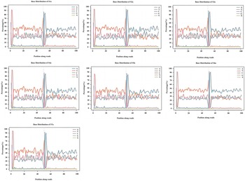
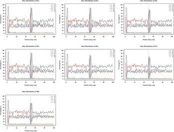
Sequencing using the RBBS method revealed no mutations or deletions in RSA sperm compared with normal sperm. We found that no differences in base distribution were observed in samples from seven normal men and eight RSA men, the DNA sequence did not change (Figures 1 and 2).
Base distribution of normal sperm, the DNA sequence did not change (n = 7 biologically independent samples).

Base distribution of male recurrent spontaneous patients, the DNA sequence did not change (n = 8 biologically independent samples).

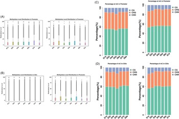
RRBS was used to explore the base depth distribution. Although RRBS only covers part of the genome, this is the region containing the promoter and CG islands. Each base distribution in promoter and CG islands was computed and then the percentage of the genome was calculated to different base depths. In Figure 3, the x-axis shows the effective sequencing depth, while the y-axis shows the percentage of the genome at the special sequencing base depth. There were three patterns of C distribution on the genome including CG, CHG, and CHH (where H represents A, C or G). The methylation levels of the C distribution patterns (CHH, CHG, CG) varied among species and even cell types in the same species. Therefore, the comparison of the methylation level of the base C in CH islands and promoters between the normal and patient groups was studied. These data suggested that the mean distribution levels of the methylated base C in promoters between the normal group and patients did not differ (Figure 3).
Methylation level distribution in the promoter (n = 4 biologically independent samples from the normal group; n = 5 biologically independent samples from recurrent spontaneous patients). (A) Methylation level distribution in the promoter in normal sperm. (B) Methylation level distribution in the promoter in sperm from male recurrent spontaneous abortion patients. (C) Percentage of mC in the promoter (left: normal group, right: patient group). (D) Percentage of mC in CGs (left: normal group, right: patient group) (n = 3 independent biological replicates).

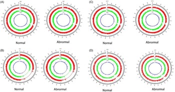
Differentially methylated regions (DMRs) are considered the marker of altered epigenetics. Usually, DMRs defined by the differentially methylated CpG sites influence gene expression and then affect biological functions. Here, the differentially methylated CpG sites were counted to identify the DMRs. In this study, at least five CpG loci were found through the differential CG methylation levels to identify DMRs, which further defined the length of chromosomes with the differentially methylated region (Figure 4).
Chromosome location of the differential methylation region in sperm from male recurrent spontaneous abortion patients (n = 4 biologically independent samples from normal people; n = 5 biologically independent samples from recurrent spontaneous patients). (A) Two differential methylation regions were located on chromosome 8. (B) One differential methylation region was located on chromosome 9. (C) One differential methylation region was located on chromosome 10. (D) One differential methylation region was located on chromosome X (n = 3 independent biological replicates).

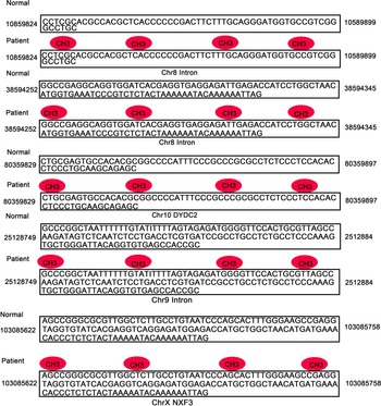
Next, we examined the differential methylation region in which the length of hypermethylated and the length of hypomethylated were analyzed. Based on the annotation depth to the gene body, the way in which methylation influences gene expression can be uncovered. The DNA methylation patterns in patient groups were located in different regions. One region was on chromosome 8 and measured 150 bp in length, while the others were on chromosomes 9, 10, and X and measured 135 bp, 68 bp, and 136 bp, respectively (Table 1). Two more genes were found to be hypermethylated in the sperm of patients, one was DYDC2 (placed in the differential methylation region of chromosome 10), and the other was NXF3 (located on chromosome X). The DMRs located on chromosomes 8 and 9 did not contain the coding sequences (Figure 5).
Number of differential methylation regions and the length of chromosomes in sperm from male recurrent spontaneous patients (n = 5 biologically independent samples). Differential methylation regions were located on chromosome 8 and their length was 150 bp, while the other located on chromosomes 9, 10, and X, and their length was 135 bp, 68 bp and 136 bp, respectively (n = 3 independent biological replicates)

The locus of the differential methylation regions on chromosomes 8, 9, 10, and X in sperm from the two groups (n = 4 biologically independent samples from the normal group, n = 5 biologically independent samples from recurrent spontaneous patients). The differential methylation regions located on the chromosomes 8 and 9 did not contain the coding sequences. Two genes were found to be hypermethylated in the DMRs, one was DYDC2 (placed in the differential methylation region of chromosome 10), the other was NXF3 (located on chromosome X) (n = 3 independent biological replicates).

To further confirm this result, we performed qRT-PCR to analyze the expression levels of the two genes. The expression level of NXF3 in the patient group was significantly lower than that in the normal group (1.36 ± 0.11 versus 3.29 ± 0.19, P < 0.001), and the expression level of DYDC2 was also significantly lower in the patient group than in the normal group (0.57 ± 0.14 versus 2.05 ± 0.32, p < 0.05) (Figure 6). These results were consistent with bisulfite sequencing.
mRNA expression levels of NXF3 and DYDC2 between the normal group (n = 19 biologically independent samples) and recurrent spontaneous patient groups (n = 19 biologically independent samples). The expression level of NXF3 in the patient group was significantly lower than that in the normal group (1.36 ± 0.11 versus 3.29 ± 0.19, P < 0.001), and the expression level of DYDC2 was also significantly lower in the patient group than in the normal group (0.57 ± 0.14 versus 2.05 ± 0.32, p < 0.05). Data are shown as the average ± standard error of the mean (SEM) (n = 3 independent biological replicates; ***P < 0.001, **P < 0.01).

Discussion
Male infertility is responsible for 50% of men’s health problems and has always been a concern for personal and social issues (Aliakbari et al., Reference Aliakbari, Taghizabet, Azizi, Rezaei-Tazangi, Samadee Gelehkolaee and Kharazinejad2022). The genetic landscape of male infertility is highly complex as semen and testis histological phenotypes are extremely heterogeneous, and at least 2000 genes are involved in spermatogenesis (Krausz and Riera-Escamilla, Reference Krausz and Riera-Escamilla2018). Genetic factors account for at least 15% of male infertility and contribute to all four of the major etiological categories of male infertility: spermatogenic quantitative defects; ductal obstruction or dysfunction; hypothalamus–pituitary axis disturbances; and spermatogenic qualitative defects (Tournaye et al., Reference Tournaye, Krausz and Oates2017). The interplay between thousands of genes, the epigenetic control of gene expression, and environmental and lifestyle factors, which influence genetic and epigenetic variants, determine the resulting male infertility phenotype. Currently, karyotyping, Y-chromosome microdeletion screening, and CFTR gene mutation tests are routinely performed to investigate possible genetic aetiology in patients with azoospermia and severe oligozoospermia. However, current testing is limited in its ability to identify a variety of genetic and epigenetic conditions that might be implicated in both idiopathic and unexplained infertility (Gunes and Esteves, Reference Gunes and Esteves2021). Diagnosing a genetic cause for infertility has obvious clinical significance as it could have implications for the reproductive health and the general health of the patient and his children. Cell-specific and timely regulation of gene expression directed by precise epigenetic modifications is essential for normal spermatogenesis (Li et al., Reference Li, Wang, Xu, Zhang and Yang2020). Therefore, in our study, whole genome sequencing was performed using the RRBS method to find differential methylation regions and candidate genes from RSA patients.
Our data showed that there is no difference in DNA base distribution between the normal group and RSA patients, implying that male recurrent spontaneous abortion did not result from gene mutation or chromosome aberration. Next, bisulfite sequencing was used to determine base depth distribution and the cytosine methylation level in promoters. Our results showed that some chromosomes (8, 9, 10, X, and Y) were hypermethylated in the sperm of male recurrent abortion patients, and identified differentially methylated sequences in DYDC2 and NXF3 genes, methylation and expression of these two genes were clearly correlated. The proportion of C+G is usually low in most genomic DNA (∼40%), but the level of methylation is very high. Hypermethylation in CpG-poor promoters without inhibition of the activity generally occurs in male gametes, subsequently resulting in the loss of CpG dinucleotides. However, CpG islands in strong methylation promoters remain during non-methylation, even under inactive conditions. The methylation state of promoters is based on gene functions and promoter sequences (Weber et al., Reference Weber, Hellmann, Stadler, Ramos, Pääbo, Rebhan and Schübeler2007). Plus, DNA methylation can impact the stability of chromosomes, and chromosomal abnormalities can affect certain developmental stages of embryos. Further analysis is needed to explore the chromosomal mechanisms affecting embryo development and implantation. Such information will help clinical assessments in prenatal diagnosis and reduce the incidence of genetically abnormal fetuses (Zhang et al., Reference Zhang, Wang, Zhao, Liu and Huang2020b).
Last, eDMR software was used to analyze the differentiation methylation region in which the gene was located. DYDC2 and NXF3 were found to be located in the differentiation methylation region. NXF3 belongs to the family of nuclear RNA export factors, which can transport mature mRNA to the cytoplasm. They have been reported to play important roles in spermatogenesis, one study found that mouse NXF3 was specifically expressed in principal cells in segment II of the caput epididymis, as well as Sertoli cells in the mouse testis, and was required to mediate transforming growth factor β (TGF-β)-induced downregulation of Tgfb3/TGF-β3 mRNA expression and protein secretion in Sertoli cells. In addition, NXF3 was also involved in TGF-β-induced transcriptional regulation of other genes associated with both Sertoli cell maturation and the restructuring of the Sertoli cell blood–testis barrier (BTB), for example GATA-binding protein 1 (Gata1), Wilms’s tumour homologue 1 (Wt1), claudin11 (Cldn11) and cyclin-dependent kinase inhibitor 1A (Cdkn1a or p21 Cip1 ). The transcriptional regulation of NXF3 was mediated through physical interaction with serine/threonine kinase receptor-associated protein (STRAP), in which NXF3 inhibited complex formation among Smad7, STRAP and activated type I TGF-β receptor (Yin et al., Reference Yin, Wang, Liang, Zhang, Liu, Li and Sun2013). DYDC2, also named Rsp2, is a 100-amino-acid (aa) protein present in various eukaryotes, which is a subunit in molecular complexes involved in X chromosome dosage compensation, histone methylation, trans-Golgi trafficking, and carcinogenesis (Gopal et al., Reference Gopal, Foster and Yang2012). In the present study, we performed RRBS, which revealed the expression of NXF3 and DYDC2 genes in semen, suggesting the possible involvement of these two genes in the pathogenesis of male infertility. We demonstrated that the increased methylation level of CpG islands in the promoter regions of the NXF3 and DYDC2 genes would affect the transcription of their genes, which in turn would inhibit the expression levels and function of the genes. The low expression of NXF3 and DYDC2 genes in the semen of the RSA group may be one of the reasons for male infertility.
In conclusion, our results showed that male infertility has a strong relationship with DNA methylation. NXF3 and DYDC2 were expressed in male spermatozoa, DNA methylation increased at the onset of male infertility, and these two genes were significantly downregulated. Although there is no difference in the mean distribution levels of the methylated base C between the normal group and the patients, men with infertility had a higher methylation level than that of the normal group. These results highlighted the potential roles of DNA methylations in male fertility. Elucidating the functions of these and other developmentally important DNA methylations will facilitate an understanding of sperm DNA methylation in human development.
Author contributions
These authors contributed equally to this work.
Financial support
This study was supported by the National Natural Science Foundation of China (81960287) and the Science and Technology Cooperation Project of Qinghai Provincial (2019-HZ-823).
Conflict of interest
The author reports no conflict of interest. The authors are responsible for the content and writing of the article.
Ethical standards
All experiments meet ethical standards.