Introduction
Molybdenum (Mo, Z = 42) is a transitional metal so named from the Neo-Latin molybdaenum, which is derived from the Ancient Greek word ‘Μόλυβδoς’, meaning ‘lead’. It is a multi-valent element whose partitioning behaviour depends on the redox state of its environment. Because the charge, size and physical properties of Mo vary depending on its oxidation state, its redox sensitivity essentially controls its geochemical behaviour. Therefore, Mo geochemistry has become an effective tool for monitoring redox conditions in, e.g. ocean basins (Dahl et al., Reference Dahl, Canfield, Rosing, Frei, Gordon, Knoll and Anbar2011), early atmospheric oxygenation (Anbar et al., Reference Anbar, Duan, Lyons, Arnold, Kendall, Creaser, Kaufman, Gordon, Scott, Garvin and Buick2007), and the impact of subducted sediments and fluids on arc magmas (Freymuth et al., Reference Freymuth, Vils, Willbold, Taylor and Elliot2015).
Molybdenum can display the oxidation states 2+, 3+, 4+, 5+, and 6+. The latter also occurs in the oxyanion (MoO4)2–. Molybdenum is the essential constituent of 87 minerals currently approved by the Commission on New Minerals, Nomenclature and Classification of the International Mineralogical Association (IMA–CNMNC), with ca. 21 of them containing the (MoO4)2– oxyanion in their crystal structure (http://cnmnc.units.it/). The small Su Seinargiu prospect in Sardinia, Italy, is a natural laboratory for the creation of Mo-bearing mineral species and probably one of the most significant locations for the study of the mineralogy of Mo in the world. More than sixty distinct mineral species have been discovered there, with the majority of them formed in response to the alteration of primary molybdenite ± bismuthinite ± bismuth assemblages (Bonaccorsi and Orlandi, Reference Bonaccorsi and Orlandi2020).
The Arabian Nubian Shield (ANS), representing a juvenile terrain (i.e. a mantle-derived crust) formed during the Pan-African Orogeny (950–550 Ma), is unique for its widespread occurrence of alkaline granites (Stoeser, Reference Stoeser1986; El-Bialy et al., Reference El-Bialy, Eliwa, Mahdy, Murata, El-Gameel, Sehsah, Omar, Kato, Fujinaga, Andresen and Thomsen2020). Some of them are highly evolved and strongly fractionated granites and host economic rare-metal deposits (Be, Nb, Ta, Zr, U, Th, Mo, Y, REE, W and Sn) (Küster, Reference Küster2009; Mahdy et al., Reference Mahdy, Iizuka, Pease, Ghoneim and Abdel–Hamid2023 and references therein). Rare-metal granite magmas were intensely affected by the circulation of magmatic–hydrothermal fluids during the ultimate stage of solidification. These magma-expelled fluids were occasionally mixed with meteoric waters (Mahdy et al., Reference Mahdy, El Kalioubi, Wohlgemuth-Ueberwasser, Shalaby and El-Afandy2015, Reference Mahdy, Ntaflos, Pease, Sami, Slobodník, Abu Steet, Abdelfadil and Fathy2020 and references therein). Fluid exsolution is associated with an exceptional increase of rare-metal concentrations and promoted concurrent pervasive granite alteration, mineral–fluid interaction, and the onset of mineral dissolution–reprecipitation processes (Abdalla, Reference Abdalla2009; Abdelfadil et al., Reference Abdelfadil, Mahdy, Ondrejka and Putiš2022). Therefore, rare-metal granites in the Arabian Nubian Shield can potentially become the source of rare and/or new mineral species. Surprisingly, only a single new mineral species has been recorded in such granites, hydroxyplumbopyrochlore, (Pb1.5□0.5)Nb2O6(OH), discovered in the Jabal Sayid peralkaline granitic complex, Saudi Arabia (Li et al., Reference Li, Li, Fan, Fan, Zhong, Jahdali, Qin, Jehani, Wang and Nahdi2020).
In this paper, we provided the full description of a new mineral species associated with rare-metal F-rich rhyolite–granite intrusions from the Um Safi area of Marsa Alam District, Central Eastern Desert, Egypt. Marsaalamite-(Y), phonetically [ˈmæɾsæˈʕælæm.aɪt], has the ideal formula Y(MoO4)(OH) and is named after the area where the mineral was discovered, the Marsa Alam District (i.e. urban and rural parts), Al-Bahr Al-Ahmer Governorate, Egypt. The mineral name includes the Levinson suffix ‘-(Y)’ to indicate the dominance of yttrium over the other lanthanides, in agreement with the nomenclature of REE mineral species (Bayliss and Levinson, Reference Bayliss and Levinson1988). Although hydroxyl REE molybdates are uncommon in nature, they have attracted considerable attention recently owing to their promising applications, especially in optics because framework structures of molybdates containing trivalent and rare earth ions can be attractive hosts for phosphors (Dorzhieva et al., Reference Dorzhieva, Sofich, Bazarov, Shendrik and Bazarova2021).
The new mineral and its name have been approved by the IMA–CNMNC under the number IMA2024–050 (Mahdy et al., Reference Mahdy, Ondrejka, Bačík, Biagioni, Sejkora, Uher, Števko, H.-J. and Mikuš2024b). The holotype specimen, represented by a one-inch polished section (NGH-1 sample), is deposited in the Museo di Storia Naturale dell’Università di Pisa, Via Roma 79, I-56011 Calci, Pisa, Italy, under the catalogue number 20074. A cotype specimen, also constituting a one-inch polished section (NGH-2B sample), is deposited in the collection of the National History Museum, Vajanského nábrežie 2, P.O. BOX 13, 810 06 Bratislava, Slovak Republic, under the catalogue number M-20414.
Occurrence
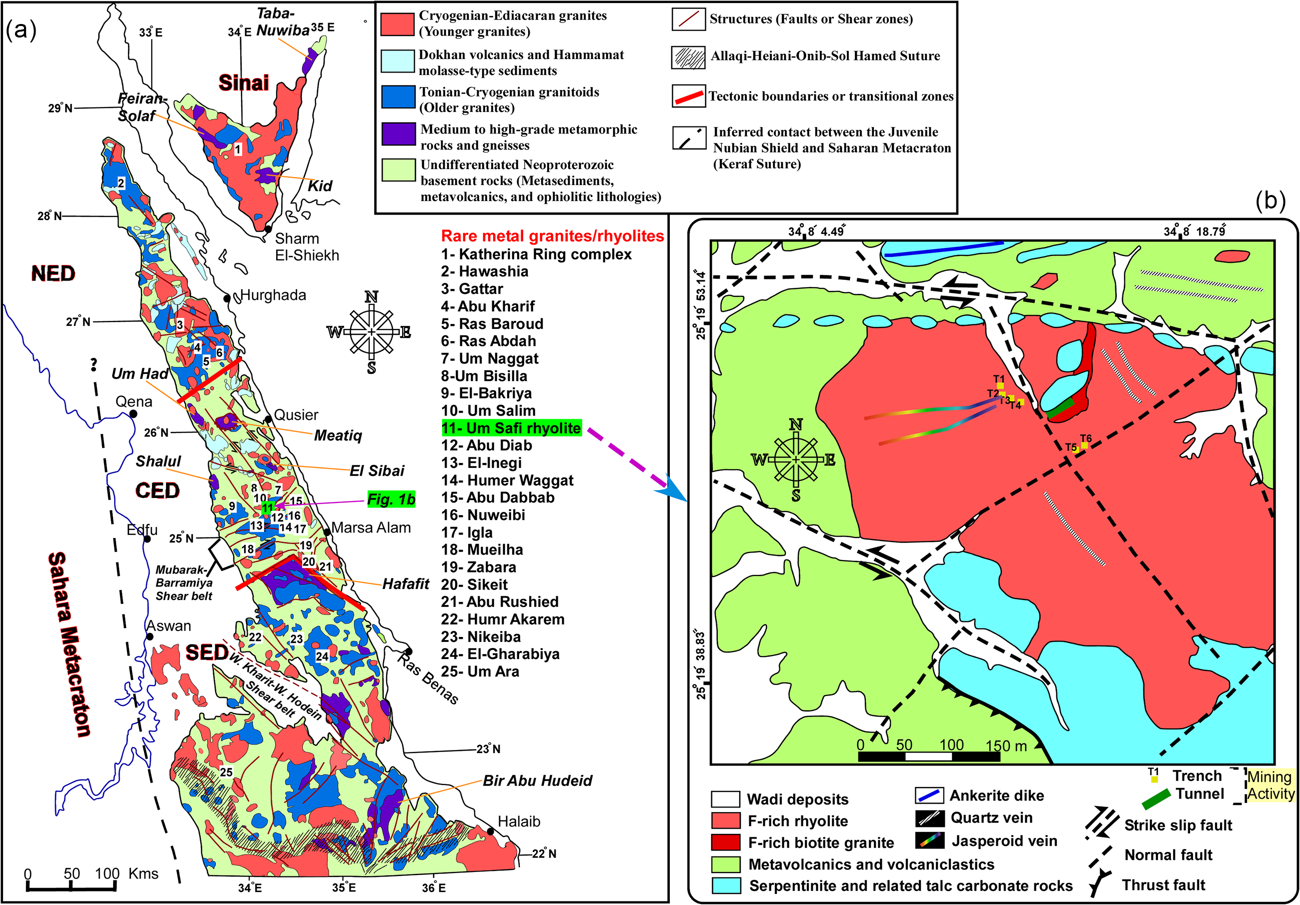
Marsaalamite-(Y) was discovered in the highly evolved (F-rich) greisenised, A-type granite of the Um Safi area of Marsa Alam District, Central Eastern Desert, Egypt. The Central Eastern Desert is the central segment of the Eastern Desert of Egypt that represents the northern segment of the Arabian Nubian Shield (Fig. 1a; Johnson et al., Reference Johnson, Andresen, Collins, Fowler, Fritz, Ghebreab, Kusky and Stern2011). The Arabian Nubian Shield is primarily composed of juvenile continental crust, which originated during the amalgamation of Gondwana and represents the northern segment of the East African Orogen (Johnson and Woldehaimanot, Reference Johnson and Woldehaimanot2003). The rock exposures of the Arabian Nubian Shield in Egypt encompass two segments, the Eastern Desert and Sinai (Fig. 1a). The Eastern Desert of Egypt is tectonically divided into three parts (Stern and Hedge, Reference Stern and Hedge1985) based on the lithology, deformation and igneous activity: Northern Eastern Desert (NED); Central Eastern Desert (CED); and Southern Eastern Desert (SED). The Central Eastern Desert is typically known for the occurrence of numerous highly evolved rare-metal granites (rich in Ta, Nb, Be, Sn, Zr, Th, Y, U, W, Mo and REE; Mahdy et al., Reference Mahdy, Ntaflos, Pease, Sami, Slobodník, Abu Steet, Abdelfadil and Fathy2020, Reference Mahdy, Iizuka, Pease, Ghoneim and Abdel–Hamid2023).
(a) Geological map of the Eastern Desert and Sinai of Egypt as the northernmost part of the Arabian Nubian Shield (ANS) showing tectonic features, rock units, distribution of rare-metal granites and location of the Um Safi area (modified after Mahdy et al., Reference Mahdy, Iizuka, Pease, Ghoneim and Abdel–Hamid2023). (b) Geological map of the Um Safi area showing the exposures of greisenised granite, F-rich rhyolite and country rocks (modified after Abouelnaga et al., Reference Abouelnaga, El-Shayeb, Mahmoud, Gaafar and Donia2015). NED = Northern Eastern Desert; CED = Central Eastern Desert; SED = Southern Eastern Desert.
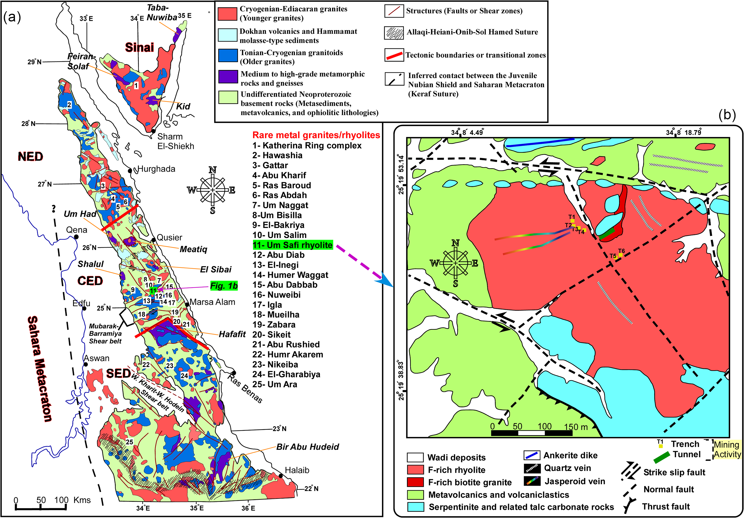
The Um Safi area is bounded by latitudes 25°20ʹ10″–25°19ʹ25″ N and longitudes 34°7ʹ40″–34°8ʹ40″ E. The exposed country rocks of the granites include mainly serpentinites and related talc-carbonate rocks, and island-arc volcanic assemblages, such as meta-andesites, volcanoclastic and volcano-sedimentary rocks (Fig. 1b). Serpentinites are the most distinctive lithological units and essential components in ophiolitic complexes of the Arabian Nubian Shield. Usually, the serpentinite blocks are altered to talc-carbonates and the island arc rocks are commonly deformed, sheared and metamorphosed up to the lower amphibolite facies. These metavolcanics in the Um Safi area are intruded by numerous sulfide-bearing quartz veins, striking predominantly NW–SE and NE–SW (Fig. 1b). Sulfur-bearing minerals occur as cavity fillings in these quartz veins and predominantly include galena and pyrite in addition to chrysocolla, anglesite and atacamite as supergene products.
The Um Safi area contains two important types of magmatic rocks: F-rich granite (smaller exposure) and F-rich rhyolite (larger exposure). The area has an elliptical shape of 620 m × 450 m (Fig. 1b). The F-rich granite is intrusive into the rhyolite (Abdalla, Reference Abdalla2001; Ibrahim, Reference Ibrahim2001). The marsaalamite-(Y)-bearing sample was collected from a granite exposure with the coordinates, latitude 25°19ʹ51″ N and longitude 34°08ʹ13″ E. The sampling location is intensively altered and sheared and contains talc serpentinite where rhyolite and granite have intersected (Fig. 1b). As a result of hydrothermal alteration, it is difficult to distinguish granite from rhyolite in the field. These rhyolite and granite rocks extend NW–SE, compatible with the main trend of faults/shear zones associated with the Najd Fault System (NFS: NW–SE left-lateral strike-slip faults and shear zones). These NW–SE shear zones were active across the Precambrian of Arabia and Egypt (Fig. 1a) at 620–580 Ma during escape tectonics in the last phase of island arc accretion (shortening) caused by the Pan-African Orogeny, and acted as pathways for ascending rare metal, post-collisional granitic magmas (e.g. Mahdy et al., Reference Mahdy, El-Arafy, El-Qassas, Metwaly, Abd El-Rahman, El-Sundolly and Said2024a). Therefore, the Um Safi F-rich rhyolite and associated F-rich granite may be genetically related, with the rhyolite representing the residual liquid of an associated, unexposed highly evolved rare-metal granite magma.
The Um Safi F-rich granite is fine to medium grained and consists of feldspars, quartz and biotite as essential mineralogical constituents. Greisenised domains within the granite are characterised by the hydrothermal alteration of feldspars into fine-grained muscovite, and of biotite into iron oxy-hydroxides and muscovite. This greisenised granite is rich in fluorite that occurs as disseminations or veinlets-filling fractures and is colourless to violet. Abundant hematitisation causes the dark pigmentation of the granitic rock. The Um Safi F-rich rhyolite is generally aphanitic and holocrystalline with subordinate glass. The highly altered zones of rhyolite are largely influenced by fluoritisation, hematitisation, sericitisation, silicification and martitisation, i.e. magnetite oxidises to hematite.
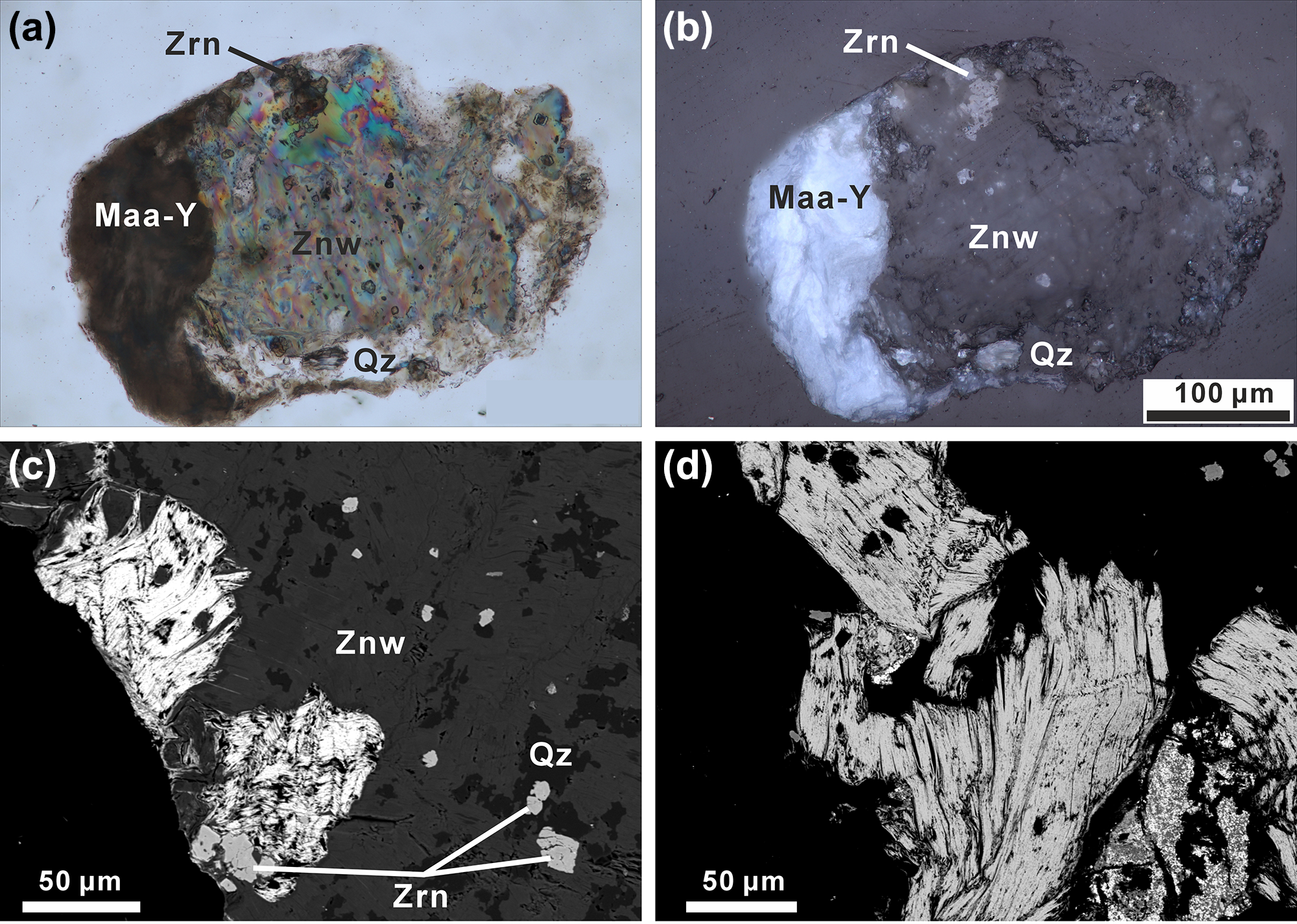
Marsaalamite-(Y) is mainly intergrown with zinnwaldite (polylithionite–siderophyllite solid solution; F = 4–6 wt.%), quartz, zircon (Fig. 2) and thorite. It is associated with arsenopyrite, As-rich thorite, baryte, bastnäsite-(Ce), cassiterite, chernovite-(Y), columbite-(Fe), fluocerite-(Ce), fluorite, iron oxy-hydroxides, löllingite, members of the hübnerite–ferberite series, members of xenotime-(Y)–thorite solid solutions, molybdenite, monazite-(Ce), pyrite, rutile, unidentified arsenates and As-bearing minerals, unidentified Th phosphate minerals, wulfenite, and Yb-rich xenotime-(Y).
Optical and back-scattered electron (BSE) microphotographs of marsaalamite-(Y) (Maa-Y) and associated minerals: (a) transmitted cross-polarised light; (b) reflected plane-polarised light. (c) Maa-Y and zinnwaldite (Znw) intergrowths and intimately associated zircon (Zrn) and quartz (Qz). (d) Mica–like sheets of Maa-Y with perfect basal cleavages, similar to textural features of mica minerals and molybdenite. Holotype specimen (NGH-1). Mineral symbols are from Warr (Reference Warr2021).

Physical and optical properties
Marsaalamite-(Y) rarely crystallised as individual grains; instead, it typically occurs as inclusions in or intergrowths with zinnwaldite (Fig. 2). It forms mica-like crystal aggregates of 0.1 to 1 mm in size. Marsaalamite-(Y) is non-magnetic, white in colour, and has an earthy lustre and white streak. It is brittle with hardness between mica and calcite (3–4 Mohs), with basal cleavage {010} and uneven micaceous fractures. The calculated density is 4.90 g.cm–3, based on the empirical formula and unit-cell parameters refined from the powder X-ray diffraction (XRD) data.
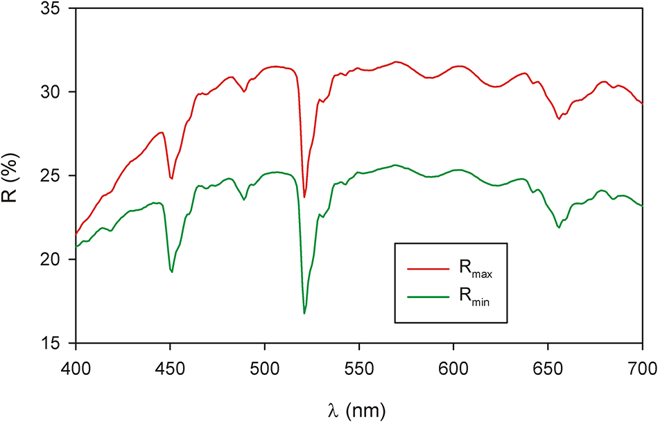
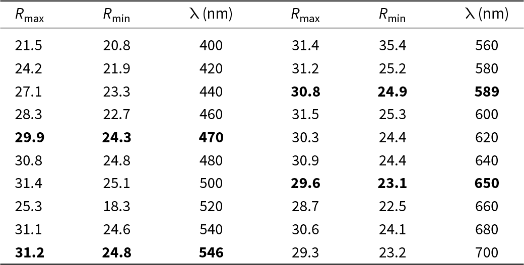
Marsaalamite-(Y) is opaque (Fig. 2a, b). In reflected light, it is greyish white and shows a distinct bireflectance, while its pleochroism and anisotropy are distinct with greyish white tints. Furthermore, no internal reflections are recognised. Reflectance values (WTiC Zeiss 370), measured in air (microspectrophotometer MSP400 Tidas at Leica microscope, objective 50×, step-size 1 nm; National Museum, Prague, Czech Republic), are listed in Table 1 (COM standard wavelengths are given in bold), and are shown in Fig. 3. The complete reflectance dataset of marsaalamite-(Y) is provided as Supplementary materials – Table S1. The character of reflectance curves for marsaalamite-(Y) with distinct minima at 451, 521 and 656 nm is uncommon. The use of other objectives (20× and 100×) provided the same results. The reflectance curves of the WTiC standard and the associated thorite show a normal appearance, excluding that analytical shortcomings are responsible for these abnormal reflectance data.
Reflectance curves for marsaalamite-(Y).

Reflectance values of marsaalamite-(Y) with COM standard wavelengths given in bold

Chemical composition
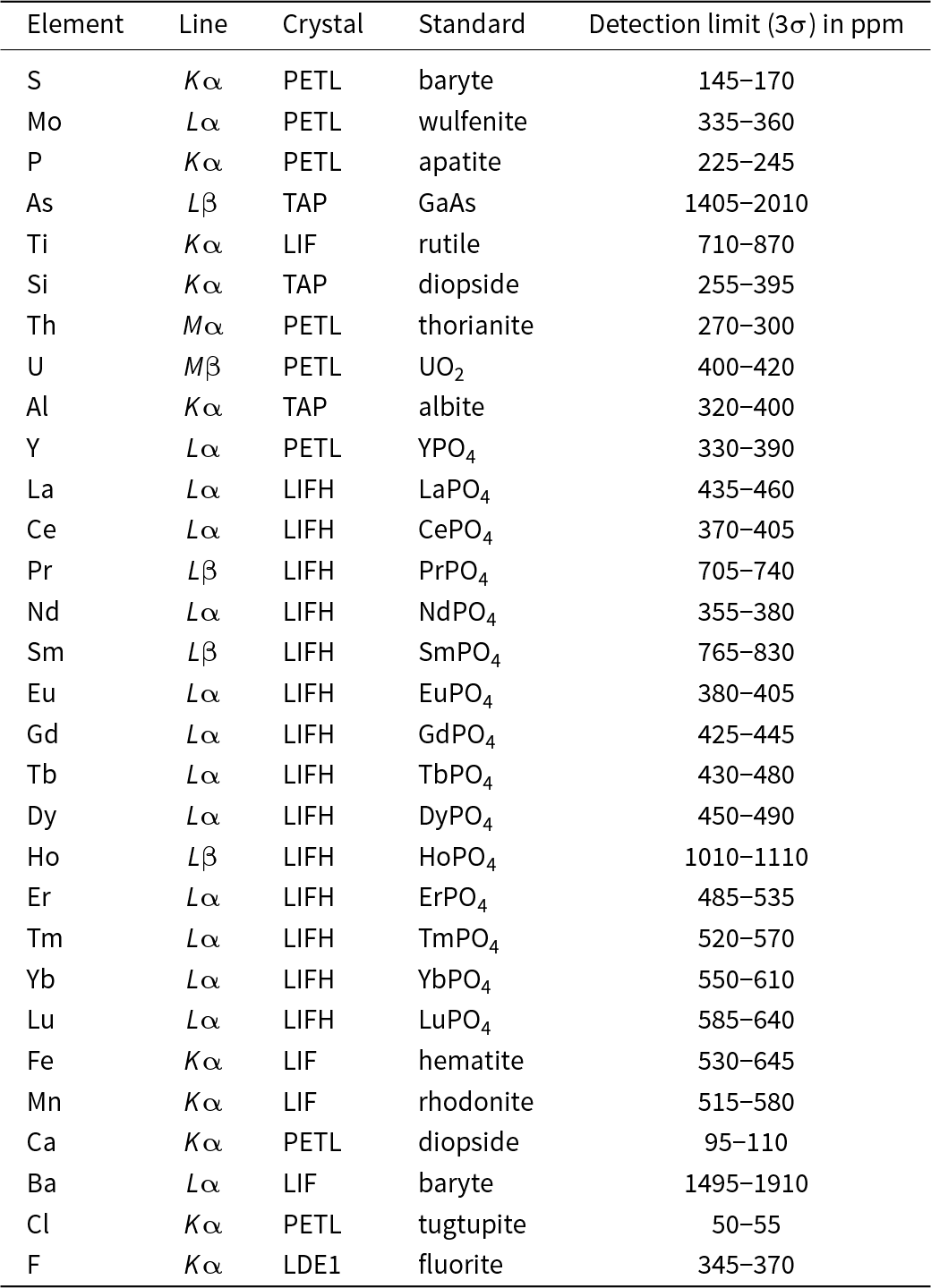
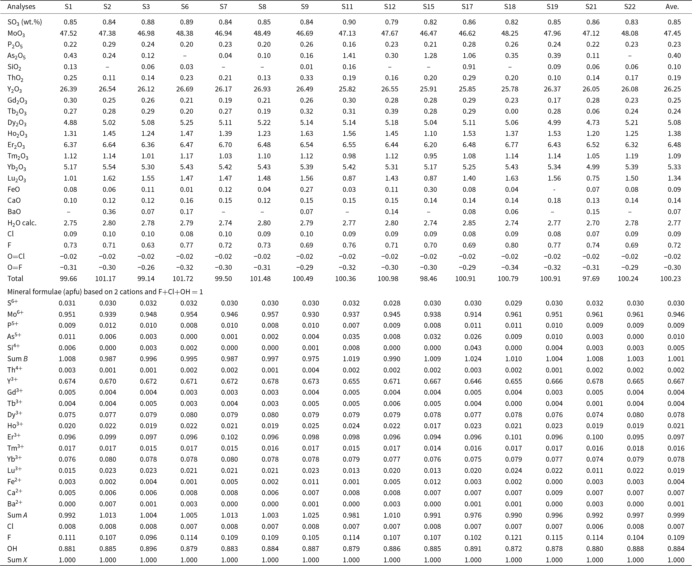
The chemical composition of marsaalamite-(Y) was determined in wavelength-dispersive (WDS) mode using a JEOL JXA-8530F electron microprobe at the Earth Science Institute, Slovak Academy of Sciences in Banská Bystrica, Slovakia. An accelerating voltage of 15 kV and a beam current of 20 nA were used. The spot beam diameter varied from 2 to 6 µm. Raw counts were converted to oxide wt.% using the ZAF matrix correction. Corrections of line interferences were performed applying the method by Åmli and Griffin (Reference Åmli and W.L1975). The detection limit for all elements is typically around 0.01 to 0.03 wt.%. The concentrations of Ti, U, Al, Mn and LREE (La to Eu) are mostly below their detection limits. Analytical conditions are summarised in Table 2 and the chemical data are reported in Table 3. The presence of (OH)– has been confirmed by Raman and infrared spectrometry (see below), and its value has been calculated from the empirical formula assuming that the (OH + F + Cl) site is completely filled, i.e. = 1 atom per formula unit (apfu).
Conditions used for the electron microprobe analyses

Chemical composition and formula of marsaalamite-(Y). Analyses numbers are abbreviated from NGH-S1, NGH-S21 etc*

* ‘–’ indicates not detected
Chemical analyses of marsaalamite-(Y) were conducted on both crystals of the holotype specimen (NGH-1) and cotype specimen (NGH-2B). The major components of marsaalamite-(Y) are Mo (46.47–48.49 wt.% MoO3, 0.91–0.96 apfu Mo), Y (25.78–26.93 wt.% Y2O3, 0.64–0.68 apfu Y), and HREE, in particular Er>Yb>Dy (6.20–6.77 wt.% Er2O3, 0.09–0.10 apfu Er; 4.99–5.54 wt.% Yb2O3, 0.07–0.08 apfu Yb; 4.73–5.25 wt.% Dy2O3, 0.07–0.08 apfu Dy). The empirical formula calculated on the basis of two cations and F+Cl+OH = 1 is (Y0.67Er0.10Dy0.08Yb0.08Ho0.02Lu0.02Tm0.02Ca0.01)Σ1.00(Mo0.95S0.03As0.01P0.01)Σ1.00O4.00[(OH)0.88F0.11Cl0.01]Σ1.00, while the simplified formula is (Y,Er,Dy,Yb)(MoO4)(OH,F). The ideal formula is Y(MoO4)(OH), which requires (in wt.%) Y2O3 42.46, MoO3 54.15, H2O 3.39, totalling to 100.
The crystals of marsaalamite-(Y) studied are chemically homogenous with no distinct variations. The chondrite-normalised patterns exhibit conspicuous HREE enrichments (Ho,Er,Tm,Yb,Lu; 14.30–16.39 wt.% HREE2O3), steep MREE (Gd,Tb,Dy; 5.07–5.85 wt.% MREE2O3), and negligible LREE (La,Ce,Pr,Nd). The REE patterns resemble those of common Y-dominated minerals.
Raman spectroscopy
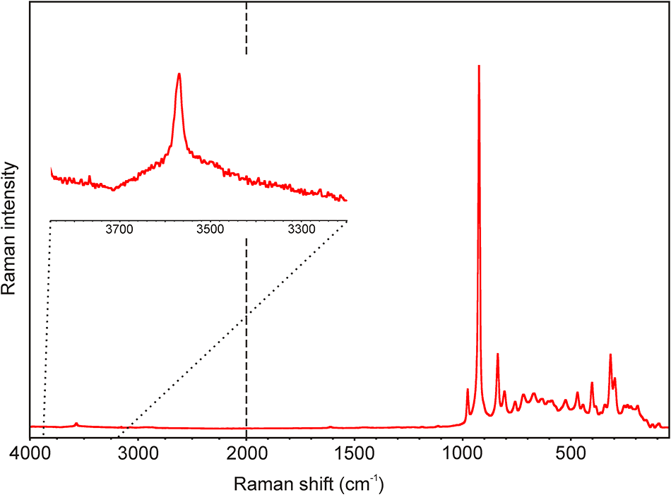
The Raman spectra of marsaalamite-(Y) were collected in the range 4000 to 50 cm–1 using a DXR-dispersive Raman Spectrometer (Thermo Scientific) mounted on a confocal Olympus microscope (National Museum, Prague, Czech Republic). The Raman signal was excited by an unpolarised red 633 nm He–Ne gas laser and detected by a CCD detector. The experimental parameters were 100× objective, 10 s exposure time, 100 exposures, 25 μm pinhole spectrograph aperture, 4 mW laser power level and 400 lines/mm grating. The spectra were repeatedly acquired from different grains to obtain a representative spectrum with the best signal-to-noise ratio. The possible thermal damage of the measured spot was excluded by visual inspection of the excited surface after measurement, by observation of possible decay of spectral features in the start of excitation and checking for thermal downshift of Raman lines. The instrument was set up by a software-controlled calibration procedure using multiple neon emission lines (wavelength calibration), multiple polystyrene Raman bands (laser-frequency calibration) and standardised white-light sources (intensity calibration). Spectral manipulations were performed using the Omnic 9 software (Thermo Scientific). The observed main bands are (in cm–1): 3570, 977, 924, 837, 806, 757, 719, 670, 632, 605, 586, 524, 469, 444, 402, 343, 316, 296, 255, 239, 222, 191, 122 and 94.
The Raman spectrum of marsaalamite-(Y) in the full range is given in Fig. 4. The rationale of its interpretation is based on the previous study of molybdates (Sejkora et al., Reference Sejkora, Čejka, Malíková, López, Xi and Frost2014, Reference Sejkora, Pauliš, Vrtiška, Pour and Dvořák2023 and references therein). A weak sharp band at 3570 cm–1 is assigned to the OH stretching vibration of the hydrogen-bonded hydroxyl groups. According to the empirical relation between energy of vibration and the corresponding bond length, i.e. the correlation equation in the form v(cm–1) = 3592–304. 109. exp(–d(O···O)/0.1321) (Libowitzky, Reference Libowitzky1999), this wavenumber corresponds to O–H···O hydrogen-bond length 3.1 Å. A very strong band at 924 cm–1 with shoulder at 977 cm–1 is assigned to the ν1 (MoO4)2– symmetric stretching vibration. The occurrence of two components of this vibration probably reflects different Mo–O lengths in the MoO4 tetrahedra. Three medium bands at 837, 806 and 757 cm–1 relate to ν3 triply degenerate antisymmetric stretching vibrations of the (MoO4)2– group. The region of stretching vibrations of the MoO4 group of marsaalamite–(Y) corresponds well to that of synthetic Y(MoO4)F (Schleid et al., Reference Schleid, Strobel, Dorhout, Nockemann, Binnemans and Hartenbach2008), with bands at 980(m), 943(vs), 866(m), 808(m) and 762(m) cm–1. Weak bands at 402 and 343 cm–1 and moderate ones at 316 and 296 cm–1 are attributed to the ν2 doubly degenerate and ν4 triply degenerate bending vibrations of the (MoO4)2– group. The series of weak bands in the range 720–440 cm–1 is probably connected with vibrations of Y(+REE)–(OH) bonds. Weak and very weak Raman bands below 260 cm–1 are associated with rotational or translational modes of the (MoO4)2– group, as well as the lattice modes.
Raman spectrum of marsaalamite-(Y) (split at 2000 cm–1).

Infrared spectroscopy
The infrared vibrational spectrum of marsaalamite-(Y) was recorded by the reflection method on a Nicolet iN10 microspectrometer (Thermo Scientific) equipped with a nitrogen cooled MCT/A detector (Nicolet CZ s.r.o., Prague, Czech Republic). Spectra over the 4000 to 670 cm–1 range were obtained from a sample area of 50 × 50 μm by the co-addition of 256 scans, with a resolution of 4 cm–1. A gold mirror was used as a reflection background. Spectra were co-added to improve the signal-to-noise ratio and were corrected using the Kramers–Kronig correction, to remove spectral distortions from diffuse reflectance measurements. Spectral manipulations were performed using the Omnic 9 software (Thermo Scientific).
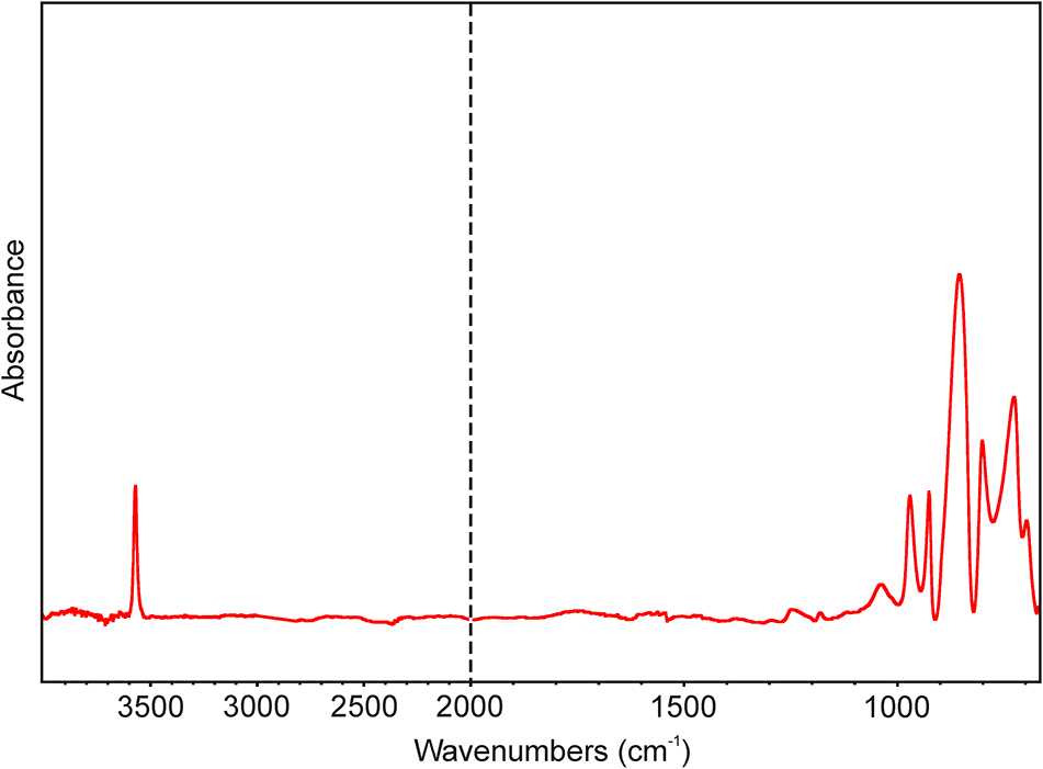
The infrared spectrum of marsaalamite-(Y) in the full range is given in Fig. 5. The main bands observed are (in cm–1): 3571, 1038, 971, 929, 845, 803, 727 and 697. A strong sharp band at 3571 cm–1 is assigned to the ν (OH) stretching vibrations of the hydrogen-bonded hydroxyl groups; its wavenumber is a good match for the corresponding Raman band (3570 cm–1). Moderate bands at 971 and 929 cm–1 are attributed to the ν1 (MoO4)2– symmetric stretching vibration. The strong band at 845 cm–1 and the medium band at 803 cm–1 are assigned to the ν3 triply degenerate antisymmetric stretching vibrations of the (MoO4)2– group. Moderate bands at 727 and 697 cm–1 are probably connected with vibrations of Y(+REE)–(OH) bonds.
Infrared spectrum of marsaalamite-(Y) (split at 2000 cm–1).

Crystallography
A single-crystal X-ray diffraction study of marsaalamite-(Y) was impossible owing to the powder-like diffraction pattern of all checked grains, in agreement with the nature of the available material.
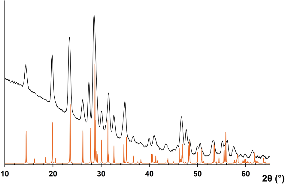
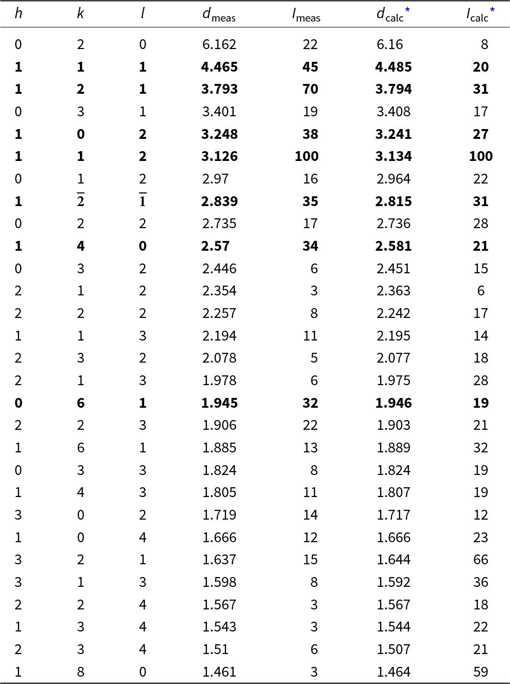
Owing to the small amount of pure material, powder X-ray diffraction data were collected using a Bruker D8 Venture single-crystal diffractometer equipped with a Photon III CCD area detector and micro-focussed CuKα radiation (Centre for Instrument Sharing of the University of Pisa, Italy), simulating a Gandolfi-like geometry. Observed X-ray diffraction lines are reported in Table 4, and Fig. 6 shows the powder X-ray diffraction pattern, along with the position of diffraction lines in isotypic Y(MoO4)F. Unit-cell parameters refined from the powder X-ray diffraction data in a monoclinic setting are, space group P21/c, a = 5.1863(7) Å, b = 12.3203(11) Å, c = 6.6953(7) Å, β = 114.173(8)°, V = 390.30(8) Å3, and Z = 4.
Powder X-ray diffraction pattern for marsaalamite-(Y) compared to the calculated pattern of Y(MoO4)F (based on the structural model of Schleid et al., Reference Schleid, Strobel, Dorhout, Nockemann, Binnemans and Hartenbach2008).

Powder X-ray diffraction data (d in Å) for marsaalamite-(Y)

Note: The seven strongest lines are given in bold.
* Calculated d and I values were obtained from Le Bail fitting of the measured powder XRD pattern in the P21/c space group.
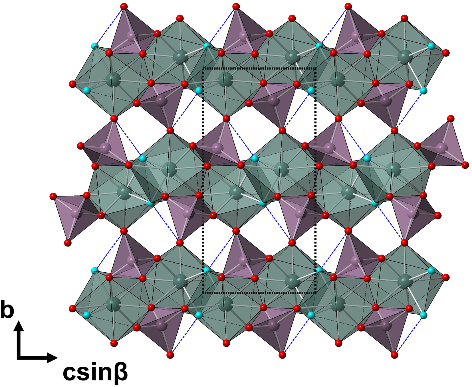
The low concentration of F and the occurrence of O–H bonds, in accord with micro-Raman and infrared data, imply that marsaalamite-(Y) is the natural (OH)-analogue of synthetic Y(MoO4)F and Y(MoO4)Cl (Schleid et al., Reference Schleid, Strobel, Dorhout, Nockemann, Binnemans and Hartenbach2008). Y(MoO4)F crystallises in a primitive cell setting with space group P21/c, whereas the lattice of Y(MoO4)Cl is face-centred with space group C2/m. Both structures contain unique crystallographic sites with Y3+ cations, which have a coordination environment of six O2– and two F– or Cl– anions. In Y(MoO4)F, the coordination environment is a square antiprism, and for Y(MoO4)Cl, a trigon-dodecahedra coordinates Y3+. The tetrahedral (MoO4)2– units of the F-compound are bound through corner-sharing to six Y-centred polyhedra, whereas those of the Cl-bearing compound are bonded to five Y-centred polyhedral, both through corner- and edge-sharing (Schleid et al., Reference Schleid, Strobel, Dorhout, Nockemann, Binnemans and Hartenbach2008). The predicted structure of marsaalamite-(Y) derived from synthetic Y(MoO4)F is shown in Fig. 7.
Structural model of marsaalamite-(Y), as seen down a, based on the structural model of synthetic Y(MoO4)F (Schleid et al., Reference Schleid, Strobel, Dorhout, Nockemann, Binnemans and Hartenbach2008). Hypothetical H-bonds are shown as dashed blue lines, assuming that (OH) groups (light blue circles) occur at the same location as F atoms; Y- and Mo-centred polyhedra are shown in grey and violet, respectively, whereas red circles represent O atoms. Dashed black lines delimit the unit cell. Drawn using CrystalMaker® software.

To distinguish between two possible unit-cell symmetries, unit-cell parameters were refined with the DiffracPlus TOPAS software using Le Bail fitting with solutions in the monoclinic and triclinic systems. The solution in the P21/c space group was the eighth best (Goodness of Fit (GoF) = 6.55), nevertheless the seven solutions can be derived from the P21/c space group by a reduction in symmetry (P2, P21) and had very similar calculated unit-cell parameters. The best solution in the face-centred space group C2 had a GoF of only 4.74 and no result was found in space group C2/m. Consequently, it is far more likely that the structure of marsaalamite-(Y) is more similar to Y(MoO4)F than to Y(MoO4)Cl. This is further expected from the similarity of Y–O (2.31–2.45 Å) and Y–F (2.24–2.31 Å) bond lengths in Y(MoO4)F. In contrast, Cl forms much longer bonds with Y (2.71–2.76 Å), causing a large distortion of YO6Cl2 polyhedra and a change in symmetry.
The Rietveld refinement of the powder XRD pattern using the structural model of synthetic Y(MoO4)F (Schleid et al., Reference Schleid, Strobel, Dorhout, Nockemann, Binnemans and Hartenbach2008) provided very similar unit-cell parameters, but with one order of magnitude larger standard uncertainties and RBragg of 99.86. The XRD pattern displays a quality insufficient for a Rietveld structural refinement. Consequently, we decided to use parameters with a smaller standard deviation obtained from Le Bail fitting in the P21/c space group, with RBragg = 3.894.
The crystallographic information file (CIF) of the synthetic Y(MoO4)F, is freely available on the Crystallography Open Database (COD, https://www.crystallography.net/cod/ , ID 4304779).
Discussion and conclusion
Origin of marsaalamite-(Y)
There is some evidence implying that marsaalamite-(Y) is probably a late-magmatic–early postmagmatic phase: (1) The Um Safi granite is a highly evolved, volatile-rich igneous system high in REE, Th, Nb, Y, Mo and As. (2) Marsaalamite-(Y) occurs as inclusions in or intergrows with F-rich zinnwaldite and is associated with zircon and thorite. (3) Crystallisation of F-rich zinnwaldite indicates increasing oxygen fugacity, fluorine and water in the environment of its formation during a late-magmatic stage and suggests extreme fractionation of the parent aqueous silicate magma, which means that marsaalamite-(Y) may be formed during a transitional magmatic–hydrothermal stage.
Like the greisenised Um Safi granite, F-rich zinnwaldite is abundant, for example, in greisenised granites and the greisen-type Sn–W–Li mineralisation from Cínovec/Zinnwald, Czech Republic/Germany (e.g. Webster et al., Reference Webster, Thomas, Förster, Seltmann and Tappen2004; Breiter et al., Reference Breiter, Ďurišová, Hrstka, Korbelová, Vaňková, Galiová, Kanický, Rambousek, Knésl, Dobeš and Dosbaba2017), or in Neogene rhyolite on the Chios Island, Greece (Mitropoulos et al., Reference Mitropoulos, Katerinopoulos and Kokkinakis1999). The exsolution of F-, Li-, and (OH)-rich mineralising hydrothermal fluids in the course of differentiation of the parental halogen-rich granitic magma triggered the greisenisation of the granite and the alteration of primary magmatic minerals. However, marsaalamite-(Y), which is intimately related to F-rich zinnwaldite, is a mineral with OH˃F, although the late exsolved mineralising fluids in the Um Safi granite were rich in F and Li.
Marsaalamite-(Y) has frequently overgrown the rim of zinnwaldite in the greisenised Um Safi granite (Fig. 2). This indicates that marsaalamite-(Y) was formed after most of the F became already fixed in zinnwaldite and other F-rich mineral phases [e.g. bastnäsite-(Ce), fluorite and fluocerite-(Ce)]. Furthermore, occurrence of marsaalamite-(Y) as inclusions in and overgrowths on zinnwaldite suggests that marsaalamite-(Y) occurred simultaneously with or immediately after the crystallisation of zinnwaldite. As a result, the formation of marsaalamite (Y) was most likely to have been controlled by fluid-induced processes rather than magmatic conditions.
Relationships to other mineral species and synthetic materials
Marsaalamite-(Y) is a member of the Nickel & Strunz subgroup 7.GB (molybdates, wolframates and niobates) with additional H2O and/or OH and the Dana subgroup 49.3 (hydrated molybdates and tungstates containing hydroxyl or halogen). It has a novel composition and it does not correspond to any unnamed mineral species.
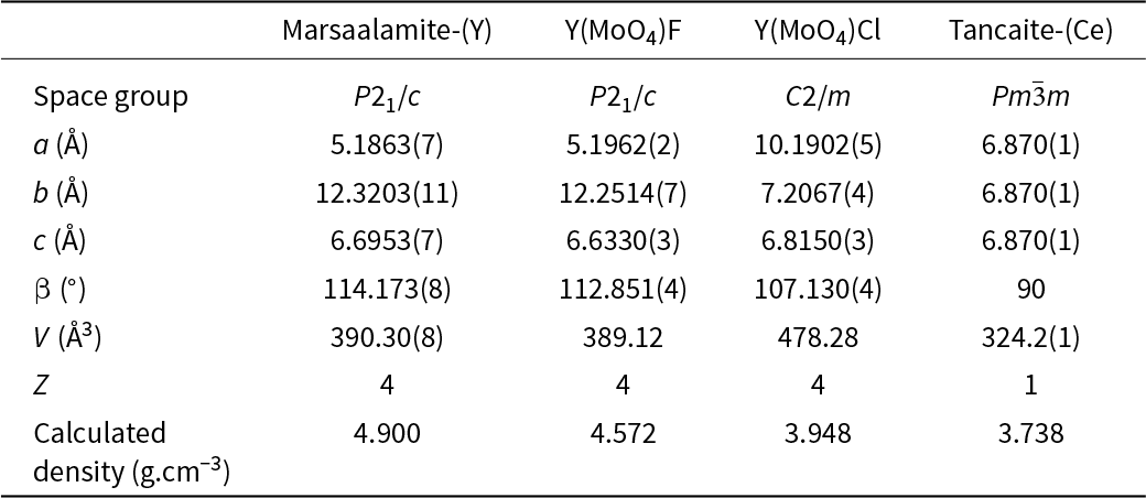
Among all known mineral species, more than 80 have molybdenum as an essential component. From these, only one other has REE as an essential component, the chemically and structurally distinct cubic tancaite-(Ce) from Su Seinargiu, Sarroch Sardinia, Italy, ideally Fe3+Ce3+(MoO4)3·3H2O (Bonaccorsi and Orlandi, Reference Bonaccorsi and Orlandi2020). In contrast, marsaalamite-(Y) is related to the crystal structure of the halide derivatives of yttrium ortho-oxomolybdate, Y(MoO4)F and Y(MoO4)Cl (Schleid et al., Reference Schleid, Strobel, Dorhout, Nockemann, Binnemans and Hartenbach2008). It is the natural (OH)-analogue of synthetic Y(MoO4)F as discussed previously. Table 5 compares these four species.
Comparison of the unit-cell parameters of marsaalamite-(Y) to synthetic Y(MoO4)F and Y(MoO4)Cl (Schleid et al., Reference Schleid, Strobel, Dorhout, Nockemann, Binnemans and Hartenbach2008) and tancaite-(Ce) (Bonaccorsi and Orlandi, Reference Bonaccorsi and Orlandi2020)

Marsaalamite-(Y) is the third mineral with a type locality in Egypt after grandfathered mineral chalconatronite (Na2Cu(CO3)2·3H2O) (Frondel and Gettens, Reference Frondel and Gettens1955) and bahariyaite (KMnO4) approved by IMA (Helmy et al., Reference Helmy, Euler, Tomaschek, Ballhaus and Friedrich2020). However, chalconatronite has been found among the corrosion products of three ancient bronze objects from Egypt, which are from the Saite-Ptolomaic Period (663–630 B.C.) and the late Coptic Period of Egypt (before the 8th century A.D.), while bahariyaite remains a highly doubtful natural occurrence and is quite possibly anthropogenic in origin.
Supplementary material
The supplementary material for this article can be found at https://doi.org/10.1180/mgm.2024.103.
Acknowledgements
The helpful comments of Peter Leverett and two anonymous reviewers, Associate Editor (Daniel Atencio) and Principal Editor (Stuart Mills) are greatly appreciated. We thank Karel Šec (Nicolet CZ s.r.o., Prague) for assistance with the FTIR measurements. The study was financially supported by the National Program of Slovakia (2023–2024) for Nasser M. Mahdy, by the Slovak Research and Development Agency under the contract no. APVV-22-0092 (2023-2026) for Martin Ondrejka, the Ministry of Culture of the Czech Republic (long-term project DKRVO 2024-2028/1.II.b; National Museum, 00023272) for Jiří Sejkora. Cristian Biagioni thanks the Centro per l’Integrazione della Strumentazione scientifica dell’Università di Pisa for the access to the single-crystal X-ray diffractometer.
Competing interests
The authors declare none.